Review
. 2021 Mar 18;22(6):3119.
doi: 10.3390/ijms22063119.

Cross-Talk among Polymorphonuclear Neutrophils, Immune, and Non-Immune Cells via Released Cytokines, Granule Proteins, Microvesicles, and Neutrophil Extracellular Trap Formation: A Novel Concept of Biology and Pathobiology for Neutrophils

Affiliations
Review

Cross-Talk among Polymorphonuclear Neutrophils, Immune, and Non-Immune Cells via Released Cytokines, Granule Proteins, Microvesicles, and Neutrophil Extracellular Trap Formation: A Novel Concept of Biology and Pathobiology for Neutrophils

Chang-Youh Tsai et al. Int J Mol Sci. .

Abstract

Polymorphonuclear neutrophils (PMNs) are traditionally regarded as professional phagocytic and acute inflammatory cells that engulf the microbial pathogens. However, accumulating data have suggested that PMNs are multi-potential cells exhibiting many important biological functions in addition to phagocytosis. These newly found novel activities of PMN include production of different kinds of cytokines/chemokines/growth factors, release of neutrophil extracellular traps (NET)/ectosomes/exosomes and trogocytosis (membrane exchange) with neighboring cells for modulating innate, and adaptive immune responses. Besides, PMNs exhibit potential heterogeneity and plasticity in involving antibody-dependent cellular cytotoxicity (ADCC), cancer immunity, autoimmunity, inflammatory rheumatic diseases, and cardiovascular diseases. Interestingly, PMNs may also play a role in ameliorating inflammatory reaction and wound healing by a subset of PMN myeloid-derived suppressor cells (PMN-MDSC). Furthermore, PMNs can interact with other non-immune cells including platelets, epithelial and endothelial cells to link hemostasis, mucosal inflammation, and atherogenesis. The release of low-density granulocytes (LDG) from bone marrow initiates systemic autoimmune reaction in systemic lupus erythematosus (SLE). In clinical application, identification of certain PMN phenotypes may become prognostic factors for severe traumatic patients. In the present review, we will discuss these newly discovered biological and pathobiological functions of the PMNs.

Keywords: antibody-dependent cellular cytotoxicity (ADCC); ectosome; exosome; low-density granulocyte; neutrophil extracellular trap (NET); polymorphonuclear myeloid-derived suppressor cell (PMN-MDSC); polymorphonuclear neutrophil; systemic lupus erythematosus (SLE); trogocytosis.


Conflict of interest statement

The authors declare no conflict of interest.

Figures

Figure 1
Figure 1
The production of cytokines/chemokines/growth factors by PMNs from normal, normal density (SLE-NDG), and low density (SLE-LDG) granulocytes of patients with SLE. The effects of these mediators on Th1/Th2 differentiation and pathological roles are summarized. IL-1ra: IL-1 receptor antagonist, NETs: neutrophil extracellular traps, CSF: colony-stimulating factor. Th: helper T; GM-CSF: granulocyte-macrophage colony stimulating factor; IFN: interferon; IL: interleukin; TGF; transforming growth factor; TNF: tumor necrosis factor. ↑ denotes increase, ↓ denotes decrease.
Figure 2
Figure 2
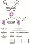
The physiological and pathophysiological roles of NADPH-dependent and NADPH-independent neutrophil extracellular traps (NETs) formation are demonstrated. Left: the web-like NETs with attached granule enzymes not only effectively trap but kill the invasive microbial pathogens as an important defense mechanism in normal physiological condition. Right: Defective NET clearance due to defective DNase-1, CRP, and C1q functions may lead to longstanding presence of the NET in the blood and tissues. These molecules may become autoantigens, damage-associated molecular patterns (DAMP)/alarmins stimulants, or coagulation system activator to induce autoimmunity, inflammatory reaction, and intravascular thrombosis. PMA: phorbol myristate acetate; LPS: lipopolysaccharide; AOSD: Adult onset Still’s disease; MS: multiple sclerosis; IBD: inflammatory bowel disease; DM: diabetes mellius; NLRP: Nucleotide-binding oligomerization domain, Leucine rich Repeat and Pyrin domain containing; APC: antigen presenting cell; ANCA: anti-neutrophil cytoplasmic antibody; SLE: systemic lupus erythematosus; RA: rheumatoid arthritis; CRP: C-reactive protein; NADPH: nicotinamide adenine dinucleotide phosphate-hydrogen ion.
Figure 3
Figure 3
Classification of neutrophil granules and their immunological functions. Different granule proteins included in the primary, secondary, and tertiary granules are summarized in brackets. The vesicles extruded from PMNs are simply divided into ectosomes and exosomes. The formation, composition, and biological/pathobiological functions of the secretary vesicles are not shown in the figure and are instead described in detail in Figure 4. MPO: myeloperoxidase; HNP: human neutrophil peptide; IL: interleukin; IFN: interferon; DC: dendritic cell; DTH: delayed-type hypersensitivity; LF: lactoferrin; CD: cluster of differentiation; CAP: cationic antimicrobial protein; LL-37: cathelicidin.
Figure 4
Figure 4
The formation, particle size, contents, and detailed immunobiological/immunopathological functions of ectosomes and exosomes extruded from PMN are demonstrated. The ectosomes are microvesicles containing granule proteins and surface membrane-expressed components such as complement receptors and CD59. The ectosomes directly detach from cell surface without deepening into cytoplasm. On the contrary, exosomes vaginate inward into the cytoplasm and take up cytoplasmic DNAs, RNAs, glycoproteins and lipids before extrusion from the cells. CR1: complement receptor type 1; MPO: myeloperoxidase; Ease: elastase; GP: glycoprotein; CD: cluster of differentiation.
Figure 5
Figure 5
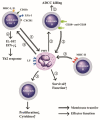
Membrane transfer among neutrophils and autologous CD4+T, allogeneic B or leukemic B cells for mediating respective effector function. (1) PMNs “trogocytose (bite up)” immunological synapse-associated molecules (HLA-I and II, CD11b, LFA-1, and CXCR1) to enhance their biological functions and survival rate. (2) PMNs trogocytose CD20-anti-CD20 complex from leukemia B cells and then kills these tumor cells by ADCC mechanism. (3) PMNs trogocytose MHC-II antigens from allogeneic B lymphocytes and then present MHC-II to allogeneic T cells to enhance proliferation and cytokine production of the allogeneic T cells, and (4) Autologous CD4+T cells trogocytose lactoferrins (LFs) expressed on PMNs to enhance IL-10 and IFN-γ production for skewing cells to Th2 immune responses. ADCC: antibody-dependent cell-mediated cytotoxicity. MHC: major histocompatibility complex; LFA: lymphocyte function-associated antigen; CD: cluster of differentiation; CXCR: cysteine-amino acid X-cysteine chemokine receptor; IFN: interferon; IL: interleukin; HLA: human leukocyte antigen.
Figure 6
Figure 6
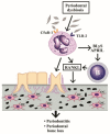
Induction of periodontitis and periodontal bone loss by PMNs initiated by periodontal dysbiosis. The periodontal dysbiotic bacteria bind to C5aR-1 and TLR-2 on PMNs. The binding stimulates BLyS and APRIL release from PMNs, which then activate B cells to produce RANKL. The RANkL binds to RANK receptors on macrophages to facilitate these cells differentiating to osteoclasts (OC). Finally, periodontitis and bone loss in the periodontal tissue occur. BLyS: B Lymphocyte Stimulator; APRIL: A PRoliferation-Inducing Ligand; RANK: Receptor Activator of Nuclear factor Kappa; RANKL: RANK ligand; TLR: Toll Like Receptor; C5aR: Complement fragment 5a receptor.
Figure 7
Figure 7
The effects of placental decidual neutrophils and neutrophil myeloid-derived suppressor cells (PMN-MDSC) on the placental angiogenesis and maternal-fetal tolerance in the first and second trimesters. NK: natural killer cells, ROS: reactive oxygen species, VEGF: vascular endothelial growth factor; CD: cluster of differentiation; IL: interleukin; FOXP3: Forkhead box P3; GARP: Glycoprotein-A Repetitions Predominant gene; CCL: cysteine-cysteine chemokine ligand.
Figure 8
Figure 8
The productive and deleterious effects of low-density granulocytes (LDG) in patients with SLE. LDGs show a tendency to form NETs and induce a number of antinuclear antibodies and anti-neutrophil cytoplasmic antibodies (ANCAs) to mediate autoimmune diseases as shown on the left side. In addition, LDGs can secrete large amount of IFN-α to facilitate CD4+T cell’s production of cytokines and to exert a lot of immunological, inflammation, and vascular effects in patients with SLE as shown on right side. IFN; interferon; LT: lymphotoxin; TNF: tumor necrosis factor; HDL: high density lipoprotein; mito-: mitochondrial.
Figure 9
Figure 9
The molecular basis of PMNs on tumor killing, tumor promotion, and immunosuppression. PMNs can mediate tumor killing by way of anti-tumor antibody or anti-immunological checkpoint antibody (anti-PD-L1) via ADCC (A). Furthermore, the ADCC killing activity can augment polymorphonuclear myeloid-derived suppressor cells (PMN-MDSC with surface markers of CD11b+-Gr-1+), which release arginase and suppressive-type exosomes containing miR-29a-3p and miR-93-5p to potently suppress NK, T cells with TCRx, Th1 and Th17 subpopulations whereas Treg is conversely activated (B). These suppressive effects on immune-related cells facilitate immunosuppression and tumor promotion. TCR: T cell receptor; PD: programmed cell death protein; PD-L: programmed cell death protein receptor; NK: natural killer cell; IFN: interferon, TNF: tumor nerosis factor; miR: microRNA; Gr: granulocytic marker; STAT: signal transducer and activator of transcription; T-bet: T box expressed in T cell transcription factor; Treg: regulatory T cell.
Figure 10
Figure 10
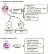
The molecular mechanism of neutrophils in inflammation resolution and wound healing. (A) Neutrophils release annexin A1 and lipoxin A4 as inflammation-resolution factors to suppress the inflammatory functions of PMNs, macrophages and mast cells. In addition, PMN-released ectosomes (Ecto), and exosomes (Exo) containing miR-223 also play important roles in suppressing inflammation. (B) Neutrophil-released hypoxia-induced factor-α (HIF-α) mediates a crucial role in tissue homeostasis and wound healing through induction of many tissue repair molecules after inflammation resolution. ECM: extracellular matrix; ICAM: intercellular adhesion molecule; miR: microRNA; M2: type 2 macrophage.
Figure 11
Figure 11
The interactions between neutrophils and platelets in hemostasis, thrombosis, systemic inflammatory vasculitis, and atherogenesis via binding between adhesion molecules (P-selectin, β2-integrin and β3-integrin) on platelets and P-selectin glycoprotein ligand-1 (PSGL-1) on neutrophils. After ligand-receptor binding, the released mediators including ROS, LTB4, and lysosomal enzymes can induce inflammatory vasculitis, whereas other factors including IL-8, VEGF and elastase can facilitate atheroma plaque development and atherogenesis in the blood vessels. LTB4: leukotriene B4; VEGF: vascular endothelial growth factor; ROS: reactive oxygen species; PL: platelet.

Similar articles

Cited by

References

    1. Kudo C., Yamashita T., Araki A., Terashita M., Watanabe T., Atsumi M., Tamura M., Sendo F. Modulation of In Vivo immune response by selective depletion of neutrophils using a monoclonal antibody, RP-3. I. Inhibition by RP-3 treatment of the priming and effector phases of delayed type hypersensitivity to sheep red blood cells in rats. J. Immunol. 1993;150:3728–3738. - PubMed
    1. Tanaka E., Sendo F. Abrogation of tumor-inhibitory MRC-OX8+ (CD8+) effector T-cell generation in rats by selective depletion of neutrophils In Vivo using a monoclonal antibody. Int. J. Cancer. 1993;54:131–136. doi: 10.1002/ijc.2910540121. - DOI - PubMed
    1. Tamura M., Sekiya S., Terashita M., Sendo F. Modulation of the in vivo immune response by selective depletion of neutrophils using a monoclonal antibody, RP-3. III. Enhancement by RP-3 treatment of the anti-sheep red blood cell plaque-forming cell response in rats. J. Immunol. 1994;153:1301–1308. - PubMed
    1. Brinkmann V., Reichard U., Goosmann C., Fauler B., Uhlemann Y., Weiss D.S., Weinrauch Y., Zychlinsky A. Neutrophil extracellular traps kill bacteria. Science. 2004;303:1532–1535. doi: 10.1126/science.1092385. - DOI - PubMed
    1. Christoffersson G., Vågesjö E., Vandooren J., Lidén M., Massena S., Reinert R.B., Brissova M., Powers A.C., Opdenakker G., Phillipson M. VEGF-A recruits a proangiogenic MMP-9-delivering neutrophil subset that induces angiogenesis in transplanted hypoxic tissue. Blood. 2012;120:4653–4662. doi: 10.1182/blood-2012-04-421040. - DOI - PMC - PubMed

LinkOut - more resources