. 2021 Mar 8;13(5):1158.
doi: 10.3390/cancers13051158.

Downregulation of Snail by DUSP1 Impairs Cell Migration and Invasion through the Inactivation of JNK and ERK and Is Useful as a Predictive Factor in the Prognosis of Prostate Cancer

Affiliations

Downregulation of Snail by DUSP1 Impairs Cell Migration and Invasion through the Inactivation of JNK and ERK and Is Useful as a Predictive Factor in the Prognosis of Prostate Cancer

Desirée Martínez-Martínez et al. Cancers (Basel). .

Abstract

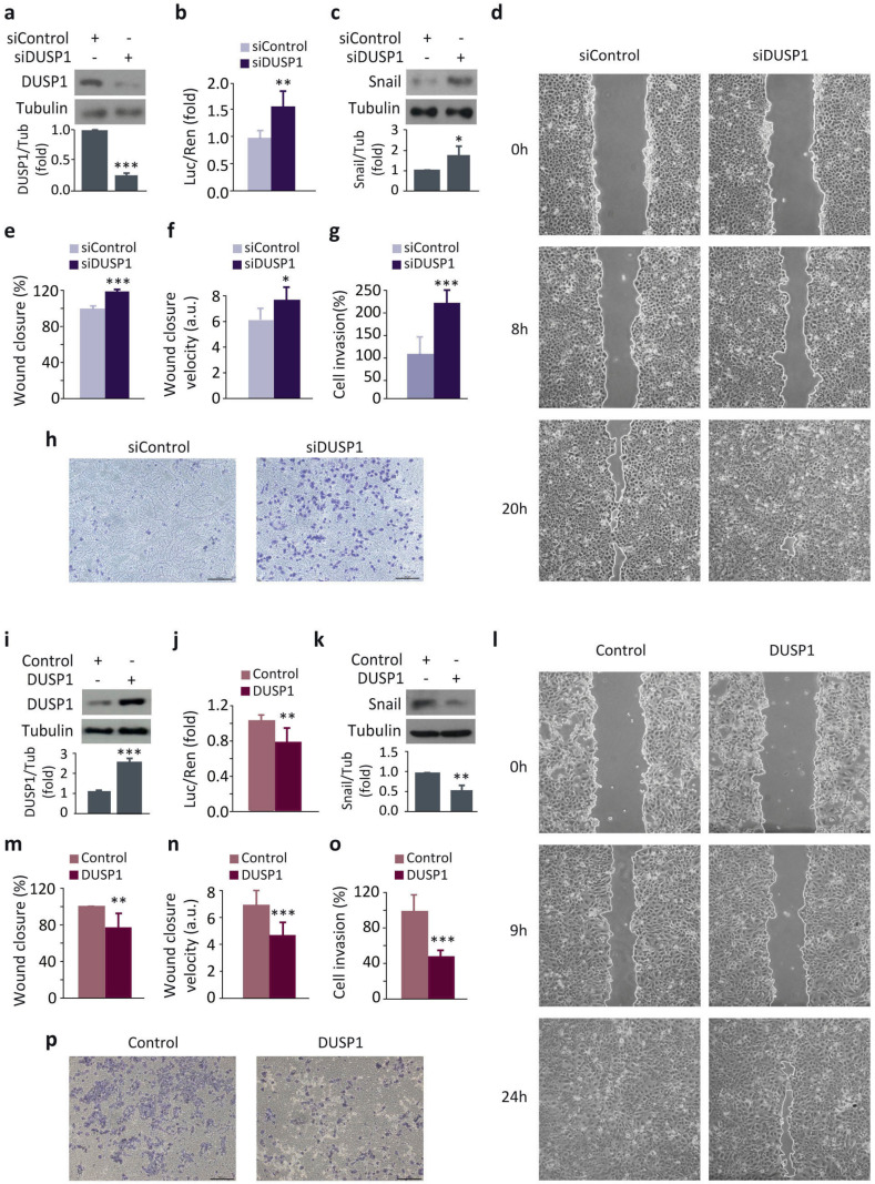
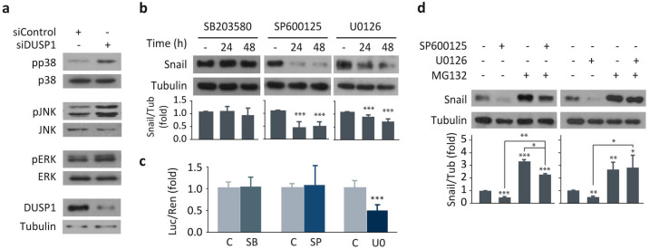
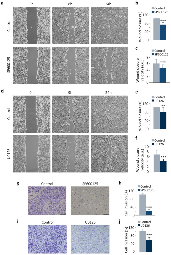
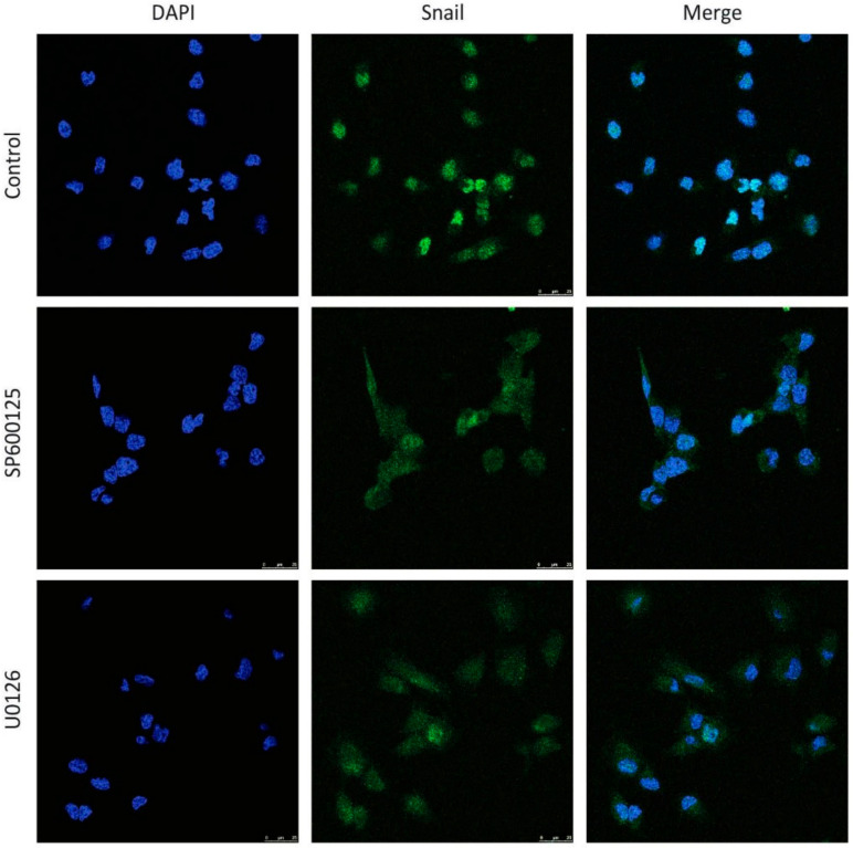
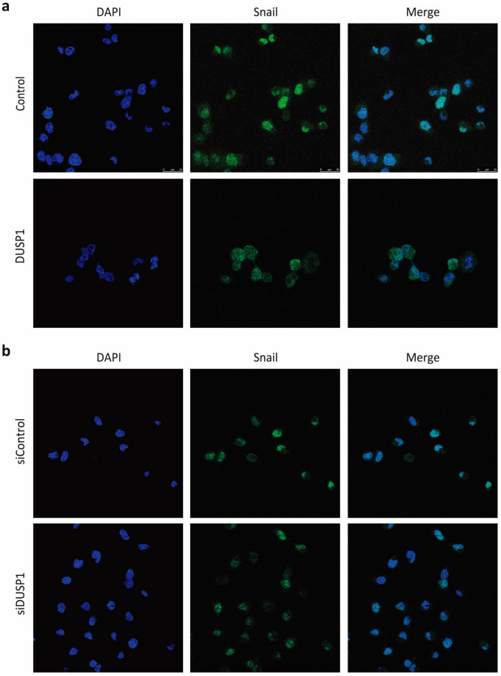
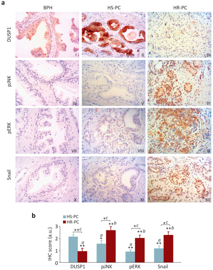
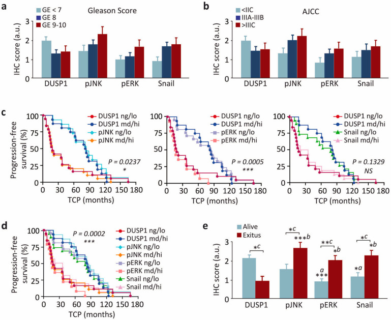
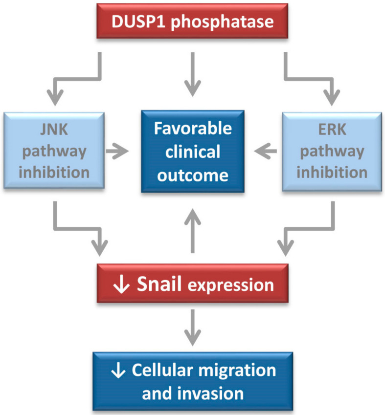
Dual specificity phosphatase 1 (DUSP1) is crucial in prostate cancer (PC), since its expression is downregulated in advanced carcinomas. Here, we investigated DUSP1 effects on the expression of mesenchymal marker Snail, cell migration and invasion, analyzing the underlying mechanisms mediated by mitogen-activated protein kinases (MAPKs) inhibition. To this purpose, we used different PC cells overexpressing or lacking DUSP1 or incubated with MAPKs inhibitors. Moreover, we addressed the correlation of DUSP1 expression with Snail and activated MAPKs levels in samples from patients diagnosed with benign hyperplasia or prostate carcinoma, studying its implication in tumor prognosis and survival. We found that DUSP1 downregulates Snail expression and impairs migration and invasion in PC cells. Similar results were obtained following the inhibition of c-Jun N-terminal kinase (JNK) and extracellular-signal-regulated kinase (ERK). In clinical samples, we evidenced an inverse correlation between DUSP1 expression and Snail levels, which are further associated with JNK and ERK activation. Consequently, the pattern DUSP1high/activated JNKlow/activated ERKlow/Snaillow is associated with an overall extended survival of PC patients. In summary, the ratio between DUSP1 and Snail expression, with additional JNK and ERK activity measurement, may serve as a potential biomarker to predict the clinical outcome of PC patients. Furthermore, DUSP1 induction or inhibition of JNK and ERK pathways could be useful to treat PC.

Keywords: DUSP1; MAPK; Snail; biomarkers; migration and invasion; patient survival; prostate cancer.


Conflict of interest statement

The authors declare no conflict of interest.

Figures

Figure 1
Figure 1
DUSP1 downregulates Snail expression and impairs cell migration and invasion in DU145 cells. (a) Cells were transfected for 48 h with the control siRNA (siControl) or the DUSP1 siRNA (siDUSP1) and expression levels of DUSP1 and Tubulin were determined by western blotting. (b) Cells were transfected for 48 h with the siControl or the siDUSP1 together with the Snail-Luc plasmid and luciferase activity was measured in cell extracts. (c) Cells were transfected as in a and expression levels of Snail and Tubulin were determined by western blotting. (df) Wound healing assay and measurement of wound closure area and velocity in cells transfected as in a. (g,h) Invasion capacity using transwell assays in cells transfected as in a. (i) Cells were transfected with a control vector (Control) or a vector encoding DUSP1 (DUSP1) and expression levels of DUSP1 and Tubulin were determined by western blotting. (j) Cells were transfected for 48 h with the Control or the DUSP1 vectors together with the Snail-Luc plasmid and luciferase activity was measured in cell extracts. (k) Cells were transfected with the Control or the DUSP1 vectors and expression levels of Snail and Tubulin were determined by western blotting. (ln) Wound healing assay and measurement of wound closure area and velocity in cells transfected as in i. (o,p) Invasion capacity using transwell assays in cells transfected as in i. For all the results, data are shown as the mean ± SEM of at least three independent experiments. For migration and invasion assays, pictures are from one representative experiment of three with similar results. Student’s t test: * 0.01 < p < 0.05; ** 0.001 < p < 0.01; *** p < 0.001.
Figure 2
Figure 2
The inhibition of JNK and ERK downregulates Snail expression in DU145 cells. (a) Cells were transfected for 48 h with the siControl or the siDUSP1 and expression levels of DUSP1, phosphorylated MAPKs (pp38, pJNK, pERK), total MAPKs and Tubulin were determined by western blotting. (b) Cells were incubated at different times in the absence or presence of 1 μM SB203580 (SB), 10 μM SP600125 (SP) or 20 μM U0126 (U0), and expression levels of Snail and Tubulin were determined by western blotting. (c) Cells were transfected with the Snail-Luc plasmid, incubated for 48 h as in b and luciferase activity was assayed in cell extracts. (d) Cells were incubated for 48 h with 10 μM SP600125 or 20 μM U0126, treated in the absence or presence of 10 μM MG132 for the last 4 h and expression levels of Snail and Tubulin were determined by western blotting. For all the results, data are shown as the mean ± SEM of at least three independent experiments. Student’s t test: * 0.01 < p < 0.05; ** 0.001 < p < 0.01; *** p < 0.001.
Figure 3
Figure 3
The inhibition of JNK and ERK decreases migration and invasion in DU145 cells. (af) Wound healing assay and measurement of wound closure area and velocity in cells incubated for 48 h with 10 μM SP600125 (ac) or 20 μM U0126 (df). (gj) Invasion capacity using transwell assays in cells incubated as above. For all the results, data are shown as the mean ± SEM of at least three independent experiments. Pictures are from one representative experiment of three with similar results. Student’s t test: ** 0.001 < p < 0.01; *** p < 0.001.
Figure 4
Figure 4
Snail subcellular location is regulated by the JNK and ERK signaling pathways. DU145 cells were incubated for 48 h with 10 μM SP600125 or 20 μM U0126 and Snail subcellular location was determined by immunofluorescence as described in Material and methods. DAPI was used to identify the nuclei. Pictures are from one representative experiment of three with similar results.
Figure 5
Figure 5
Snail subcellular location is regulated by the phosphatase DUSP1. (a) DU145 cells were transfected for 48 h with the Control or the DUSP1 vectors. (b) Cells were transfected for 48 h with the siControl or the siDUSP1. In both set of experiments, Snail subcellular location was determined by immunofluorescence as described in Material and methods. DAPI was used to identify the nuclei. Pictures are from one representative experiment of three with similar results.
Figure 6
Figure 6
JNK and ERK cooperatively regulate Snail expression, cell migration and invasion in DU145 cells. Cells were incubated in the absence (C) or presence of 10 μM SP600125 (SP, 24 h) and 20 μM U0126 (U0, 48 h). (a) Expression levels of Snail and Tubulin were determined by western blotting. (bd) Wound healing assay and measurement of wound closure area and velocity. (e,f) Invasion capacity using transwell assays. For all the results, data are shown as the mean ± SEM of at least three independent experiments. For migration and invasion assays, pictures are from one representative experiment of three with similar results. Student’s t test: * 0.01 < p < 0.05; ** 0.001 < p < 0.01; *** p < 0.001.
Figure 7
Figure 7
DUSP1 expression inversely correlates with Snail levels and activated JNK and ERK in human prostate samples. (a) Immunohistochemical analysis of expression levels of DUSP1 (I–III), phosphorylated JNK (pJNK, IV–VI), phosphorylated ERK (pERK, VII–IX) and Snail (X–XII) from human prostate cancer samples. Micrographs were taken at 200× magnification and show serial sections from the same gland stained with each one of the four used antibodies. (b) Immunohistochemical score for DUSP1, pJNK, pERK and Snail in samples from HS-PC and HR-PC. The statistical analysis was performed with One-way ANOVA and Dunnet´s multiple comparison test, and asterisks show the statistical significance of differences between the groups (a: comparison with DUSP1 from HS-PC samples; b: comparison with DUSP1 from HR-PC samples; c: HS-PC vs HR-PC for each marker), * 0.01 < p < 0.05; ** 0.001 < p < 0.01.
Figure 8
Figure 8
The relationship of DUSP1 and Snail levels and JNK and ERK activities are associated with disease progression and clinical outcome in patients with prostate cancer. (a,b) Immunohistochemical score for DUSP1, phosphorylated JNK and ERK (pJNK and pERK) and Snail in samples ranged into three categories based on their Gleason Score (a) or AJCC group staging at diagnosis (b). (c) Progression-free survival of patients showing immunohistochemical score for DUSP1/pJNK, DUSP1/pERK or DUSP1/Snail. Samples were ranged into two categories based on the staining pattern of the majority of tumor cells in the whole section (negative/low (ng/lo); moderate/high (md/hi)). (d) Progression-free survival of patients showing immunohistochemical score for DUSP1/pJNK/pERK/Snail. Samples were ranged into two categories as described in c. (e) Immunohistochemical score for DUSP1, pJNK, pERK and Snail in samples from patients either alive or dead. The statistical analysis was performed with One-way ANOVA and Dunnet´s multiple comparison test, and asterisks show the statistical significance of differences between the groups (a: comparison with DUSP1 from HS-PC samples; b: comparison with DUSP1 from HR-PC samples; c: HS-PC vs HR-PC for each marker). TCP, Time to clinical progression, * 0.01 < p < 0.05; ** 0.001 < p < 0.01; *** p < 0.001.
Figure 9
Figure 9
The phosphatase DUSP1 regulates metastasis-associated events in prostate cancer. This study demonstrate that DUSP1 overexpression downregulates Snail levels and decreases cell migration and invasion. Moreover, DUSP1 inactivates ERK and JNK pathways, whose inhibition exert similar effects on Snail expression, cell migration and invasion than overexpression of the phosphatase. In addition, JNK and ERK cooperate to regulate Snail expression, cell migration and invasion through different mechanisms. Finally, in clinical samples, the expression pattern DUSP1high/activeJNKlow/activeERKlow/Snaillow is associated with overall extended survival of patients and may serve as potential biomarker for identifying patients with favorable clinical outcome.

Similar articles

Cited by

References

    1. Bray F., Ferlay J., Soerjomataram I., Siegel R.L., Torre L.A., Jemal A. Global cancer statistics 2018: GLOBOCAN estimates of incidence and mortality worldwide for 36 cancers in 185 countries. CA Cancer J. Clin. 2018;68:394–424. doi: 10.3322/caac.21492. - DOI - PubMed
    1. Odero-Marah V., Hawsawi O., Henderson V., Sweeney J. Epithelial-Mesenchymal Transition (EMT) and Prostate Cancer. Adv. Exp. Med. Biol. 2018;1095:101–110. doi: 10.1007/978-3-319-95693-0_6. - DOI - PubMed
    1. Pei D., Shu X., Gassama-Diagne A., Thiery J.P. Mesenchymal-epithelial transition in development and reprogramming. Nat. Cell Biol. 2019;21:44–53. doi: 10.1038/s41556-018-0195-z. - DOI - PubMed
    1. Nieto M.A., Huang R.Y., Jackson R.A., Thiery J.P. Emt: 2016. Cell. 2016;166:21–45. doi: 10.1016/j.cell.2016.06.028. - DOI - PubMed
    1. Thiery J.P., Acloque H., Huang R.Y., Nieto M.A. Epithelial-mesenchymal transitions in development and disease. Cell. 2009;139:871–890. doi: 10.1016/j.cell.2009.11.007. - DOI - PubMed

LinkOut - more resources