The frontier of live tissue imaging across space and time
- PMID: 33798422
- PMCID: PMC8034393
- DOI: 10.1016/j.stem.2021.02.010
The frontier of live tissue imaging across space and time
Abstract
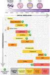
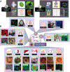
What you see is what you get-imaging techniques have long been essential for visualization and understanding of tissue development, homeostasis, and regeneration, which are driven by stem cell self-renewal and differentiation. Advances in molecular and tissue modeling techniques in the last decade are providing new imaging modalities to explore tissue heterogeneity and plasticity. Here we describe current state-of-the-art imaging modalities for tissue research at multiple scales, with a focus on explaining key tradeoffs such as spatial resolution, penetration depth, capture time/frequency, and moieties. We explore emerging tissue modeling and molecular tools that improve resolution, specificity, and throughput.
Copyright © 2021 Elsevier Inc. All rights reserved.
Conflict of interest statement
Declaration of interests The authors declare no competing interests.
Figures


Similar articles
-
Assessment and statistical modeling of the relationship between remotely sensed aerosol optical depth and PM2.5 in the eastern United States.Res Rep Health Eff Inst. 2012 May;(167):5-83; discussion 85-91. Res Rep Health Eff Inst. 2012. PMID: 22838153
-
In Vivo Observations of Rapid Scattered Light Changes Associated with Neurophysiological Activity.In: Frostig RD, editor. In Vivo Optical Imaging of Brain Function. 2nd edition. Boca Raton (FL): CRC Press/Taylor & Francis; 2009. Chapter 5. In: Frostig RD, editor. In Vivo Optical Imaging of Brain Function. 2nd edition. Boca Raton (FL): CRC Press/Taylor & Francis; 2009. Chapter 5. PMID: 26844322 Free Books & Documents. Review.
-
Cellular mechanisms of epithelial stem cell self-renewal and differentiation during homeostasis and repair.Wiley Interdiscip Rev Dev Biol. 2020 Jan;9(1):e361. doi: 10.1002/wdev.361. Epub 2019 Aug 29. Wiley Interdiscip Rev Dev Biol. 2020. PMID: 31468728 Review.
-
Tissue stem cells: definition, plasticity, heterogeneity, self-organization and models--a conceptual approach.Cells Tissues Organs. 2002;171(1):8-26. doi: 10.1159/000057688. Cells Tissues Organs. 2002. PMID: 12021488
-
Modulation of human multipotent and pluripotent stem cells using surface nanotopographies and surface-immobilised bioactive signals: A review.Acta Biomater. 2016 Nov;45:31-59. doi: 10.1016/j.actbio.2016.08.054. Epub 2016 Sep 3. Acta Biomater. 2016. PMID: 27596488 Review.
Cited by
-
Connecting past and present: single-cell lineage tracing.Protein Cell. 2022 Nov;13(11):790-807. doi: 10.1007/s13238-022-00913-7. Epub 2022 Apr 19. Protein Cell. 2022. PMID: 35441356 Free PMC article. Review.
-
Visualizable and Lubricating Hydrogel Microspheres Via NanoPOSS for Cartilage Regeneration.Adv Sci (Weinh). 2023 May;10(15):e2207438. doi: 10.1002/advs.202207438. Epub 2023 Mar 27. Adv Sci (Weinh). 2023. PMID: 36973540 Free PMC article.
-
Using Ex Vivo Live Imaging to Investigate Cell Divisions and Movements During Mouse Dental Renewal.J Vis Exp. 2023 Oct 27;(200):10.3791/66020. doi: 10.3791/66020. J Vis Exp. 2023. PMID: 37955380 Free PMC article.
-
Growth factor dependency in mammary organoids regulates ductal morphogenesis during organ regeneration.Sci Rep. 2022 May 3;12(1):7200. doi: 10.1038/s41598-022-11224-6. Sci Rep. 2022. PMID: 35504930 Free PMC article.
-
Imagining the future of optical microscopy: everything, everywhere, all at once.Commun Biol. 2023 Oct 28;6(1):1096. doi: 10.1038/s42003-023-05468-9. Commun Biol. 2023. PMID: 37898673 Free PMC article. Review.
References
-
- Baptista PM, Siddiqui MM, Lozier G, Rodriguez SR, Atala A, and Soker S (2011). The use of whole organ decellularization for the generation of a vascularized liver organoid. Hepatology (Baltimore, Md) 53, 604–617. - PubMed
Publication types
MeSH terms
Grants and funding
LinkOut - more resources
Full Text Sources
Other Literature Sources

