

Clinical Spectrum and Outcomes of Ocular and Periocular Complications following External-Beam Radiotherapy for Inoperable Malignant Maxillary Sinus Tumors
- PMID: 33796515
- PMCID: PMC7989770
- DOI: 10.1159/000511011
Clinical Spectrum and Outcomes of Ocular and Periocular Complications following External-Beam Radiotherapy for Inoperable Malignant Maxillary Sinus Tumors
Abstract
Purpose: To highlight the clinical spectrum, management, and outcomes of ocular/periocular complications following high-dose external-beam radiotherapy (EBRT) for inoperable malignant maxillary sinus-involving tumors (MMST).
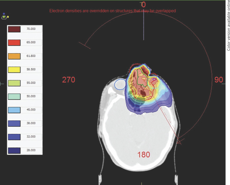
Methods: A retrospective, interventional case series. All patients who were diagnosed with inoperable MMST (with orbital involvement) and treated with high-dose fractionated EBRT (65 Gy in 30 fractions) at James Cook University Hospital, UK, were included.
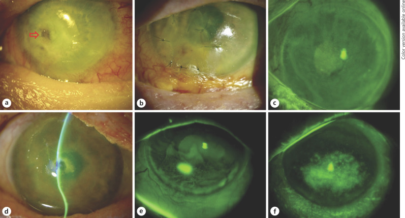
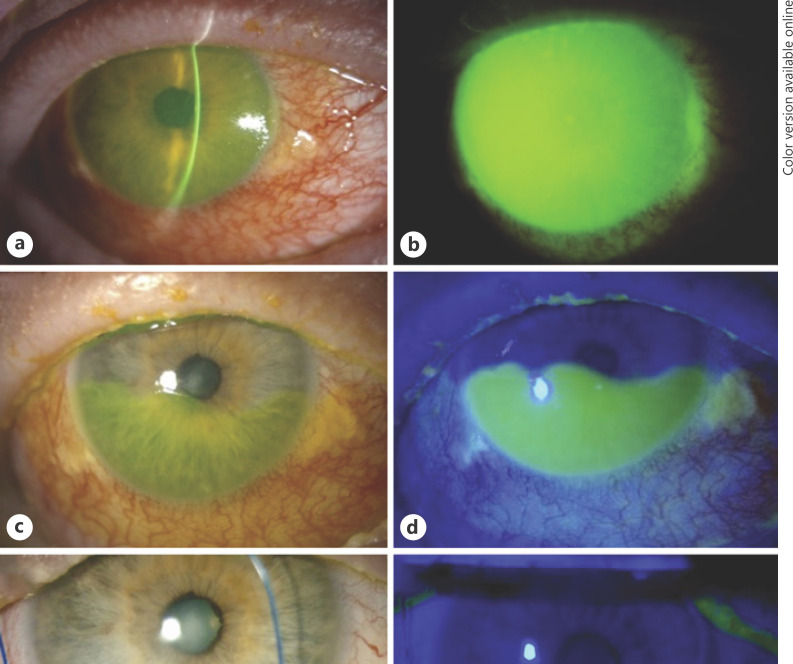
Results: Seven patients with advanced MMST (T4aN0M0-T4bN2cM0) were included and were followed up for 23.8 ± 10.2 months. Severe lid margin disease, dry eye, and neurotrophic keratopathy were universally observed. Other complications included cicatricial conjunctivitis (71%), corneal perforation (57%), limbal stem cell deficiency (LSCD; 43%), glaucoma (29%), and superimposed candida keratitis (14%). Amniotic membrane transplant (AMT; 71%), tarsorrhaphy (43%), tectonic keratoplasty (29%), and evisceration (14%) were warranted. Intact corneal epithelium was observed in all patients and good corrected-distance visual acuity (≥20/60) was observed in 3 (43%) patients at final follow-up.
Conclusion: High-dose EBRT for inoperable MMST can lead to a wide array of severe ocular/periocular complications. AMT serves as a potentially useful treatment modality to restore the ocular surface integrity after severe radiation keratopathy. We advocate active monitoring for any evolving ophthalmic complications during and after EBRT to enable timely intervention.
Keywords: Amniotic membrane; Limbal stem cell deficiency; Maxillary tumor; Neurotrophic keratopathy; Paranasal sinus tumor; Radiation keratopathy; Radiotherapy.
Copyright © 2020 by S. Karger AG, Basel.
Conflict of interest statement
The authors have no conflicts of interest to declare.
Figures
Similar articles
-
Limbal stem cell transplantation: an evidence-based analysis.Ont Health Technol Assess Ser. 2008;8(7):1-58. Epub 2008 Oct 1. Ont Health Technol Assess Ser. 2008. PMID: 23074512 Free PMC article.
-
[Amniotic membrane graft in ocular surface disease. Prospective study with 31 cases].J Fr Ophtalmol. 2001 Oct;24(8):798-812. J Fr Ophtalmol. 2001. PMID: 11894530 French.
-
Adjuvant Role of Amniotic Membrane Transplantation in Acute Ocular Stevens-Johnson Syndrome: A Randomized Control Trial.Ophthalmology. 2016 Mar;123(3):484-91. doi: 10.1016/j.ophtha.2015.10.027. Epub 2015 Dec 11. Ophthalmology. 2016. PMID: 26686968 Clinical Trial.
-
Ocular Sequelae of Stevens-Johnson Syndrome: A Comprehensive Approach.Cornea. 2020 Nov;39 Suppl 1:S3-S6. doi: 10.1097/ICO.0000000000002532. Cornea. 2020. PMID: 33031215 Review.
-
[Current and experimental treatment approaches for neurotrophic keratopathy].Ophthalmologe. 2019 Feb;116(2):127-137. doi: 10.1007/s00347-018-0843-5. Ophthalmologe. 2019. PMID: 30707284 Review. German.
Cited by
-
Profile of biological characterizations and clinical application of corneal stem/progenitor cells.World J Stem Cells. 2022 Nov 26;14(11):777-797. doi: 10.4252/wjsc.v14.i11.777. World J Stem Cells. 2022. PMID: 36483848 Free PMC article. Review.
-
Amniotic membrane transplantation for infectious keratitis: a systematic review and meta-analysis.Sci Rep. 2021 Jun 21;11(1):13007. doi: 10.1038/s41598-021-92366-x. Sci Rep. 2021. PMID: 34155280 Free PMC article.
-
Risk Factors, Clinical Outcomes, and Prognostic Factors of Bacterial Keratitis: The Nottingham Infectious Keratitis Study.Front Med (Lausanne). 2021 Aug 11;8:715118. doi: 10.3389/fmed.2021.715118. eCollection 2021. Front Med (Lausanne). 2021. PMID: 34458289 Free PMC article.
-
Minimally invasive corneal neurotization for neurotrophic keratopathy: The potential effect of age, denervation chronicity and lesion location.Am J Ophthalmol Case Rep. 2023 Jan 21;29:101804. doi: 10.1016/j.ajoc.2023.101804. eCollection 2023 Mar. Am J Ophthalmol Case Rep. 2023. PMID: 36718433 Free PMC article.
-
Neurotrophic Keratopathy in Systemic Diseases: A Case Series on Patients Treated With rh-NGF.Front Med (Lausanne). 2022 May 30;9:920688. doi: 10.3389/fmed.2022.920688. eCollection 2022. Front Med (Lausanne). 2022. PMID: 35707524 Free PMC article.
References
-
- Ansa B, Goodman M, Ward K, Kono SA, Owonikoko TK, Higgins K, et al. Paranasal sinus squamous cell carcinoma incidence and survival based on Surveillance, Epidemiology, and End Results data, 1973 to 2009. Cancer. 2013 Jul;119((14)):2602–10. - PubMed
-
- McCary WS, Levine PA. Management of the eye in the treatment of sinonasal cancers. Otolaryngol Clin North Am. 1995 Dec;28((6)):1231–8. - PubMed
-
- Barrett A, Dobbs J, Morris S, Roques T. Practical Radiotherapy Planning. 4th ed. CRC Press; 2009. p. p. 186.
LinkOut - more resources
Full Text Sources
