Review
. 2021 Apr 6;148(7):dev192344.
doi: 10.1242/dev.192344. Print 2021 Apr 1.

Diversity and robustness of bone morphogenetic protein pattern formation

Affiliations
Review

Diversity and robustness of bone morphogenetic protein pattern formation

Aasakiran Madamanchi et al. Development. .

Abstract

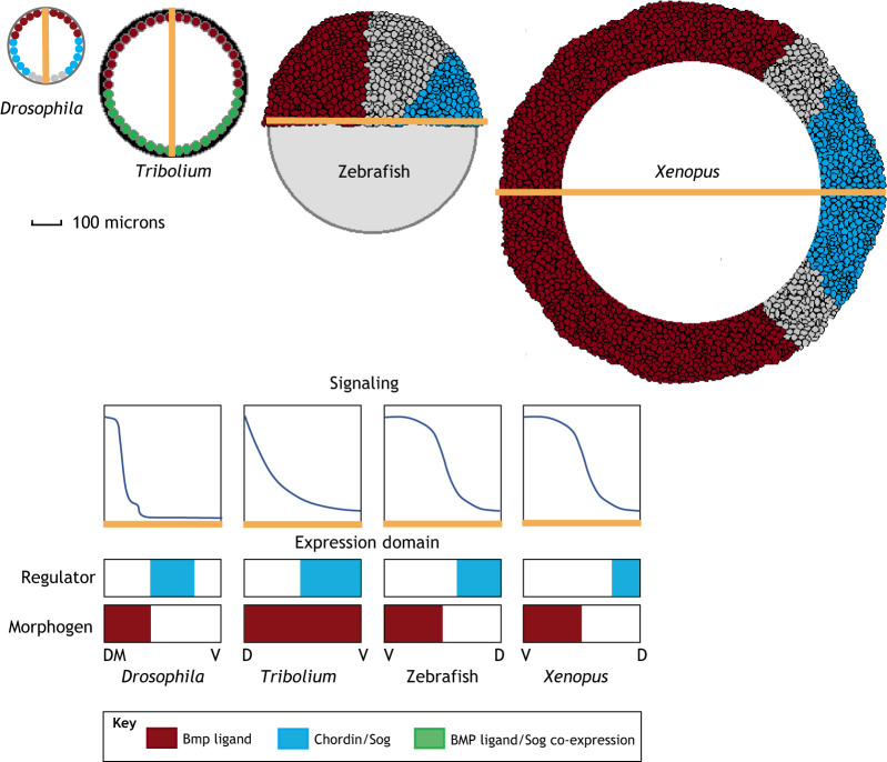
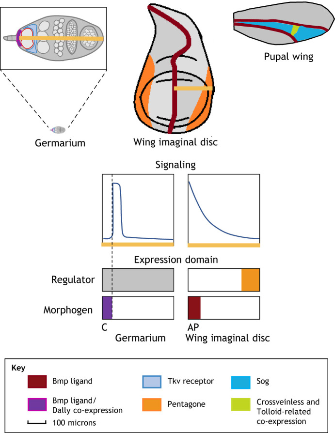
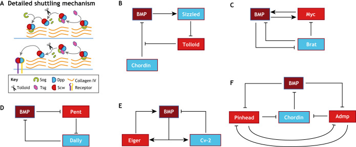
Pattern formation by bone morphogenetic proteins (BMPs) demonstrates remarkable plasticity and utility in several contexts, such as early embryonic development, tissue patterning and the maintenance of stem cell niches. BMPs pattern tissues over many temporal and spatial scales: BMP gradients as short as 1-2 cell diameters maintain the stem cell niche of the Drosophila germarium over a 24-h cycle, and BMP gradients of several hundred microns establish dorsal-ventral tissue specification in Drosophila, zebrafish and Xenopus embryos in timescales between 30 min and several hours. The mechanisms that shape BMP signaling gradients are also incredibly diverse. Although ligand diffusion plays a dominant role in forming the gradient, a cast of diffusible and non-diffusible regulators modulate gradient formation and confer robustness, including scale invariance and adaptability to perturbations in gene expression and growth. In this Review, we document the diverse ways that BMP gradients are formed and refined, and we identify the core principles that they share to achieve reliable performance.

Keywords: Axis formation; BMP patterning; Drosophila; Morphogen gradient; Zebrafish.


Conflict of interest statement

Competing interestsThe authors declare no competing or financial interests.

Figures

Fig. 1.
Fig. 1.
Signaling gradient profiles and expression domains of BMP patterned embryos. Top schematics show Dpp and Sog expression domains in the Drosophila embryo (240 μm; transverse section). Dpp expression domain and co-expression of Dpp and Sog in the Tribolium castaneum embryo (480 μm; transverse section). Expression domains of bmp ventrally and chordin dorsally in the zebrafish embryo (700 μm; late blastula) and the Xenopus embryo (1.2 mm; early gastrula, transverse section). Lower schematics show qualitative graphs of BMP signaling gradients and the expression domains for the morphogen and negative regulator. The yellow x-axis in the signaling graphs corresponds to the yellow line in the images of the top schematic. D, dorsal; DM, dorsal midline; V, ventral.
Fig. 2.
Fig. 2.
Morphogen gradient formation mechanisms. (A) Morphogen gradient concept. (B) Gradient formation via active transport mechanisms: cytonemes (top) and migrasome/transcytosis (bottom). Cytonemes are cellular projections which can emanate from cells towards the morphogen source. Cytonemes carry Type II Bmp receptors which can take ligand back to the cell where they can signal in a receptor complex. Migrasome/transcytosis shows vesicle-based transport of ligand away from the source. (C) Gradient formation by free (passive) diffusion. Ligand diffuses from areas of high concentration near the source to areas of lower concentration. Pre-steady state ligand concentration is depicted. (D) Gradient formation via regulated diffusion. Extracellular matrix, immobile regulators and diffusible extracellular regulators all act to regulate diffusion. Note: receptors and ligands not to scale.
Fig. 3.
Fig. 3.
Signaling gradient profiles and expression domains of BMP-patterned organs of different scales. Drosophila germarium (top left): BMP/Dpp and Dally are co-expressed in cap cells (purple). Type IV Collagen Vkg (not pictured) is expressed throughout the GSC niche. Tkv is highly expressed in somatic escort cells. GSC cells are outlined in red (image modified from Sun et al., 2010). Drosophila third instar imaginal wing disc (top middle): BMP/Dpp is expressed in a narrow stripe at the AP boundary. Pentagone is expressed at the periphery. Crossvein formation in Drosophila pupal wing disc (top right): BMP/Dpp is expressed in longitudinal veins, Sog is expressed throughout the pupal wing. Crossveinless and Tolloid-related are expressed in the future posterior cross vein location where they can act to promote BMP/Dpp signaling by liberating ligand from Sog-Dpp complexes. Lower schematics show qualitative graphs of BMP signaling gradients and the expression domains for the morphogen and negative regulator. Germarium regulator depicted as a gray box to reflect ubiquitous presence of multiple regulators including Type IV Collagen Vkg. Signaling and expression domain graphs not shown for pupal wing disc as active transport does not take place over a single axis. AP, anterior posterior boundary; C, cap cells.
Fig. 4.
Fig. 4.
Diversity of regulatory mechanisms. (A) Highly mobile regulators can engage in shuttling processes, which have concentrating effects. Often ligand gradients end up smaller than their expression domain. Shuttling mechanisms establish peak signaling opposite of the regulator, regardless of where morphogen is expressed. (B) Poorly diffusive regulators have primarily inhibitory effects as they bind ligand and block signaling. This can act in a source-sink function as the ligand diffuses towards the ‘sink’ of immobile regulators. (C) Countergradients involve highly mobile regulators, as in shuttling. However, countergradient regulators do not have any pro-signaling functions.
Fig. 5.
Fig. 5.
Shuttling and extracellular regulation. (A) BMP/Dpp shuttling as observed in Drosophila DV patterning. Sog binds Dpp/Scw ligand heterodimer and forms a Type IV Collagen bound complex that prevents signaling in areas of high Sog concentration. Tsg disrupts Sog-Dpp/Scw binding to Collagen and enables diffusion. Tsg also acts as a scaffold to promote Tolloid-mediated Sog cleavage and Dpp/Scw liberation. In areas with high Sog levels, the liberated ligand heterodimer typically reforms a Sog complex and begins another round of shuttling. In high Tolloid and low Sog levels areas, the ligand heterodimer is free to signal. Iterative rounds of complex formation and cleavage have the effect of moving ligand away from Sog expression, leading to a concentrated high peak at the dorsal midline. (B-F) Network diagrams of extracellular BMP regulation in diverse contexts. (B) BMP influences its own extracellular regulation in embryonic axis formation. BMP signaling leads to upregulation of the ventral protein Sizzled, which competitively inhibits Tolloid and prevents Tolloid-mediated Chordin cleavage. (C) In the Drosophila germarium, downstream BMP signaling target, Myc, provides positive feedback by upregulating BMP ligand uptake. Brat creates a bistable switch for differentiation by inhibiting BMP signal transduction and Myc activity. (D) In the Drosophila wing disc, Pent, a secreted factor that is negatively regulated by BMP signaling, supports BMP signaling by directing the internalization of Dally, a negative regulator of BMP signaling. (E) In the Drosophila embryo, downstream BMP signaling products Eiger and Cv-2 provide positive and negative feedback, respectively, to BMP signaling. These feedback mechanisms act to fine tune the BMP gradient and confer spatial bistability. (F) In zebrafish, Pinhead and Admp both act to support BMP signaling by promoting Chordin degradation. Pinhead and Admp are both downregulated by each other and by BMP signaling. The reciprocal repression circuit of Admp and Pinhead provides robustness in BMP gradient formation.

Similar articles

Cited by

References

    1. Affolter, M. and Basler, K. (2007). The Decapentaplegic morphogen gradient: from pattern formation to growth regulation. Nat. Rev. Genet. 8, 663-674. 10.1038/nrg2166 - DOI - PubMed
    1. Akiyama, T. and Gibson, M. C. (2015a). Decapentaplegic and growth control in the developing Drosophila wing. Nature 527, 375-378. 10.1038/nature15730 - DOI - PubMed
    1. Akiyama, T. and Gibson, M. C. (2015b). Morphogen transport: theoretical and experimental controversies. Wiley Interdiscip. Rev. Dev. Biol. 4, 99-112. 10.1002/wdev.167 - DOI - PubMed
    1. Akiyama-Oda, Y. and Oda, H. (2006). Axis specification in the spider embryo: dpp is required for radial-to-axial symmetry transformation and sog for ventral patterning. Development 133, 2347-2357. 10.1242/dev.02400 - DOI - PubMed
    1. Antebi, Y. E., Linton, J. M., Klumpe, H., Bintu, B., Gong, M., Su, C., McCardell, R. and Elowitz, M. B. (2017). Combinatorial Signal Perception in the BMP Pathway. Cell 170, 1184-1196.e1124. 10.1016/j.cell.2017.08.015 - DOI - PMC - PubMed

Publication types

MeSH terms

LinkOut - more resources