Asthmatic condition induced the activity of exosome secretory pathway in rat pulmonary tissues
- PMID: 33794910
- PMCID: PMC8015058
- DOI: 10.1186/s12950-021-00275-7
Asthmatic condition induced the activity of exosome secretory pathway in rat pulmonary tissues
Abstract
Background: The recent studies highlighted the critical role of exosomes in the regulation of inflammation. Here, we investigated the dynamic biogenesis of the exosomes in the rat model of asthma.
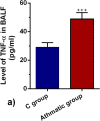
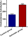
Results: Our finding showed an increase in the expression of IL-4 and the suppression of IL-10 in asthmatic lung tissues compared to the control samples (p < 0.05). Along with the promotion of IL-4, the protein level of TNF-α was induced, showing an active inflammatory status in OVA-sensitized rats. According to our data, the promotion of asthmatic responses increased exosome biogenesis indicated by increased CD63 levels and acetylcholine esterase activity compared to the normal condition (p < 0.05).
Conclusion: Data suggest that the stimulation of inflammatory response in asthmatic rats could simultaneously increase the paracrine activity of pulmonary cells via the exosome biogenesis. Exosome biogenesis may correlate with the inflammatory response.
Keywords: Asthma; CD63; Exosome biogenesis; Inflammatory cytokines; Rats.
Conflict of interest statement
The authors have no competing interests to declare.
Figures


Similar articles
-
Type 2 diabetes mellitus stimulated pulmonary vascular inflammation and exosome biogenesis in rats.Cell Biochem Funct. 2023 Jan;41(1):78-85. doi: 10.1002/cbf.3764. Epub 2022 Nov 6. Cell Biochem Funct. 2023. PMID: 36335538
-
Bone marrow mesenchymal stem cells and condition media diminish inflammatory adhesion molecules of pulmonary endothelial cells in an ovalbumin-induced asthmatic rat model.Microvasc Res. 2019 Jan;121:63-70. doi: 10.1016/j.mvr.2018.10.005. Epub 2018 Oct 18. Microvasc Res. 2019. PMID: 30343002
-
[Research on feasibility of in vitro inflammatory wound microenvironment simulated by using inflammatory wound tissue homogenate of mice].Zhonghua Shao Shang Za Zhi. 2020 Nov 20;36(11):1024-1034. doi: 10.3760/cma.j.cn501120-20200720-00351. Zhonghua Shao Shang Za Zhi. 2020. PMID: 33238685 Chinese.
-
[Effects of fasudil on the expression of Rho kinase-1 and airway inflammation in a mouse model of asthma.].Zhonghua Jie He He Hu Xi Za Zhi. 2009 Nov;32(11):847-9. Zhonghua Jie He He Hu Xi Za Zhi. 2009. PMID: 20079297 Chinese.
-
Proinflammatory role of epithelial cell-derived exosomes in allergic airway inflammation.J Allergy Clin Immunol. 2013 Apr;131(4):1194-203, 1203.e1-14. doi: 10.1016/j.jaci.2012.12.1565. Epub 2013 Feb 14. J Allergy Clin Immunol. 2013. PMID: 23414598
Cited by
-
MSC-derived exosomes protect auditory hair cells from neomycin-induced damage via autophagy regulation.Biol Res. 2024 Jan 13;57(1):3. doi: 10.1186/s40659-023-00475-w. Biol Res. 2024. PMID: 38217055 Free PMC article.
-
Suppression of gastric cancer cell proliferation by miR-494-3p inhibitor-loaded engineered exosomes.Heliyon. 2024 May 7;10(10):e30803. doi: 10.1016/j.heliyon.2024.e30803. eCollection 2024 May 30. Heliyon. 2024. PMID: 38770297 Free PMC article.
-
Therapeutic potential of exosomes/miRNAs in polycystic ovary syndrome induced by the alteration of circadian rhythms.Front Endocrinol (Lausanne). 2022 Nov 16;13:918805. doi: 10.3389/fendo.2022.918805. eCollection 2022. Front Endocrinol (Lausanne). 2022. PMID: 36465652 Free PMC article. Review.
-
Biogenesis and function of exosome lncRNAs and their role in female pathological pregnancy.Front Endocrinol (Lausanne). 2023 Sep 7;14:1191721. doi: 10.3389/fendo.2023.1191721. eCollection 2023. Front Endocrinol (Lausanne). 2023. PMID: 37745705 Free PMC article. Review.
-
Application of Single Extracellular Vesicle Analysis Techniques.Int J Nanomedicine. 2023 Sep 20;18:5365-5376. doi: 10.2147/IJN.S421342. eCollection 2023. Int J Nanomedicine. 2023. PMID: 37750091 Free PMC article. Review.
References
Grants and funding
LinkOut - more resources
Full Text Sources
Other Literature Sources
Miscellaneous

