. 2021 Apr 1;11(2):1653-1677.
doi: 10.1002/cphy.c190046.

Perinatal Hypoxemia and Oxygen Sensing

Affiliations

Perinatal Hypoxemia and Oxygen Sensing

Gary C Mouradian Jr et al. Compr Physiol. .

Abstract

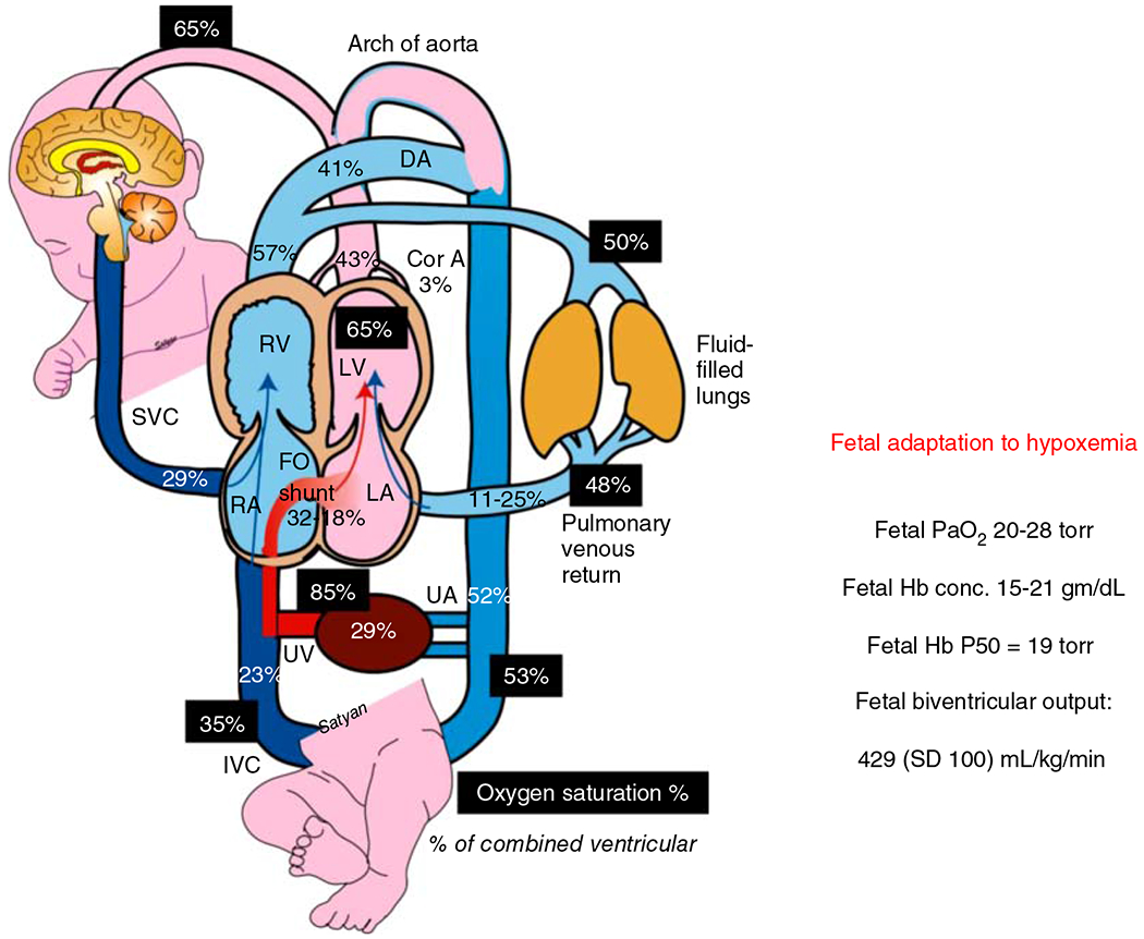
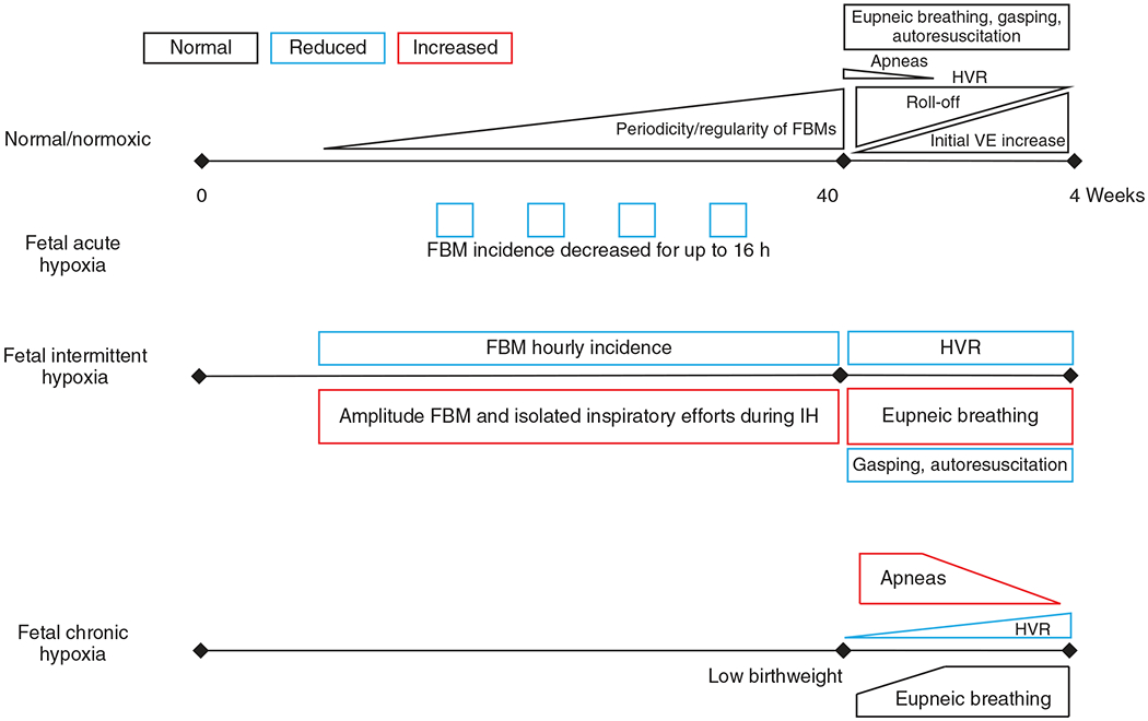
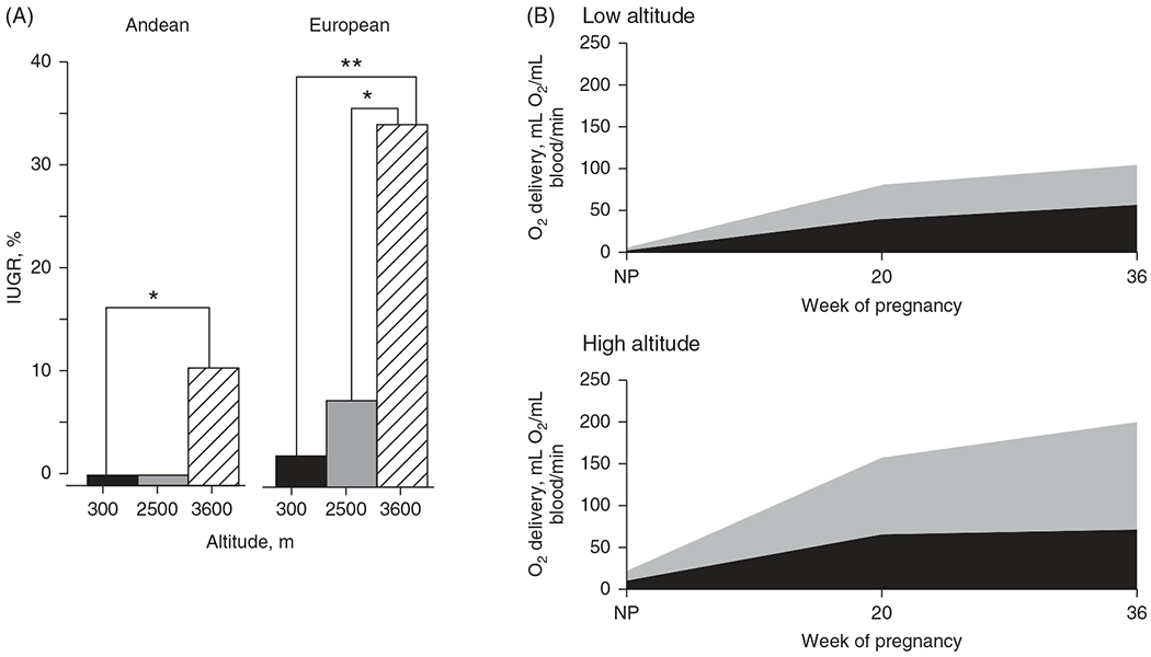
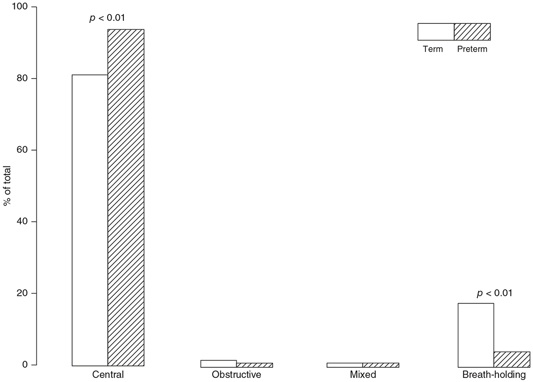
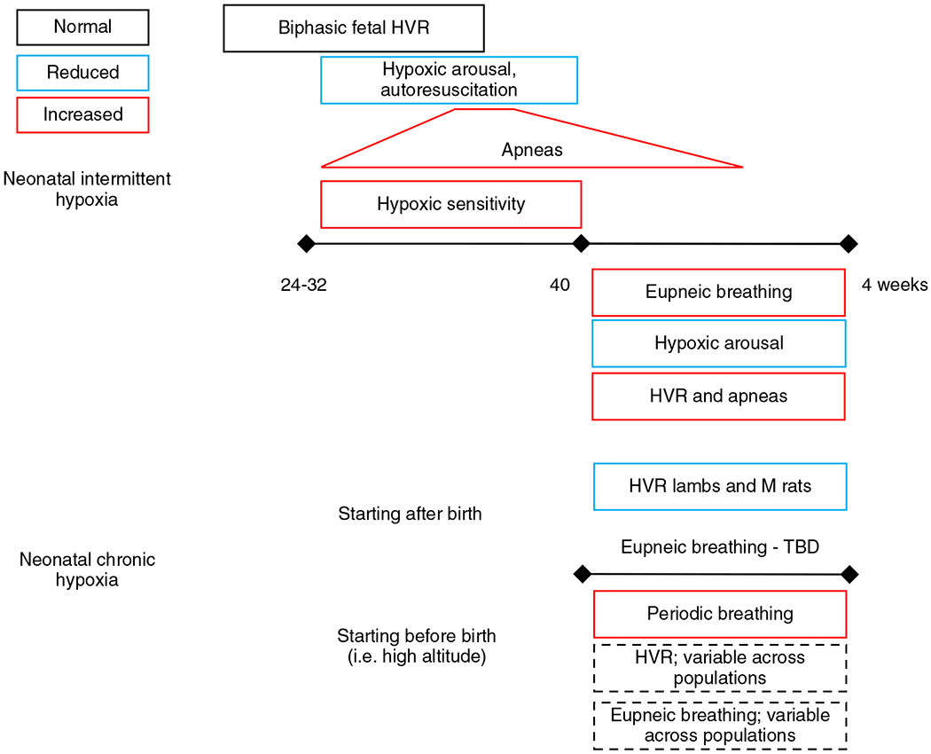
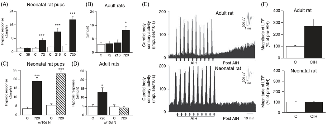
The development of the control of breathing begins in utero and continues postnatally. Fetal breathing movements are needed for establishing connectivity between the lungs and central mechanisms controlling breathing. Maturation of the control of breathing, including the increase of hypoxia chemosensitivity, continues postnatally. Insufficient oxygenation, or hypoxia, is a major stressor that can manifest for different reasons in the fetus and neonate. Though the fetus and neonate have different hypoxia sensing mechanisms and respond differently to acute hypoxia, both responses prevent deviations to respiratory and other developmental processes. Intermittent and chronic hypoxia pose much greater threats to the normal developmental respiratory processes. Gestational intermittent hypoxia, due to maternal sleep-disordered breathing and sleep apnea, increases eupneic breathing and decreases the hypoxic ventilatory response associated with impaired gasping and autoresuscitation postnatally. Chronic fetal hypoxia, due to biologic or environmental (i.e. high-altitude) factors, is implicated in fetal growth restriction and preterm birth causing a decrease in the postnatal hypoxic ventilatory responses with increases in irregular eupneic breathing. Mechanisms driving these changes include delayed chemoreceptor development, catecholaminergic activity, abnormal myelination, increased astrocyte proliferation in the dorsal respiratory group, among others. Long-term high-altitude residents demonstrate favorable adaptations to chronic hypoxia as do their offspring. Neonatal intermittent hypoxia is common among preterm infants due to immature respiratory systems and thus, display a reduced drive to breathe and apneas due to insufficient hypoxic sensitivity. However, ongoing intermittent hypoxia can enhance hypoxic sensitivity causing ventilatory overshoots followed by apnea; the number of apneas is positively correlated with degree of hypoxic sensitivity in preterm infants. Chronic neonatal hypoxia may arise from fetal complications like maternal smoking or from postnatal cardiovascular problems, causing blunting of the hypoxic ventilatory responses throughout at least adolescence due to attenuation of carotid body fibers responses to hypoxia with potential roles of brainstem serotonin, microglia, and inflammation, though these effects depend on the age in which chronic hypoxia initiates. Fetal and neonatal intermittent and chronic hypoxia are implicated in preterm birth and complicate the respiratory system through their direct effects on hypoxia sensing mechanisms and interruptions to the normal developmental processes. Thus, precise regulation of oxygen homeostasis is crucial for normal development of the respiratory control network. © 2021 American Physiological Society. Compr Physiol 11:1653-1677, 2021.


Figures

Figure 1
Figure 1
Adaptation of the fetus to low oxygen environment present in utero. Fetal oxygen supply is provided by maternal blood bathing the chorionic villi in the placenta. The umbilical vein carries the oxygenated blood to inferior vena cava (IVC) and eventually across the foramen ovale to left atrium and left ventricle to be pumped into coronary and cerebral circulations. The oxygen saturation of fetal blood in different sites are indicated by dark shaded boxes. The percent of cardiac output distributed to each organ is indicated in plain text. The mean values for fetal biventricular output, range of normal Hb concentrations and fetal PaO2 are indicated in the text box to the right, along with the HbP50 for fetal Hb (197, 257, 268). Reused, with permission, from Richard A. Polin and William W Fox, 2016, Fetal and Neonatal Physiology, Ed: Polin, Abman, Rowitch, Benitz and Fox, 5th edition, Lakshminrusimha and Steinhorn “Pathophysiology of PPHN,” pp 1576-1587. Copyright Satyan Lakshminrusimha.
Figure 2
Figure 2
Effects of ibotenic acid lesioning within or sparing the thalamic parafascicular (Pf) nuclear complex in neonatal lambs on the depression phase of the hypoxic ventilatory response. Lesioning the Pf removes the ventilatory depression at 10 and 15 min (decline 10 and 15, respectively) expressed as a change in ventilation from prelesion values (upper left) or as a percent of the augmentation phase (lower left) (A). Lesions sparing the Pf have no effect on the hypoxic ventilatory decline (B). *P < 0.005, **P < 0.03 compared with augmentation phase. †P < 0.005 versus prelesion at same time, ‡ P < 0.05 versus thalamic lesions sparing Pf at same time. Adapted, with permission, from Koos BJ, et al., 2016 (164).
Figure 3
Figure 3
The thalamic parafascicular nuclear region mediates hypoxic depression of fetal breathing movements in fetal sheep. Hypoxia-induced suppression of breathing and associated eye movements is completely removed following lesioning of the thalamic parafascicular nuclear region by ibotenic (IBO) acid (A). Adapted, with permission, from Koos BJ, 2002 (163). In a similar experiment using exogenous adenosine, the known neurochemical mediating hypoxia-induced suppression of fetal breathing suppresses breathing and associated eye movements before IBO injection but is significantly impaired following IBO lesioning of the thalamic parafascicular nuclear region (B). Adapted, with permission, from Koos BJ, et al., 2000 (162).
Figure 4
Figure 4
Schematic summary of in-text descriptions of the effects fetal normoxia and fetal acute, intermittent, and chronic hypoxia have on breathing. During normal development, the periodicity and regularity of FBM activity steadily rises and approaches postnatal breathing regularity at birth. Upon birth, few apneas are present and quickly diminishes with age. The hypoxic ventilatory response (HVR) begins with a significant secondary role-off pertaining to a decrease in metabolic rate following an initial increase in ventilation (VE). This biphasic HVR quickly matures (within weeks of birth) to a sustained increase in ventilation as the metabolic roll-off diminishes in effect. Fetal acute hypoxia causes a decrease in FBM incidence. Fetal intermittent hypoxia, also called gestational intermittent hypoxia, decreases FBM hourly incidence but increases FBM amplitude and inspiratory efforts during the hypoxic episodes. Postnatally, fetal intermittent hypoxia reduces the HVR and capacity for gasping and autoresuscitation and an increase in eupneic breathing. Fetal chronic hypoxia is typically observed in high-altitude births where infants are born with lower weight. Infants also have increased apneas at birth associated with diminished and delayed development of the HVR. Eupneic breathing is fairly normal aside from the apneas. See text for references.
Figure 5
Figure 5
Rate of IUGR at altitude and differences in uterine blood flow during pregnancy between nonnative (European) and native (Andean) high-altitude populations. The rate of IUGR increases with altitude and at high-altitude Europeans have a fivefold greater occurrence of IUGR compared to Andeans after adjusting for other fetal growth factors (shown in the graph are unadjusted values; A). A potential explanation for the IUGR rate disparity at altitude between Andeans and Europeans is the compensatory twofold greater increase in uteroplacental oxygen delivery in Andean compared to European women at 36 weeks’ gestation indicating potential genetic adaptions across generations (B). NP, nonpregnant. *P < 0.05, **P < 0.01. Reused, with permission, from Julian CG, 2011 (138).
Figure 6
Figure 6
Distribution of apnea types lasting 3 to 15 s in eight term (gestational age: 39.5±0.3 weeks) and eight preterm (gestational age: 34.3±0.4 weeks) infants measured between birth and 56 weeks old. A total of 783 and 4086 apneas were recorded in term and preterm infant groups, respectively. Reused, with permission, from Lee D, et al., 1987 (170). © 1987, Springer Nature.
Figure 7
Figure 7
Schematic summary of in-text descriptions of the effects neonatal intermittent and chronic hypoxia have on breathing. Infants born premature (24-32 weeks) retain a biphasic fetal hypoxic ventilatory response (HVR) which is associated with decreased hypoxic arousal and autorescuscitation but increased number of apneas (which plateaus around 10 weeks after birth). The increased apneas are a reflection of apnea of prematurity but also enhanced hypoxic sensitivity which can cause ventilatory overshoots and trigger the CO2 apneic threshold. Term infants exposed to neonatal intermittent hypoxia have increased eupneic breathing, HVR, and more apneas though arousal from hypoxia is reduced. Exposure to chronic neonatal hypoxia commencing after birth is associated with reduced HVR [in lambs and male (M) rats]. The effects on eupneic breathing are equivocal and thus remain to be fully elucidated. Infants that continue to be exposed to chronic hypoxia after birth (i.e. high-altitude births) demonstrate increased periodic breathing but the effects on the HVR and eupneic breathing are variable across high altitude populations. See text for references.
Figure 8
Figure 8
Effects of intermittent hypoxic episodes on carotid body hypoxic sensitivity and long-term facilitation in neonatal versus adult rats. Progressive increases in intermittent hypoxic episodes (36, 72, 216, and 720) cause progressive hypoxic sensitization of the carotid bodies in neonates (A) whereas hypoxic sensitization requires 720 episodes in adults and is not as robust as in neonates (B). The hypoxic sensitization in the neonatal (C) but not the adult (D) rats is sustained after their return to normoxia for 10 days. Intermittent hypoxia causes long-term facilitation in adult rats (E) but not in neonatal rats (F) despite having enhanced carotid body hypoxic sensitivity. Reused, with permission, from Pawar A, et al., 2008 (243).
Figure 9
Figure 9
Schematic of the hypothesized cellular and molecular mechanisms of central oxygen sensing by the astrocyte. Hypoxia sensed from perfusing blood by the astrocyte inhibits the mitochondria and thus stimulating mitochondrial reactive oxygen species (ROS) production, leading to an increase in lipid peroxidation. PLC-IP3 signaling releases intracellular calcium stores and releases ATP onto nearby pre-BötC neurons that express two ATP receptors, P2YR and P2XR. In parallel, hypoxia may cause opening of the connexin (Cx) hemichannel leading to release of ATP and lactate, the latter with unknown stimulatory effects on the preBötC neurons. Release of ATP causes further release of ATP in autocrine and paracrine manners, increasing respiratory rate and sympathetic activity through preBötC neurons. Reused, with permission, from Gourine AV and Funk GD, 2017 (106). © 1985, The American Physiological Society.

Similar articles

Cited by

References

    1. Abu-Shaweesh JM, Martin RJ. Neonatal apnea: What’s new? Pediatr Pulmonol 43 (10): 937–944, 2008. - PubMed
    1. Adzick NS, Harrison MR, Glick PL, Villa RL, Finkbeiner W. Experimental pulmonary hypoplasia and oligohydramnios: Relative contributions of lung fluid and fetal breathing movements. J Pediatr Surg 19(6): 658–665, 1984. - PubMed
    1. Alcorn D, Adamson TM, Maloney JE, Robinson PM. Morphological effects of chronic bilateral phrenectomy or vagotomy in the fetal lamb lung. J Anat 130 (Pt 4): 683–695, 1980. - PMC - PubMed
    1. Al-Matary A, Kutbi I, Qurashi M, Khalil M, Alvaro R, Kwiatkowski K, Cates D, Rigatto H. Increased peripheral chemoreceptor activity may be critical in destabilizing breathing in neonates. Semin Perinatol 28 (4): 264–272, 2004. - PubMed
    1. Alvaro R, Rigatto H. Control of breathing in newborns. In: Bracci R, Buonocore G, Weindling M, editors. Neonatology. Milano: Springer, 2012.