

doi: 10.1085/jgp.202112879.
Fast inactivation of Nav1.3 channels by FGF14 proteins: An unconventional way to regulate the slow firing of adrenal chromaffin cells
Affiliations
- PMID: 33792614
- PMCID: PMC8020463
- DOI: 10.1085/jgp.202112879
Item in Clipboard
Fast inactivation of Nav1.3 channels by FGF14 proteins: An unconventional way to regulate the slow firing of adrenal chromaffin cells
J Gen Physiol.
.
Abstract
Using Nav1.3 and FGF14 KO mice, Martinez-Espinosa et al. provide new findings on how intracellular FGF14 proteins interfere with the endogenous fast inactivation gating and regulate the “long-term inactivation” of Nav1.3 channels that sets Nav channel availability and spike adaptation during sustained stimulation in adrenal chromaffin cells.
Figures
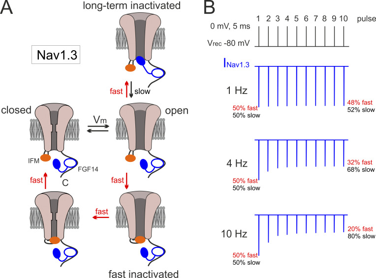
Schematic model for the Nav1.3/FGF14 protein interactions that regulate fast and slow recovery from inactivation and determine Nav availability during pulse trains of varying frequency in rodent chromaffin cells.
(A) A simplified schematic diagram of Nav1.3 channel transitions between closed, open, fast, and long-term inactivated states derived from Dover et al. (2010), Milescu et al. (2010), Venkatesan et al. (2014), and the two articles from Martinez-Espinosa et al. (2021a, . The endogenous IFM inactivation particle in the DIII–DIV cytoplasmic loop (red oval) and the N terminus of the FGF14 protein (blue oval), with its core domain (white oval) tethered to the C terminus of the channel, compete for access within the pore to inactivate the open channel during step depolarization. The two inactivation particles compete for docking within the open pore (fast and long-term inactivated states). The onset of fast and long-term inactivation is comparably fast (a few milliseconds; up and down red arrows in right panels). Recovery from long-term inactivation is slow (50–400 ms) and may occur while the channel is open (black arrow) or closed (not shown). Recovery from fast inactivation is fast and proceeds through the closed state of the channel (horizontal and up red arrows; Kuo and Bean, 1994). The closed and fast inactivated states preceding the two indicated to the left are not shown for simplicity (see Goldfarb, 2012). (B) Nav1.3 currents (blue traces) recorded from mouse chromaffin cells during voltage-clamp commands with a 10-pulse train of increasing frequency. Nav current amplitudes are drawn after having interpolated the data of Figs. 8, 10, and S1 of Martinez-Espinosa et al. (2021a). Steps of 5 ms to 0 mV from −80 mV holding potentials (Vh) were applied at 1, 4, or 10 Hz. Nav1.3 current amplitudes decrease progressively during the 10-pulse trains. At 1 Hz the amplitude attenuation is nearly detectable, while at 10 Hz it is remarkable. The percentage of occupancies in the fast and slow recovery pathways calculated by Martinez-Espinosa et al. (2021a, are indicated in red (fast recovery) and black (slow recovery). After terminating the first depolarizing step, Nav channels are equally distributed (50%) in both the fast and slow pathway. With increasing frequency, Nav availability is strongly attenuated. Nav channels accumulate in the slow recovery pathway during the pulse train: 52% at 1 Hz, 68% at 4 Hz, and 80% at 10 Hz.
Comment on
- doi: 10.1085/jgp.202012784
- doi: 10.1085/jgp.202012785
Similar articles
-
Reduced availability of voltage-gated sodium channels by depolarization or blockade by tetrodotoxin boosts burst firing and catecholamine release in mouse chromaffin cells.J Physiol. 2015 Feb 15;593(4):905-27. doi: 10.1113/jphysiol.2014.283374. Epub 2015 Jan 26. J Physiol. 2015. PMID: 25620605 Free PMC article.
-
Up-regulation of NaV1.7 sodium channels expression by tumor necrosis factor-α in cultured bovine adrenal chromaffin cells and rat dorsal root ganglion neurons.Anesth Analg. 2014 Feb;118(2):318-324. doi: 10.1213/ANE.0000000000000085. Anesth Analg. 2014. PMID: 24445633
-
Impaired chromaffin cell excitability and exocytosis in autistic Timothy syndrome TS2-neo mouse rescued by L-type calcium channel blockers.J Physiol. 2019 Mar;597(6):1705-1733. doi: 10.1113/JP277487. Epub 2019 Jan 28. J Physiol. 2019. PMID: 30629744 Free PMC article.
-
Calcium-activated potassium channels in adrenal chromaffin cells.Ion Channels. 1996;4:261-301. doi: 10.1007/978-1-4899-1775-1_7. Ion Channels. 1996. PMID: 8744211 Review.
-
Cav1.3 and Cav1.2 channels of adrenal chromaffin cells: emerging views on cAMP/cGMP-mediated phosphorylation and role in pacemaking.Biochim Biophys Acta. 2013 Jul;1828(7):1608-18. doi: 10.1016/j.bbamem.2012.11.013. Epub 2012 Nov 15. Biochim Biophys Acta. 2013. PMID: 23159773 Review.
Cited by
-
Slow recovery from fast inactivation of Nav1.3 channels: a common gating mechanism shared in sweet- and sour-sensing cells.Pflugers Arch. 2021 Jun;473(6):855-857. doi: 10.1007/s00424-021-02575-6. Epub 2021 May 10. Pflugers Arch. 2021. PMID: 33970334 Free PMC article. No abstract available.
References
-
- Cummins, T.R., Aglieco F., Renganathan M., Herzog R.I., Dib-Hajj S.D., and Waxman S.G.. 2001. Nav1.3 sodium channels: rapid repriming and slow closed-state inactivation display quantitative differences after expression in a mammalian cell line and in spinal sensory neurons. J. Neurosci. 21:5952–5961. 10.1523/JNEUROSCI.21-16-05952.2001 - DOI - PMC - PubMed
Publication types
MeSH terms
LinkOut - more resources
Full Text Sources
Other Literature Sources
Research Materials
