. 2021 Mar 31;6(1):129.
doi: 10.1038/s41392-021-00510-w.

PUMA facilitates EMI1-promoted cytoplasmic Rad51 ubiquitination and inhibits DNA repair in stem and progenitor cells

Affiliations

PUMA facilitates EMI1-promoted cytoplasmic Rad51 ubiquitination and inhibits DNA repair in stem and progenitor cells

Jin Wook Kang et al. Signal Transduct Target Ther. .

Abstract

Maintenance of genetic stability via proper DNA repair in stem and progenitor cells is essential for the tissue repair and regeneration, while preventing cell transformation after damage. Loss of PUMA dramatically increases the survival of mice after exposure to a lethal dose of ionizing radiation (IR), while without promoting tumorigenesis in the long-term survivors. This finding suggests that PUMA (p53 upregulated modulator of apoptosis) may have a function other than regulates apoptosis. Here, we identify a novel role of PUMA in regulation of DNA repair in embryonic or induced pluripotent stem cells (PSCs) and immortalized hematopoietic progenitor cells (HPCs) after IR. We found that PUMA-deficient PSCs and HPCs exhibited a significant higher double-strand break (DSB) DNA repair activity via Rad51-mediated homologous recombination (HR). This is because PUMA can be associated with early mitotic inhibitor 1 (EMI1) and Rad51 in the cytoplasm to facilitate EMI1-mediated cytoplasmic Rad51 ubiquitination and degradation, thereby inhibiting Rad51 nuclear translocation and HR DNA repair. Our data demonstrate that PUMA acts as a repressor for DSB DNA repair and thus offers a new rationale for therapeutic targeting of PUMA in regenerative cells in the context of DNA damage.


Conflict of interest statement

The authors declare no competing interests.

Figures
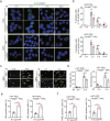
Fig. 1
Fig. 1
Knockout of PUMA enhances IR-induced DNA repair in PSCs and HPCs. a Representative images of γH2AX foci in PUMA wild type (WT) and KO PSCs and HPCs after IR. Scale bars, 10 μm. PSCs and HPCs were treated with 2 Gy IR and then fixed at 0, 2, 8, or 24 h for γH2AX staining. b Quantification of γH2AX foci in a. The percentage of positive cells (≥10 γH2AX foci) is shown. c Representative images of comet tails in PUMA WT and KO PSCs or HPCs at indicated time points after treated with 2 Gy of IR. Scale bars, 250 μm. d Quantification of comet tail intensity in c. e, f Quantification of HR (e) or NHEJ (f)-positive cells in PUMA WT and KO PSCs or HPCs. At 48 h after I-SceI transfection, PUMA WT or KO PSC and HPC cells with stable expression of a pDR-GFP or pEJ5-GFP reporter were treated with 2 Gy IR and then harvested at 8 h for analysis, using flow cytometry to examine recombination induced by I-SceI digestion. Dead cells were excluded by PI staining. Data are representative of three independent experiments with similar results. Error bars, SD. *P < 0.05, **P < 0.01
Fig. 2
Fig. 2
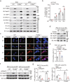
Knockout of PUMA elevates Rad51 in PSCs or HPCs. a Immunoblotting (IB) of Rad51, 53BP1, and PUMA expression in PUMA WT and KO PSCs or HPCs after IR. PSCs and HPCs were treated with 2 Gy IR, and then collected at 0 or 8 h post-IR for IB analysis. Actin was used as a control. b Quantification of positive cells with Rad51 foci in PUMA WT and KO PSCs at indicated time points after IR. c Representative images of Rad51 foci in PUMA WT and KO PSCs after IR. Scale bars, 10 μm. d Effects of PUMA re-expression on expression of Rad51 and 53BP1 in PUMA KO PSCs after IR. Lentivirus-mediated PUMA was infected into PUMA KO PSCs, and then selected the clones of transfected cells expressing a similar level of PUMA as that PUMA WT cells. e Effect of re-expression of PUMA on HR repair in PUMA KO PSCs. f IB of expression of Rad51 and 53BP1 in PSCs or HPCs with a Rad51 shRNA. g Effect of Rad51 knockdown on PUMA-mediated HR repair. Data are representative of three independent experiments with similar results. Error bars, SD. *P < 0.05, **P < 0.01, ***P < 0.001
Fig. 3
Fig. 3
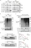
PUMA binds to cytosolic Rad51 and promotes its ubiquitination and stability. a Immunoprecipitation (IP) and IB of PUMA interaction with Rad51 in PSCs or HPCs. PSC and HPC cells were treated with 2 Gy of IR, and then collected at 0 or 8 h for IP and IB analyses. β-Actin was used as a loading control. b IP and IB of Rad51 ubiquitination in PSCs treated with or without 2 Gy IR. His-tagged Ub was transfected into PUMA KO PSCs, and then cells were lyzed for IP and IB analyses. c Effects of re-expression of PUMA in PUMA KO PSCs on Rad51 ubiquitination. PUMA cDNA (0, 2, or 4 μg) was transfected into PUMA KO PSCs. At 48 h after transfection, cells were treated with or without of 30 μM MG132 for 4 h. d Effects of PUMA knockout on Rad51 stability. At 8 h after IR, cells were treated with cycloheximide (CHX, 20 mg/ml) for the indicated time points. e Quantification of Rad51 protein levels in d. Data are representative of three independent experiments with similar results. Error bars, SD. **P < 0.01
Fig. 4
Fig. 4
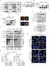
PUMA interacts with cytoplasmic Rad51 and EMI1. a IP and IB of PUMA interaction with EMI1, FBH1, and UCHL3 in PSCs and HPCs with or without IR. b IP and IB of PUMA interaction with EMI1 in HEK293T cells. c In vitro GST pull-down analysis. Purified GST-PUMA or GST proteins were mixed with cell extracts from PSCs. d Representative images of colocalization of PUMA and EMI1 in PSCs at 8 h post-IR. DAPI was used to show nucleus. An anti-EMI1 antibody (JG35-83, #NBP2-76833, Novus Biologicals) was used. Scale bars, 5 μm. e IP and IB of EMI1 interaction with cytosolic PUMA and Rad51 in PSCs with or without IR. f Effects of EMI1 KO with a sgRNA on PUMA-mediated Rad51 ubiquitination in PSCs and HPCs after IR. g Representative images of γH2AX foci in PUMA WT and KO PSCs or HPCs transduced with or without an EMI1 sgRNA after IR. Scale bars, 10 μm. h Quantification of γH2AX foci in g. The percentage of positive cells (≥5 γH2AX foci) was shown. Data are representative of three independent experiments with similar results. Error bars, SD. *P < 0.05, ***P < 0.001
Fig. 5
Fig. 5
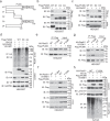
PUMA promotes Rad51 ubiquitination by EMI1. a Schematics of PUMA WT and truncated constructs. b IP and IB of the interaction of PUMA mutants with EMI1 in HEK293T cells. Flag-tagged PUMA WT or the truncation mutants with HA-EMI1 was transfected into HEK293T cells. c IP and IB of the association of PUMA mutants with Rad51 in HEK293T cells. d Effects of ectopic expression of the D3 or D4 mutant of PUMA on EMI1-mediated Rad51 ubiquitination. Flag-tagged PUMA WT, the truncation mutants, or an empty vector with His-Ub, HA-Rad51, and V5-EMI1 was transfected into HEK293T cells. e IP and IB of PUMA interaction with Rad51 F129A mutant. f PUMA increases EMI1 association with Rad51 WT, but not the F129A mutant. V5-EMI1 and HA-Rad51 WT or F129A mutant with or without PUMA were co-expressed in HEK293T cells. g Effect of Rad51 F129A mutation on PUMA interaction with EMI1. h PUMA promotes EMI1-mediated the ubiquitination of cytosolic Rad51 WT, but not the F129A mutant. Data are representative of three independent experiments with similar results. Error bars, SD. *P < 0.05, **P < 0.01
Fig. 6
Fig. 6
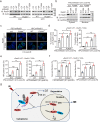
Knockout of PUMA promotes IR-induced cytoplasm-to-nucleus translocation and focus formation of Rad51, HR repair, and cell survival. a IB of re-expression of shRNA-resistant Rad51 WT or the F129A mutant in PUMA WT and KO PSC/shRad51 and HPC/shRad51 cells. b Protein expression levels of Rad51 WT and F129A mutant in the cytosol and the nucleus in PSC/shRad51 cells after IR. Lamin B1 and GAPDH are the markers for nuclear and cytosolic fraction. c Representative images of effects of re-expression of the F129A mutant on Rad51 focus formation in PUMA WT and KO PSC/shRad51 or HPC/shRad51 cells after IR. DAPI was used to show nucleus. Scale bars, 10 μm. d Quantification of positive cells with Rad51 foci in c. e Effects of re-expression of the F129A mutant on HR repair. f Effects of re-expression of the F129A mutant on cell survival after IR. Cell survival analysis was performed according to the MTT assay method with a Cell Titer 96 Aqueous Cell Proliferation Assay kit. g A working model. IR-induced PUMA interacts with cytoplasmic Rad51 and EMI1, which facilitates EMI1-mediated Rad51 ubiquitination and degradation, thereby leading to the inhibition of Rad51-mediated promoted DNA repair and enhancement of IR-induced cell death. Data are representative of three independent experiments with similar results. Error bars, SD. *P < 0.05, **P < 0.01

Similar articles

Cited by

References

    1. Blanpain C, Mohrin M, Sotiropoulou PA, Passegue E. DNA-damage response in tissue-specific and cancer stem cells. Cell Stem Cell. 2011;8:16–29. doi: 10.1016/j.stem.2010.12.012. - DOI - PubMed
    1. Frosina G. The bright and the dark sides of DNA repair in stem cells. J. Biomed. Biotechnol. 2010;2010:845396. doi: 10.1155/2010/845396. - DOI - PMC - PubMed
    1. Mohrin M, et al. Hematopoietic stem cell quiescence promotes error-prone DNA repair and mutagenesis. Cell Stem Cell. 2010;7:174–185. doi: 10.1016/j.stem.2010.06.014. - DOI - PMC - PubMed
    1. Yu J, et al. PUMA induces the rapid apoptosis of colorectal cancer cells. Mol. Cell. 2001;7:673–682. doi: 10.1016/S1097-2765(01)00213-1. - DOI - PubMed
    1. Han J, et al. Expression of bbc3, a pro-apoptotic BH3-only gene, is regulated by diverse cell death and survival signals. Proc. Natl Acad. Sci. USA. 2001;98:11318–11323. doi: 10.1073/pnas.201208798. - DOI - PMC - PubMed

Publication types

MeSH terms