Placental superoxide dismutase 3 mediates benefits of maternal exercise on offspring health
- PMID: 33770509
- PMCID: PMC8103776
- DOI: 10.1016/j.cmet.2021.03.004
Placental superoxide dismutase 3 mediates benefits of maternal exercise on offspring health
Abstract
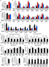
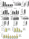
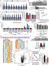
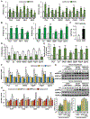
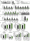
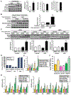
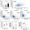
Poor maternal diet increases the risk of obesity and type 2 diabetes in offspring, adding to the ever-increasing prevalence of these diseases. In contrast, we find that maternal exercise improves the metabolic health of offspring, and here, we demonstrate that this occurs through a vitamin D receptor-mediated increase in placental superoxide dismutase 3 (SOD3) expression and secretion. SOD3 activates an AMPK/TET signaling axis in fetal offspring liver, resulting in DNA demethylation at the promoters of glucose metabolic genes, enhancing liver function, and improving glucose tolerance. In humans, SOD3 is upregulated in serum and placenta from physically active pregnant women. The discovery of maternal exercise-induced cross talk between placenta-derived SOD3 and offspring liver provides a central mechanism for improved offspring metabolic health. These findings may lead to novel therapeutic approaches to limit the transmission of metabolic disease to the next generation.
Keywords: AMPK; DNA methylation; TET; glucose metabolism; maternal exercise; placenta; pregnancy; superoxide dismutase 3; vitamin D.
Copyright © 2021 Elsevier Inc. All rights reserved.
Conflict of interest statement
Declaration of interests The authors declare no competing interests.
Figures
Comment in
-
Working out how maternal exercise benefits offspring.Nat Rev Endocrinol. 2021 Jun;17(6):320. doi: 10.1038/s41574-021-00494-1. Nat Rev Endocrinol. 2021. PMID: 33846589 No abstract available.
Similar articles
-
Maternal Exercise-Induced SOD3 Reverses the Deleterious Effects of Maternal High-Fat Diet on Offspring Metabolism Through Stabilization of H3K4me3 and Protection Against WDR82 Carbonylation.Diabetes. 2022 Jun 1;71(6):1170-1181. doi: 10.2337/db21-0706. Diabetes. 2022. PMID: 35290440 Free PMC article.
-
Exercise prevents the adverse effects of maternal obesity on placental vascularization and fetal growth.J Physiol. 2019 Jul;597(13):3333-3347. doi: 10.1113/JP277698. Epub 2019 May 28. J Physiol. 2019. PMID: 31115053 Free PMC article.
-
Role of metformin in epigenetic regulation of placental mitochondrial biogenesis in maternal diabetes.Sci Rep. 2020 May 20;10(1):8314. doi: 10.1038/s41598-020-65415-0. Sci Rep. 2020. PMID: 32433500 Free PMC article.
-
Maternal High-Fat Diet Impairs Placental Fatty Acid β-Oxidation and Metabolic Homeostasis in the Offspring.Front Nutr. 2022 Apr 14;9:849684. doi: 10.3389/fnut.2022.849684. eCollection 2022. Front Nutr. 2022. PMID: 35495939 Free PMC article.
-
Placental function in maternal obesity.Clin Sci (Lond). 2020 Apr 30;134(8):961-984. doi: 10.1042/CS20190266. Clin Sci (Lond). 2020. PMID: 32313958 Free PMC article. Review.
Cited by
-
DNA Methylation in the Hypothalamic Feeding Center and Obesity.J Obes Metab Syndr. 2023 Dec 30;32(4):303-311. doi: 10.7570/jomes23073. Epub 2023 Dec 21. J Obes Metab Syndr. 2023. PMID: 38124554 Free PMC article. Review.
-
Metabolism, Mitochondrial Dysfunction, and Redox Homeostasis in Pulmonary Hypertension.Antioxidants (Basel). 2022 Feb 21;11(2):428. doi: 10.3390/antiox11020428. Antioxidants (Basel). 2022. PMID: 35204311 Free PMC article. Review.
-
Use of Electron Paramagnetic Resonance (EPR) to Evaluate Redox Status in a Preclinical Model of Acute Lung Injury.Mol Imaging Biol. 2024 Jun;26(3):495-502. doi: 10.1007/s11307-023-01826-5. Epub 2023 May 16. Mol Imaging Biol. 2024. PMID: 37193807 Free PMC article.
-
Nutrient regulation of development and cell fate decisions.Development. 2023 Oct 15;150(20):dev199961. doi: 10.1242/dev.199961. Epub 2023 Jun 1. Development. 2023. PMID: 37260407 Free PMC article. Review.
-
Grandmaternal exercise improves metabolic health of second-generation offspring.Mol Metab. 2022 Jun;60:101490. doi: 10.1016/j.molmet.2022.101490. Epub 2022 Apr 7. Mol Metab. 2022. PMID: 35398278 Free PMC article.
References
-
- Agarwal P, Morriseau TS, Kereliuk SM, Doucette CA, Wicklow BA, and Dolinsky VW (2018). Maternal obesity, diabetes during pregnancy and epigenetic mechanisms that influence the developmental origins of cardiometabolic disease in the offspring. Critical reviews in clinical laboratory sciences 55, 71–101. - PubMed
-
- Bray NL, Pimentel H, Melsted P, and Pachter L (2016). Near-optimal probabilistic RNA-seq quantification. Nature biotechnology 34, 525–527. - PubMed
-
- Call JA, Donet J, Martin KS, Sharma AK, Chen X, Zhang J, Cai J, Galarreta CA, Okutsu M, Du Z, et al. (2017). Muscle-derived extracellular superoxide dismutase inhibits endothelial activation and protects against multiple organ dysfunction syndrome in mice. Free radical biology & medicine 113, 212–223. - PMC - PubMed
Publication types
MeSH terms
Substances
Grants and funding
LinkOut - more resources
Full Text Sources
Other Literature Sources
Medical
Molecular Biology Databases
Miscellaneous

