Roles of Selenoproteins in Brain Function and the Potential Mechanism of Selenium in Alzheimer's Disease
- PMID: 33762907
- PMCID: PMC7982578
- DOI: 10.3389/fnins.2021.646518
Roles of Selenoproteins in Brain Function and the Potential Mechanism of Selenium in Alzheimer's Disease
Abstract
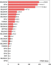
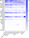
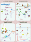
Selenium (Se) and its compounds have been reported to have great potential in the prevention and treatment of Alzheimer's disease (AD). However, little is known about the functional mechanism of Se in these processes, limiting its further clinical application. Se exerts its biological functions mainly through selenoproteins, which play vital roles in maintaining optimal brain function. Therefore, selenoproteins, especially brain function-associated selenoproteins, may be involved in the pathogenesis of AD. Here, we analyze the expression and distribution of 25 selenoproteins in the brain and summarize the relationships between selenoproteins and brain function by reviewing recent literature and information contained in relevant databases to identify selenoproteins (GPX4, SELENOP, SELENOK, SELENOT, GPX1, SELENOM, SELENOS, and SELENOW) that are highly expressed specifically in AD-related brain regions and closely associated with brain function. Finally, the potential functions of these selenoproteins in AD are discussed, for example, the function of GPX4 in ferroptosis and the effects of the endoplasmic reticulum (ER)-resident protein SELENOK on Ca2+ homeostasis and receptor-mediated synaptic functions. This review discusses selenoproteins that are closely associated with brain function and the relevant pathways of their involvement in AD pathology to provide new directions for research on the mechanism of Se in AD.
Keywords: Alzheimer’s disease; Ca2+ homeostasis; brain function; neurotransmission; selenoprotein.
Copyright © 2021 Zhang and Song.
Conflict of interest statement
The authors declare that the research was conducted in the absence of any commercial or financial relationships that could be construed as a potential conflict of interest.
Figures


Similar articles
-
Dietary selenium sources differentially regulate selenium concentration, mRNA and protein expression of representative selenoproteins in various tissues of yellow catfish Pelteobagrus fulvidraco.Br J Nutr. 2022 Feb 28;127(4):490-502. doi: 10.1017/S000711452100194X. Epub 2021 Jun 4. Br J Nutr. 2022. PMID: 34085611
-
Selenium Restores Synaptic Deficits by Modulating NMDA Receptors and Selenoprotein K in an Alzheimer's Disease Model.Antioxid Redox Signal. 2021 Oct 10;35(11):863-884. doi: 10.1089/ars.2019.7990. Epub 2020 Jul 7. Antioxid Redox Signal. 2021. PMID: 32475153
-
Selenomethionine alleviates environmental heat stress induced hepatic lipid accumulation and glycogen infiltration of broilers via maintaining mitochondrial and endoplasmic reticulum homeostasis.Redox Biol. 2023 Nov;67:102912. doi: 10.1016/j.redox.2023.102912. Epub 2023 Oct 4. Redox Biol. 2023. PMID: 37797371 Free PMC article.
-
Selenium, Selenoproteins, and Heart Failure: Current Knowledge and Future Perspective.Curr Heart Fail Rep. 2021 Jun;18(3):122-131. doi: 10.1007/s11897-021-00511-4. Epub 2021 Apr 9. Curr Heart Fail Rep. 2021. PMID: 33835398 Free PMC article. Review.
-
Endoplasmic reticulum-resident selenoproteins and their roles in glucose and lipid metabolic disorders.Biochim Biophys Acta Mol Basis Dis. 2024 Aug;1870(6):167246. doi: 10.1016/j.bbadis.2024.167246. Epub 2024 May 18. Biochim Biophys Acta Mol Basis Dis. 2024. PMID: 38763408 Review.
Cited by
-
Role of micronutrients in Alzheimer's disease: Review of available evidence.World J Clin Cases. 2022 Aug 6;10(22):7631-7641. doi: 10.12998/wjcc.v10.i22.7631. World J Clin Cases. 2022. PMID: 36158513 Free PMC article. Review.
-
Low-Density Lipoprotein Receptor-Related Protein 8 at the Crossroad between Cancer and Neurodegeneration.Int J Mol Sci. 2022 Aug 10;23(16):8921. doi: 10.3390/ijms23168921. Int J Mol Sci. 2022. PMID: 36012187 Free PMC article. Review.
-
Multi-omics approach reveals dysregulated genes during hESCs neuronal differentiation exposure to paracetamol.iScience. 2023 Aug 28;26(10):107755. doi: 10.1016/j.isci.2023.107755. eCollection 2023 Oct 20. iScience. 2023. PMID: 37731623 Free PMC article.
-
Epigenome-wide DNA methylation in leukocytes and toenail metals: The normative aging study.Environ Res. 2023 Jan 15;217:114797. doi: 10.1016/j.envres.2022.114797. Epub 2022 Nov 12. Environ Res. 2023. PMID: 36379232 Free PMC article.
-
Characterization and Quantification of Selenoprotein P: Challenges to Mass Spectrometry.Int J Mol Sci. 2021 Jun 11;22(12):6283. doi: 10.3390/ijms22126283. Int J Mol Sci. 2021. PMID: 34208081 Free PMC article. Review.
References
-
- Abasi M., Massumi M., Riazi G., Amini H. (2012). The synergistic effect of beta-boswellic acid and Nurr1 overexpression on dopaminergic programming of antioxidant glutathione peroxidase-1-expressing murine embryonic stem cells. Neuroscience 222 404–416. 10.1016/j.neuroscience.2012.07.009 - DOI - PubMed
-
- Bernal J. (2000). “Thyroid hormones in brain development and function,” in Endotext, eds Feingold K.R., Anawalt B., Boyce A., Chrousos G., De Herder W.W., Dungan K. (South Dartmouth, MA: ). - PubMed
Publication types
LinkOut - more resources
Full Text Sources
Other Literature Sources
Miscellaneous

