

Intra-Arterial, but Not Intrathecal, Baclofen and Codeine Attenuates Cough in the Cat
- PMID: 33746778
- PMCID: PMC7973226
- DOI: 10.3389/fphys.2021.640682
Intra-Arterial, but Not Intrathecal, Baclofen and Codeine Attenuates Cough in the Cat
Abstract
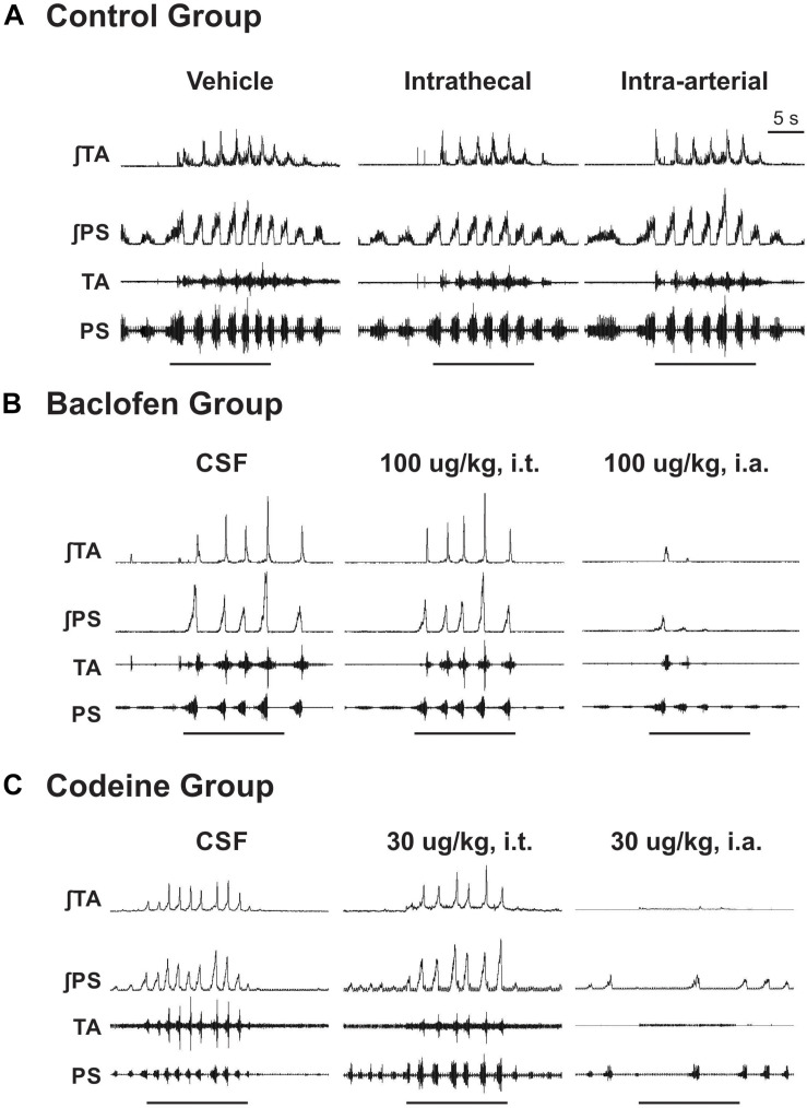
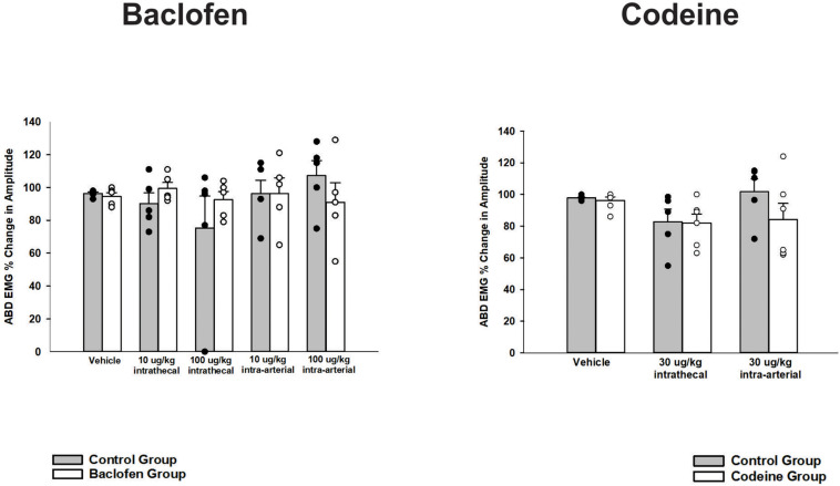
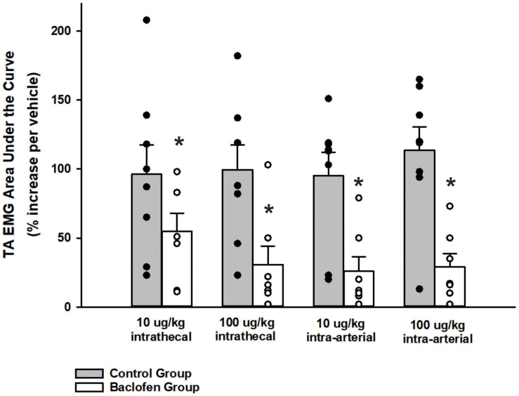
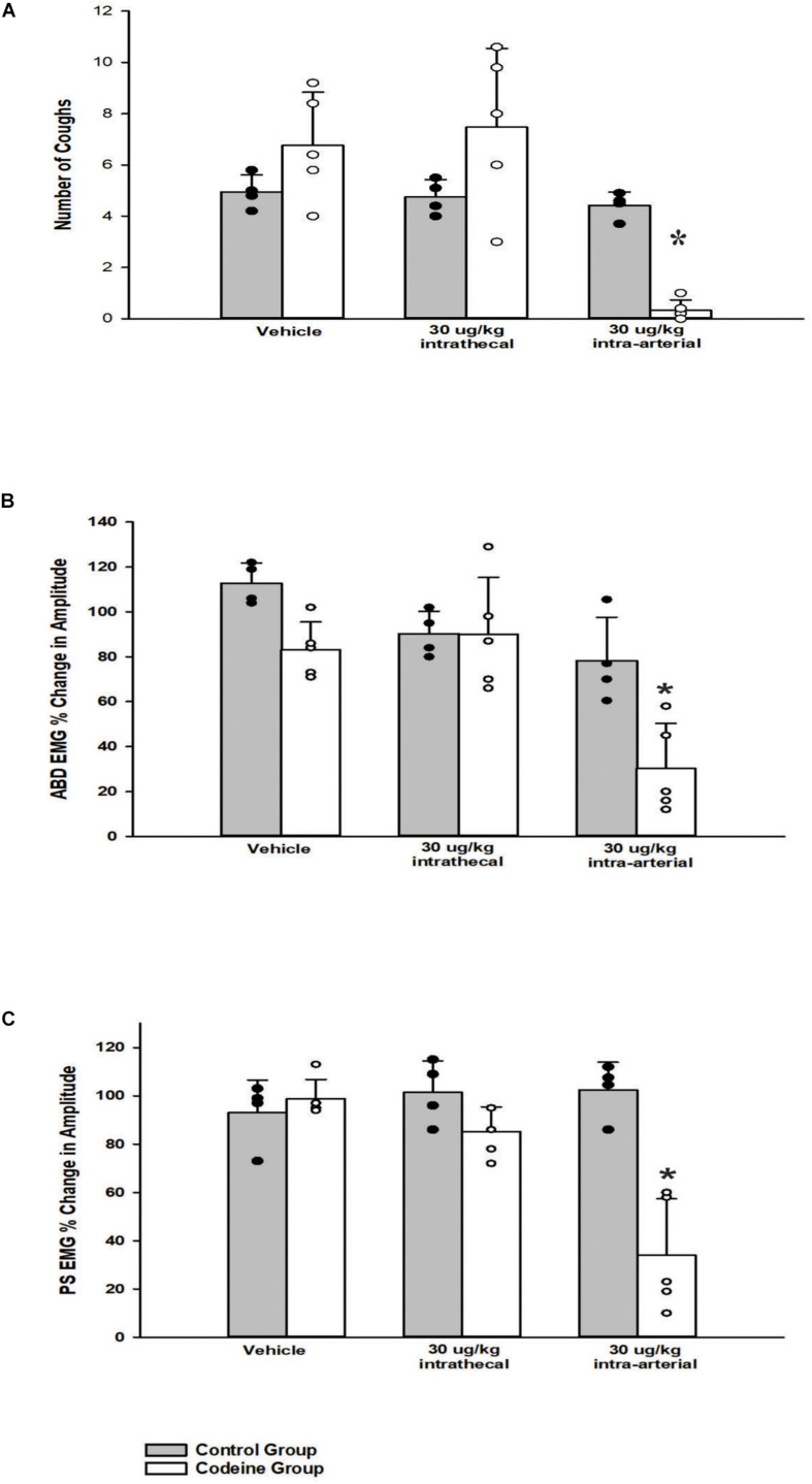
Centrally-acting antitussive drugs are thought to act solely in the brainstem. However, the role of the spinal cord in the mechanism of action of these drugs is unknown. The purpose of this study was to determine if antitussive drugs act in the spinal cord to reduce the magnitude of tracheobronchial (TB) cough-related expiratory activity. Experiments were conducted in anesthetized, spontaneously breathing cats (n = 22). Electromyograms (EMG) were recorded from the parasternal (PS) and transversus abdominis (TA) or rectus abdominis muscles. Mechanical stimulation of the trachea or larynx was used to elicit TB cough. Baclofen (10 and 100 μg/kg, GABA-B receptor agonist) or codeine (30 μg/kg, opioid receptor agonist) was administered into the intrathecal (i.t.) space and also into brainstem circulation via the vertebral artery. Cumulative doses of i.t. baclofen or codeine had no effect on PS, abdominal muscle EMGs or cough number during the TB cough. Subsequent intra-arterial (i.a.) administration of baclofen or codeine significantly reduced magnitude of abdominal and PS muscles during TB cough. Furthermore, TB cough number was significantly suppressed by i.a. baclofen. The influence of these drugs on other behaviors that activate abdominal motor pathways was also assessed. The abdominal EMG response to noxious pinch of the tail was suppressed by i.t. baclofen, suggesting that the doses of baclofen that were employed were sufficient to affect spinal pathways. However, the abdominal EMG response to expiratory threshold loading was unaffected by i.t. administration of either baclofen or codeine. These results indicate that neither baclofen nor codeine suppress cough via a spinal action and support the concept that the antitussive effect of these drugs is restricted to the brainstem.
Keywords: GABA-B receptor agonist; airway protection; antitussives; baclofen; codeine; cough; intrathecal; opioid.
Copyright © 2021 Olsen, Rose, Golder, Wang, Hammond and Bolser.
Conflict of interest statement
The authors declare that the research was conducted in the absence of any commercial or financial relationships that could be construed as a potential conflict of interest.
Figures

Similar articles
-
Peripheral and central sites of action of GABA-B agonists to inhibit the cough reflex in the cat and guinea pig.Br J Pharmacol. 1994 Dec;113(4):1344-8. doi: 10.1111/j.1476-5381.1994.tb17145.x. Br J Pharmacol. 1994. PMID: 7889290 Free PMC article.
-
Influence of central antitussive drugs on the cough motor pattern.J Appl Physiol (1985). 1999 Mar;86(3):1017-24. doi: 10.1152/jappl.1999.86.3.1017. J Appl Physiol (1985). 1999. PMID: 10066718
-
Influence of baclofen on laryngeal and spinal motor drive during cough in the anesthetized cat.Laryngoscope. 2013 Dec;123(12):3088-92. doi: 10.1002/lary.24143. Epub 2013 May 13. Laryngoscope. 2013. PMID: 23670824 Free PMC article.
-
Current and future centrally acting antitussives.Respir Physiol Neurobiol. 2006 Jul 28;152(3):349-55. doi: 10.1016/j.resp.2006.01.015. Epub 2006 Mar 6. Respir Physiol Neurobiol. 2006. PMID: 16517221 Free PMC article. Review.
-
Role of opioidergic and serotonergic mechanisms in cough and antitussives.Pulm Pharmacol. 1996 Oct-Dec;9(5-6):349-56. doi: 10.1006/pulp.1996.0046. Pulm Pharmacol. 1996. PMID: 9232674 Review.
Cited by
-
Cough hypersensitivity and chronic cough.Nat Rev Dis Primers. 2022 Jun 30;8(1):45. doi: 10.1038/s41572-022-00370-w. Nat Rev Dis Primers. 2022. PMID: 35773287 Free PMC article. Review.
References
-
- Aylward M., Maddock J., Davies D. E., Protheroe D. A., Leideman T. (1984). Dextromethorphan and codeine: comparison of plasma kinetics and antitussive effects. Eur. J. Respir. Dis. 65 283–291. - PubMed
-
- Bajic J., Zuperku E. J., Tonkovic-Capin M., Hopp F. A. (1992). Expiratory bulbospinal neurons of dogs. I. Control of discharge patterns by pulmonary stretch receptors. Am. J Physiol. 262 R1075–R1086. - PubMed
-
- Basmajian J. V., Stecko G. A. (1962). New Bipolar Electrode for Electromyography. J. Appl. Physiol. 17 849–849. 10.1152/jappl.1962.17.5.849 - DOI
Grants and funding
LinkOut - more resources
Full Text Sources
Other Literature Sources
Miscellaneous