. 2021 Apr 5;56(7):1014-1029.e7.
doi: 10.1016/j.devcel.2021.02.025. Epub 2021 Mar 17.

Negative elongation factor regulates muscle progenitor expansion for efficient myofiber repair and stem cell pool repopulation

Affiliations

Negative elongation factor regulates muscle progenitor expansion for efficient myofiber repair and stem cell pool repopulation

Daniel C L Robinson et al. Dev Cell. .

Abstract

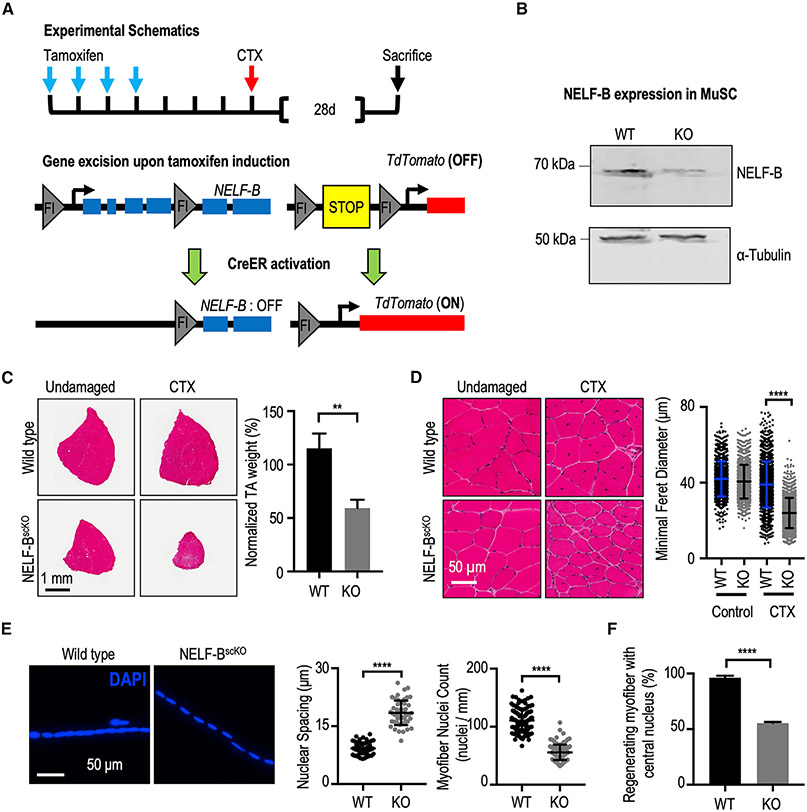
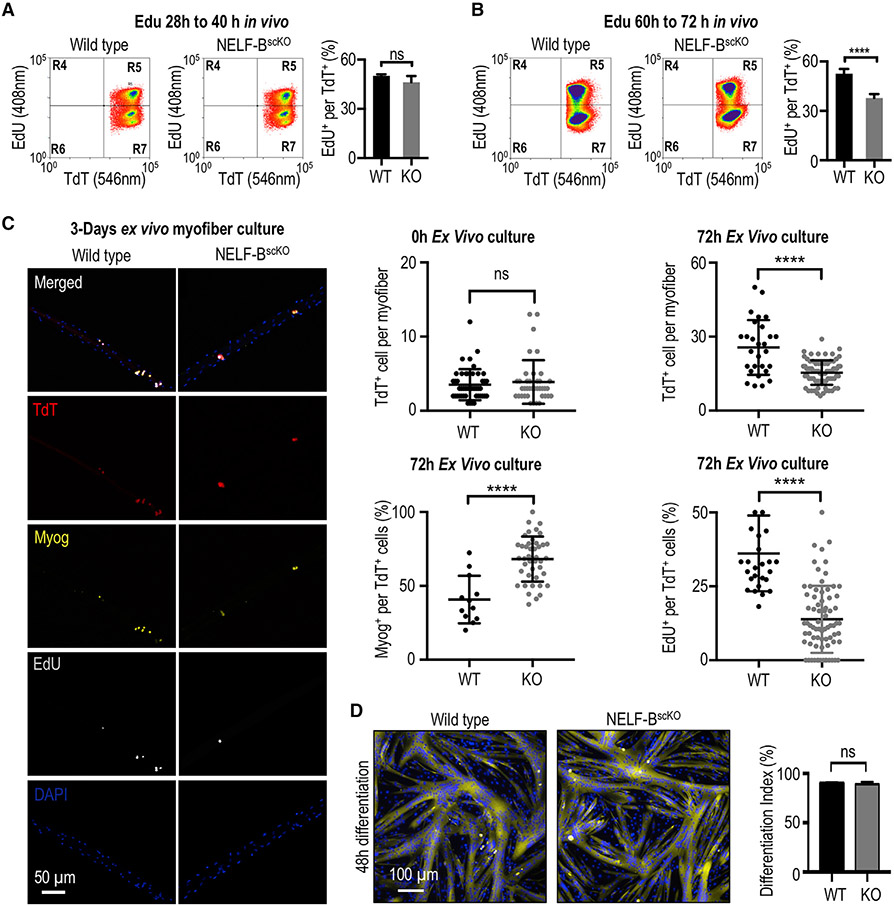
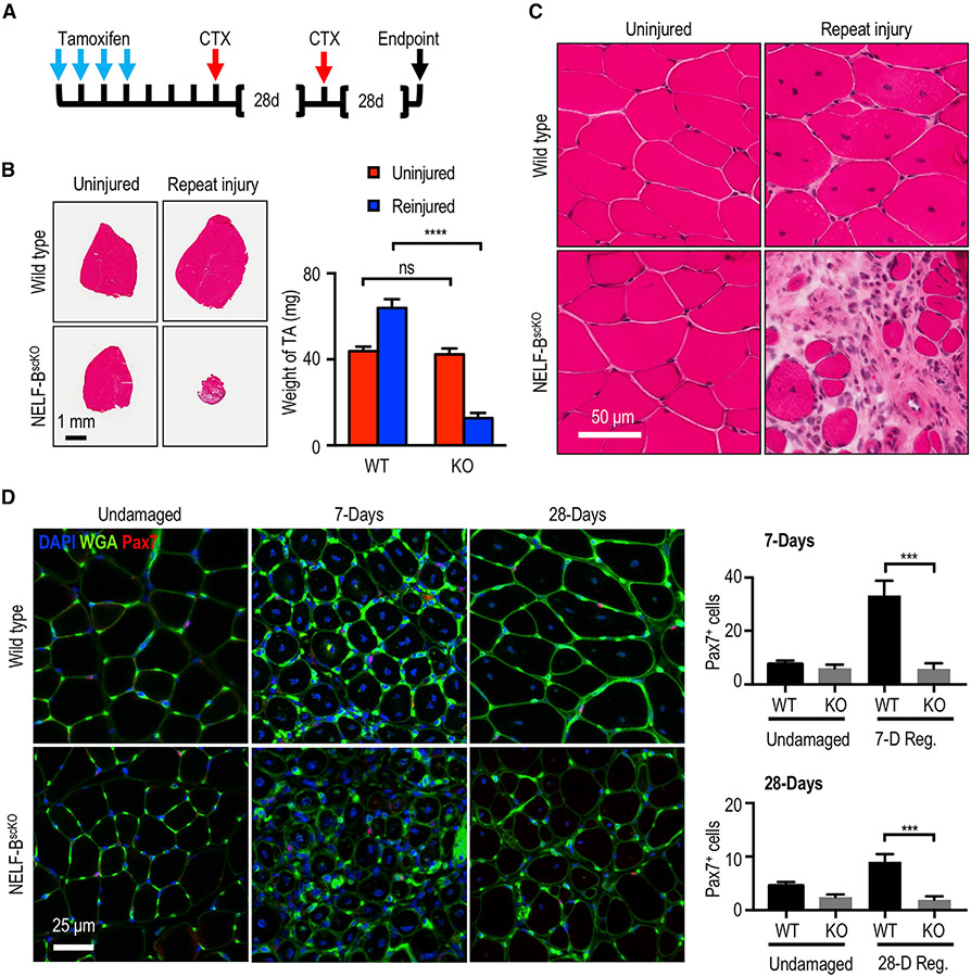
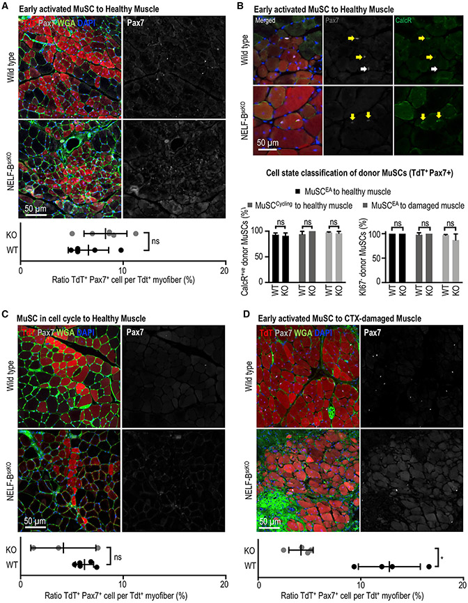
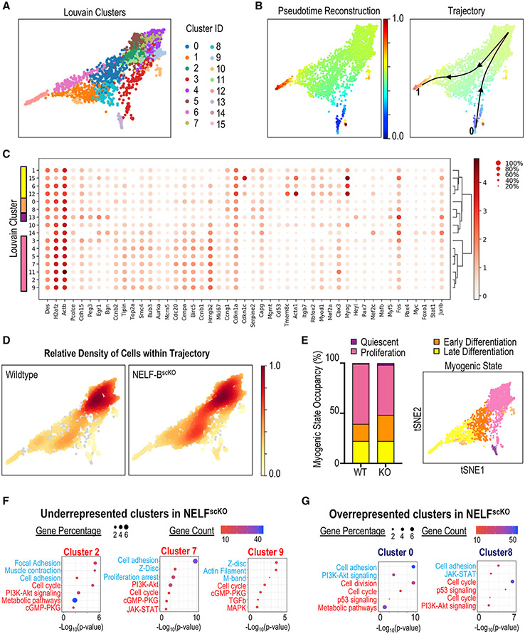
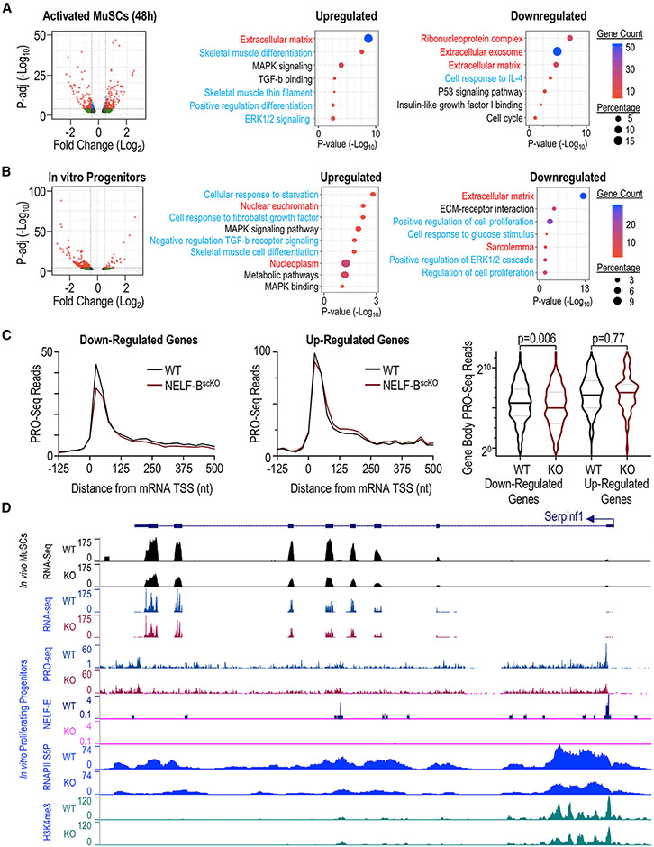
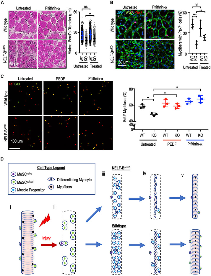
Negative elongation factor (NELF) is a critical transcriptional regulator that stabilizes paused RNA polymerase to permit rapid gene expression changes in response to environmental cues. Although NELF is essential for embryonic development, its role in adult stem cells remains unclear. In this study, through a muscle-stem-cell-specific deletion, we showed that NELF is required for efficient muscle regeneration and stem cell pool replenishment. In mechanistic studies using PRO-seq, single-cell trajectory analyses and myofiber cultures revealed that NELF works at a specific stage of regeneration whereby it modulates p53 signaling to permit massive expansion of muscle progenitors. Strikingly, transplantation experiments indicated that these progenitors are also necessary for stem cell pool repopulation, implying that they are able to return to quiescence. Thus, we identified a critical role for NELF in the expansion of muscle progenitors in response to injury and revealed that progenitors returning to quiescence are major contributors to the stem cell pool repopulation.

Keywords: NELF; PEDF signaling; muscle regeneration; muscle stem cells; nascent transcript stability; p53 signaling; promoter proximal pausing; stem cell niche; stem cell self-renewal; transcriptional regulation.


Conflict of interest statement

Declaration of interests The authors declare no competing interests.

Figures

Figure 1.
Figure 1.. NELF is required for efficient MuSC-mediated myofiber repair after injury
(A) Schematic representation of the recombination strategy. Tamoxifen administered in vivo activates CreERT2 to excise floxed genomic sequences. This is followed with intramuscular injection(s) of cardiotoxin to induce skeletal muscle damage. (B) Western blot of a MuSCs whole cell extract isolated from WT (Pax7CreER/TdTscKO) and NELF-BscKO (NELF-BscKOPax7CreERTdTscKO) mice shows a 90% deletion efficiency of NELF-B. (C) Hematoxylin and eosin stain of TA muscle cross-sections at 28 days post-injury (CTX), scale bar = 1 mm. The weight of the regenerated TA is presented relative to that of the undamaged contralateral leg and shows a reduction in size in the NELF-BscKO mice compared with WT controls (±SE, p-value < 0.01, n = 3). (D) Magnified view of the regenerated and undamaged TA cross-sections shown in (C) demonstrate a reduced minimal Feret’s myofiber diameter in the regenerated NELF-BscKO myofibers (±SE, p-value < 0.0001, n = 3), scale bar = 50 μm. (E) Single myofibers isolated from the regenerated EDL at 28 days post-injury were stained with DAPI to visualize nuclei from NELF-BscKO (n = 50 myofibers from 3 biological replicates) and WT (n = 57 myofibers from 3 biological replicates) mice, scale bar = 50 μm. Internuclear spacing is measured as the distance between the center of adjacent nuclei, whereas the myonuclei count was determined over the length of the myofiber (±SE, p-value < 0.0001). (F) The relative abundance of newly regenerated myofibers in which the centrally located nucleus is captured in a 10 μm cross-section was calculated with tissues shown in (C) (±SE, p-value < 0.0001, n = 3).
Figure 2.
Figure 2.. NELF is required for the massive expansion of muscle progenitors in response to injury
(A) FACS-acquired quantification of EdU+ MuSCs (identified as the TdT+/ITGA7+ population) derived from the skeletal muscle 40 h post-injury (±SE, n = 4). (B) FACS-acquired quantification of EdU+ MuSCs (identified as the TdT+/ITGA7+ population) derived from the skeletal muscle 72 h post-injury to monitor in vivo myoblast proliferation (±SE, p-value < 0.0001, n = 5). (C) Single myofibers isolated from the EDL muscle from NELF-BscKO (n = 47 from 3 biological replicates) or WT (n = 46 from 3 biological replicates) mice were cultured (0 or 72 h) and stained with antibodies as indicated, scale bar = 50 μm. Quantification of TdT+, EdU+, and Myog+ cells are shown (±SE, *p-value < 0.0001). (D) Primary myoblasts isolated from NELF-BscKO and WT controls were plated at high density and induced to undergo terminal differentiation, scale bar = 100 μm. The differentiation index was calculated as the percentage of nuclei present in multinucleated myotubes (±SE, n = 3).
Figure 3.
Figure 3.. Loss of NELF in the regenerating muscle leads to a depletion of the MuSC pool
(A) Experimental schematic showing the sequential injury approach where mice that had undergone tamoxifen-induced recombination were subjected to two rounds of CTX treatment at 28-day intervals. (B) Hematoxylin and eosin stain of the regenerated TA muscle after the repeat injury (±SE, p-value< 0.001, n = 3) was used to visualize regeneration and the weight of the regenerated muscle was determined relative to the uninjured contralateral TA muscle (±SE, n = 3), scale bar = 1 mm. (C) Magnification of the hematoxylin and eosin staining for the repeat-injured TA from (B) showing fibrosis and interstitial cells, scale bar = 50 μm. (D) Immunofluorescence characterization of MuSCs (Pax7+) in the regenerated TA muscle 7 or 28 days after injury (±SE, p-value < 0.001, n = 4), scale bar = 25 μm.
Figure 4.
Figure 4.. NELF is not required for muscle progenitors to return to quiescence and repopulated the MuSC niche
Engraftment experiments were performed using MuSCs derived from WT and NELF-BscKO into recipient mice. In all instances, 12,000 donor MuSCs (TdT+ ITGA7+) derived from WT or NELF-BscKOs were transplanted into NSG (NOD.Cg-Prkdcscid Il2rgtm1Wjl/SzJ) mice, which were irradiated to deplete the endogenous MuSC population. The TA muscles were recovered 28 days after transplantation and were subjected to immunofluorescence analysis using the antibodies indicated. The niche replenishment ratio was calculated by normalizing the number of donor MuSCs (TdT+ Pax7+) to the number of TdT+ myofibers. (A) Freshly isolated MuSCs from the healthy muscle of WT and NELF-BscKO donors were transplanted into uninjured, irradiated recipient NSG muscle(±SE, n = 5), scale bar = 50 μm. (B) The ability of transplanted MuSCs to return to the quiescent state was examined through co-staining of TdT+ cells in the niche with Pax7 and CalcR, or Pax7 and Ki67 (see Figure S4F) (±SE, n = 3), scale bar = 50 μm. (C) Activated MuSCs isolated from the regenerating muscle of WT and NELF-BscKO donors (40 h after CTX injection) into uninjured, irradiated recipient NSG muscle (±SE, n = 3), scale bar = 50 μm. (D) Freshly isolated MuSCs from the healthy muscle of WT and NELF-BscKO donors transplanted into irradiated, recipient NSG muscle at 48 h after CTX damage, scale bar = 50 μm. A paired, two-tailed t test was used to compare the contralateral-matched TA muscles for TdT+ donor cell contribution with the Pax7+ MuSC niche (±SE, p-value < 0.05, n = 3).
Figure 5.
Figure 5.. Single-cell transcriptome analysis of muscle progenitors in regeneration
Myogenic progenitor cells (TdT+, ITGA7+) were isolated from CTX injured hind-limb muscles at 72 h post-injury, sorted based on fluorescence, and subjected to scRNA-Seq using the 10x genomic platform. (A) Myogenic cells were clustered using the PAGA algorithm, which identified 16 different Louvain clusters on combined cell analysis for NELF-BscKO and WT samples. (B) Pseudotime trajectory re-construction using the PAGA algorithm shows the cell trajectory across 16 clusters from the Pax7+ MuSCs (blue), through to the Myog+ myocytes (red). (C) Dotplot analysis of the 16 clusters on selected genes shows differences in Louvain cluster gene signatures. (D) Density mapping of WT and NELF-BscKO myoblasts on the PAGA trajectory reveals differential population occupancy in clusters 0 and 8 (up in NELF-BscKO) as well as clusters 2, 7, and 9 (down in NELF-BscKO). (E) Cells were classified into different myogenic cell states based on global gene signatures representing quiescence (cluster 13), proliferation (cluster 2,3,4,5,7,9,11), early differentiation (cluster 0,8), and late differentiation (cluster 1,6,12,15). (F and G) Gene Ontology analysis was performed on differentially expressed genes identified in the 5 clusters that were either underrepresented (F) or overrepresented (G) in NELF-BscKO mice. Representative upregulated (blue) and downregulated (red) GO terms are shown as a function of Log10(p-value).
Figure 6.
Figure 6.. Transcriptome analysis of muscle progenitors in regeneration
RNA-seq analysis was performed on fluorescence-sorted NELF-BscKO and WT myogenic progenitors (TdT+ ITGA7+) using and adjusted p-value < 0.01 as a cutoff at different timepoints, represented as volcano plots. Gene Ontology analysis performed on differentially expressed genes shows terms that are upregulated and downregulated in NELF-BscKO samples as a function of Log10(p-value). Terms are represented based on their origin from biological process (blue), cell component (red), or Kegg pathway (black). (A) Myogenic progenitors freshly isolated from skeletal muscle at 48 h post-injury show 305 upregulated and 343 downregulated genes. (B) In vitro cultured myoblasts show 441 downregulated genes and 102 upregulated genes. (C) Precision Run-On sequencing mapped onto RNA-sequencing results show that downregulated genes in the NELF-BscKO mice have decreased nascent transcript levels at the promoter-proximal region as well as the gene body (p-value = 0.006). Genes upregulated in NELF-BscKO mice show no significant change in nascent transcript levels compared with WT controls (p-value = 0.77). (D) A representative UCSC browser track showing the reduced gene expression of SerpinF1 for RNA-seq performed at 48-h post-injury on RNA collected from cultured myoblasts. PRO-seq shows a loss of nascent transcript emerging from the transcription start site (TSS) upon a NELF-BscKO, and Cut&Tag analysis showing a loss of NELF-E binding at the promoter region upon a NELF-BscKO and a reduced occupancy of RNAPII (S5P).
Figure 7.
Figure 7.. Rescue experiments and proposed model explaining role of NELF in myogenesis
(A) Pifithrin-α (25 μM or vehicle) in Captisol (20% w/v in saline) was administered to mice by intraperitoneal injections (2 mg/kg of pifithrin-α) at 40, 64, and 88 h after CTX injury of the TA muscle. Following regeneration (7 days), histological analysis of regenerating muscle with hematoxylin and eosin staining was used to visualize the regeneration (±SE, p-value < 0.01, n = 3 with 500 myofiber measurements per biological replicate), scale bar = 50 μm. (B) Immunofluorescent characterization of the Pax7+ population in the regenerated TA cross-sections after pifithrin-α treatment (±SE, **p-value < 0.01 or ***p-value < 0.001, n = 3), scale bar 50 μm. (C) Myoblasts were isolated from induced WT and NELF-BscKO mice and treated in culture with PEDF (500 ng/mL) or pifithrin-α (25 μM). Proliferating cells were then pulsed with EdU (10 μM) for 4 h (n = 3) scale bar = 100 μm. (D) Proposed model for the implication of NELF in skeletal muscle regeneration. When healthy skeletal muscle is injured (i), MuSCs are activated and beginning to undergo population expansion (ii). Reduced proliferation upon a NELF-BscKO results in lower myogenic progenitor cell presence than in WT conditions (iii). As regeneration progresses (iv), myogenic progenitors will either commit to terminal differentiation to form new skeletal muscle, or return to quiescence to repopulate the stem cell niche (v).

Similar articles

Cited by

References

    1. Adelman K, and Lis JT (2012). Promoter-proximal pausing of RNA polymerase II: emerging roles in metazoans. Nat. Rev. Genet 13, 720–731. - PMC - PubMed
    1. Adelman K, Marr MT, Werner J, Saunders A, Ni Z, Andrulis ED, and Lis JT (2005). Efficient release from promoter-proximal stall sites requires transcript cleavage factor TFIIS. Mol. Cell 17, 103–112. - PubMed
    1. Amemiya HM, Kundaje A, and Boyle AP (2019). The ENCODE blacklist: identification of problematic regions of the genome. Sci. Rep 9, 9354. - PMC - PubMed
    1. Amleh A, Nair SJ, Sun J, Sutherland A, Hasty P, and Li R (2009). Mouse cofactor of BRCA1 (Cobra1) is required for early embryogenesis. PLoS One 4, e5034. - PMC - PubMed
    1. Aoi Y, Smith ER, Shah AP, Rendleman EJ, Marshall SA, Woodfin AR, Chen FX, Shiekhattar R, and Shilatifard A (2020). NELF regulates a promoter-proximal step distinct from RNA Pol II pause-release. Mol. Cell 78, 261–274.e5. - PMC - PubMed

Publication types

MeSH terms