

Fate Mapping of Cancer Cells in Metastatic Lymph Nodes Using Photoconvertible Proteins
- PMID: 33704727
- PMCID: PMC8924909
- DOI: 10.1007/978-1-0716-1205-7_26
Fate Mapping of Cancer Cells in Metastatic Lymph Nodes Using Photoconvertible Proteins
Abstract
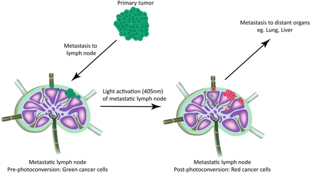
The lymph node microenvironment is extremely dynamic and responds to immune stimuli in the host by reprogramming immune, stromal, and endothelial cells. In normal physiological conditions, the lymph node will initiate an appropriate immune response to clear external threats that the host may experience. However, in metastatic disease, cancer cells often colonize local lymph nodes, disrupt immune function, and even leave the lymph node to create additional metastases. Understanding how cancer cells enter, colonize, survive, proliferate, and interact with other cell types in the lymph node is challenging. Here, we describe the use of photoconvertible fluorescent proteins to label and trace the fate of cancer cells once they enter the lymph node.
Keywords: Circulating tumor cells; Confocal microscopy; Dendra2; Intravital imaging; Lymph node; Metastasis; Photoconvertible proteins; Photodiode.
Figures
Similar articles
-
Lymph node metastases can invade local blood vessels, exit the node, and colonize distant organs in mice.Science. 2018 Mar 23;359(6382):1403-1407. doi: 10.1126/science.aal3622. Epub 2018 Mar 22. Science. 2018. PMID: 29567713 Free PMC article.
-
Spatiotemporally controlled induction of gene expression in vivo allows tracking the fate of tumor cells that traffic through the lymphatics.Int J Cancer. 2020 Aug 15;147(4):1190-1198. doi: 10.1002/ijc.32766. Epub 2019 Nov 21. Int J Cancer. 2020. PMID: 31675122
-
Lymph node blood vessels provide exit routes for metastatic tumor cell dissemination in mice.Science. 2018 Mar 23;359(6382):1408-1411. doi: 10.1126/science.aal3662. Science. 2018. PMID: 29567714
-
Lymphatic metastasis.Cancer Metastasis Rev. 1983;2(3):307-17. doi: 10.1007/BF00048483. Cancer Metastasis Rev. 1983. PMID: 6367969 Review.
-
The lymph node microenvironment and its role in the progression of metastatic cancer.Semin Cell Dev Biol. 2015 Feb;38:98-105. doi: 10.1016/j.semcdb.2015.01.008. Epub 2015 Jan 22. Semin Cell Dev Biol. 2015. PMID: 25620792 Free PMC article. Review.
Cited by
-
Volume imaging to interrogate cancer cell-tumor microenvironment interactions in space and time.Front Immunol. 2023 May 16;14:1176594. doi: 10.3389/fimmu.2023.1176594. eCollection 2023. Front Immunol. 2023. PMID: 37261345 Free PMC article. Review.
References
-
- Cady B (2007) Regional lymph node metastases, a singular manifestation of the process of clinical metastases in cancer: contemporary animal research and clinical reports suggest unifying concepts. Cancer Treat Res 135:185–201 - PubMed
-
- Kawada K, Taketo MM (2011) Significanceand mechanism of lymph node metastasis in cancer progression. Cancer Res 71 (4):1214–1218 - PubMed
MeSH terms
Substances
Grants and funding
LinkOut - more resources
Full Text Sources
Other Literature Sources
