. 2021 Apr 26;95(10):e00015-21.
doi: 10.1128/JVI.00015-21. Epub 2021 Mar 10.

Oncolytic Foamy Virus - generation and properties of a nonpathogenic replicating retroviral vector system that targets chronically proliferating cancer cells

Affiliations

Oncolytic Foamy Virus - generation and properties of a nonpathogenic replicating retroviral vector system that targets chronically proliferating cancer cells

Karol M Budzik et al. J Virol. .

Abstract

Nonpathogenic retroviruses of the Spumaretrovirinae subfamily can persist long-term in the cytoplasm of infected cells, completing their lifecycle only after the nuclear membrane dissolves at the time of cell division. Since the targeting of slowly dividing cancer cells remains an unmet need in oncolytic virotherapy we constructed a replication competent Foamy Virus vector (oFV) from the genomes of two chimpanzee Simian Foamy Viruses (PAN1 and PAN2) and inserted a GFP transgene in place of the bel-2 open reading frame. oFV-GFP infected and propagated with slow kinetics in multiple human tumor cell lines, inducing a syncytial cytopathic effect. Infection of growth arrested MRC5 cells was not productive, but oFV genomes persisted in the cytoplasm and the productive viral lifecycle resumed when cell division was later restored. In vivo, the virus propagated extensively in intraperitoneal ovarian cancer xenografts, slowing tumor growth, significantly prolonging survival of the treated mice and sustaining GFP transgene expression for at least 45 days. Our data indicate that oFV is a promising new replication-competent viral and gene delivery platform for efficient targeting of the most fundamental trait of cancer cells, their ability to sustain chronic proliferation.Significance:The infectivity of certain retroviruses is limited to dividing cells, which makes them attractive tools for targeting cancer cell proliferation. Previously developed replication-competent gammaretroviral vectors spread efficiently in rapidly dividing cancer cells, but not in cancer cells that divide more slowly. In contrast to rapidly proliferating transplantable mouse tumors, slow proliferation is a hallmark of human cancers and may have contributed to the clinical failure of the preclinically promising Murine Leukemia Virus vector Toca511 which failed to show efficacy in a phase 3 clinical trial in patients with glioblastoma. The studies presented in our manuscript show that oncolytic Foamy Virus (oFV) vectors are capable of persisting unintegrated in quiescent cells and resuming their life cycle once the cells start dividing again. This property of oFVs, together with their lack of pathogenicity and their ability to catalyze the fusion of infected cancer cells, makes them an attractive platform for further investigation.


Figures

FIG 1
FIG 1
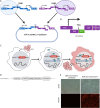
Generation of the oFV vectors and indicator cell lines for monitoring FV replication. (A) Infectious molecular clone for the chimeric oFV virus comprised of genome segments from PAN1 and PAN2 that were cloned into pcDNA3.1 and ligated together. (B) Engineering of the GFP-carrying virus. A part of bel2 was replaced with emerald GFP cDNA and linked to tas via a T2A self-cleaving peptide. (C) Indicator cells contain a reporter gene driven by the U3 SFV promoter, which is activated by Tas during FV infection (see text for details). (D) Structure of the lentiviral vector used for generation of indicator cell lines. SFV U3, promoter-containing U3 region of the SFV LTR; Ubiq. prom., ubiquitin promoter; PGK prom, phosphoglycerate kinase promoter; puromycin, puromycin resistance cassette; neomycin, neomycin resistance cassette; WPRE, woodchuck hepatitis virus posttranscriptional regulatory element. (E) Indicator BHK-U3-mCherry cells express mCherry only when infected with oFV.
FIG 2
FIG 2
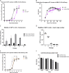
Transgene insertion attenuates the oFV vector, but the GFP transgene is stable for up to 5 passages. (A) Indicator BHK-U3-mCherry cells were infected at an MOI of 0.01 with oFV or oFV-GFP, and the spread of the viruses was assessed by sampling the cells every other day using flow cytometry analysis to determine the percentage of mCherry- or GFP-positive cells at each time point. Results of two independent experiments, each with two biological replicates, are presented. (B) BHK-U3-mCherry cells were infected at an MOI of 3 with oFV or oFV-GFP, and the titers of progeny virus were measured at different time points postinfection. Results of two independent experiments, each with two biological replicates, are presented. (C) oFV-GFP was passaged in U251-U3-mCherry cells 5 times. The first passage was done at an MOI of 0.5. For every subsequent passage, 250 μl of media from the previous passage was used for infection of fresh U251-U3-mCherry cells. Four and 8 days into each passage, the infected cells were analyzed by flow cytometry to determine the percentage of GFP-positive cells. Presented are results of 2 independent experiments with 1 biological replicate. (D) oFV-GFP was passaged in U251-U3-mCherry cells 5 times as described above. At the end of each passage, the infected cells were collected, their genomic DNA isolated, and env and GFP copy number relative to β-actin was determined using qPCR. Result of an experiment with 3 biological replicates is shown as GFP-to-env copy number ratio. (E) Indicator BHK-U3-mCherry cells were infected at an MOI of 0.01 with oFV or the parental PAN1 and PAN2 viruses, and their spread was assessed by sampling the cells every other day using flow cytometry analysis to determine the percentage of mCherry- or GFP-positive cells at each time point. Result of two independent experiments, each with two biological replicates, are presented. (F) Titers of cell-free and cell-associated progeny PNA1, PAN2, oFV, and oFV-GFP virions measured on day 8 postinfection at an MOI of 0.01. Results of two independent experiments, each with two biological replicates, are presented.
FIG 3
FIG 3
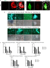
oFV and oFV-GFP cause intercellular fusion and have a broad cancer tropism. (A) Indicator U251-U3-mCherry cells were infected with oFV or oFV-GFP, and the syncytia induced by the virus were imaged with confocal microscopy (white, 4′,6-diamidino-2-phenylindole [DAPI]; red, mCherry; green, GFP). (B) Various human cancer cell lines were infected with oFV-GFP at an MOI of 0.5 and imaged for the expression of GFP on indicated days postinfection. U251, glioblastoma; Mia Paca, pancreatic adenocarcinoma; CDB1, cholangiocarcinoma; A549, lung adenocarcinoma; SKOV, ovarian carcinoma; H226, mesothelioma; HT-29, colorectal carcinoma; PC3, prostate adenocarcinoma. Extent of syncytium formation varies between different cell lines, with U251, H226, A549, and PC3 fusing well. (C) Various human cancer cell lines were infected with oFV at an MOI of 0.5 or 1, and viability was measured 5 and 6 or 8 days postinfection. Results of 2 independent experiments with 3 biological replicates are shown.
FIG 4
FIG 4
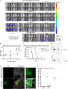
oFV and oFV-GFP have an oncolytic activity in vivo, and infection leads to intratumoral transgene expression. (A) We implanted 2.5 million SKOV-3-Fluc cells intraperitoneally in athymic nude mice and treated them with a single, intraperitoneal dose of 1 × 107 IU of oFV, oFV-GFP, or PBS control 7 days post-tumor implantation. The tumor burden was measured at indicated time points by bioluminescence imaging with IVIS Lumina; 10 mice per group were used. Both oFV and oFV-GFP exhibited significant oncolytic potential in vivo. D, dorsal view; V, ventral view. (B) Quantification of the ventral bioluminescence signal in panel A. (C) Survival of the SKOV-3-luc-tumor-bearing mice after oFV vector therapy; *, P < 0.0001. (D) Tumors from PBS- and oFV-GFP treated mice which eventually succumbed to the disease were explanted. Total DNA was isolated from the explants and PCR analyzed using primers that bind within the oFV env gene and primers that bind upstream and downstream of the transgene insertion site within the remaining sequences of bel2. +C, positive control (poFV-GFP plasmid); NTC, no-template control. (E) Immunohistochemistry staining for GFP in sections of an oFV-GFP-infected tumor harvested 45 days postinfection. (F) Total DNA was isolated from oFV-GFP-infected tumors harvested from mice that succumbed to the disease on day 45 (tumor 1), 56 (tumors 2, 3, and 4), 65 (tumor 5), or 72 (tumor 6) postinfection, and GFP and env copy number relative to β-actin were measured using qPCR.
FIG 5
FIG 5
oFV-GFP spreads in slowly dividing cancer cells faster than MLV-GFP and, unlike MLV-GFP, can latently persist in quiescent cells. (A and B) Multistep growth curves of MLV-GFP and oFV-GFP in rapidly dividing U251 (A) and slowly dividing H226 (B) cancer cell lines; cells were infected at an MOI of 0.01, and the spread of the virus was followed by sampling the infected cells every 2 (U251) or every 3 (H226) days. Quantification of GFP-positive cells with flow cytometry; results of 2 independent experiments with 2 biological replicates are presented. (C) MRC5 cells were serum starved (0.1% FBS) for 4 days or cultured in 10% FBS media and stained with propidium iodide to determine the percentage of cells in different phases of the cell cycle and confirm the quiescent state of the cells. Analysis of the proportion of cells in different phases of the cell cycle was performed with ModFit LT V3.3.11. (D) Comparison of the ability of MLV-GFP and oFV-GFP to productively infect cells that are cycling, quiescent, or quiescent and induced to divide 12 h before infection or 8, 16, 32, or 64 h postinfection. All groups were infected with MLV-GFP or oFV-GFP at an MOI of 1. Results of 2 independent experiments with 3 biological replicates are shown.
FIG 6
FIG 6
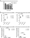
oFV-GFP exhibits stronger cytotoxicity and transgene expression that MLV-GFP. (A) Viability of U251 cells infected with MLV-GFP or oFV-GFP. Unlike MLV, oFV kills infected cells (statistical significance determined using the Holm-Sidak method; n = 3). (B)s Comparison of GFP expression in MRC5 and U251 cells infected with MLV-GFP and oFV-GFP 3 days postinfection at an MOI of 1 using qRT-PCR Statistical significance determined using the Mann-Whitney test; 2 independent experiments with 3 biological replicates. (C) Comparison of GFP expression in MRC5 and U251 cells infected with MLV-GFP and oFV-GFP 3 days postinfection at an MOI of 1 using flow cytometry. Statistical significance was determined using the Mann-Whitney test; n = 6.

Similar articles

Cited by

References

    1. Rethwilm A, Bodem J. 2013. Evolution of foamy viruses: the most ancient of all retroviruses. Viruses 5:2349–2374. 10.3390/v5102349. - DOI - PMC - PubMed
    1. Rethwilm A. 1995. Regulation of foamy virus gene expression. Curr Top Microbiol Immunol 193:1–24. 10.1007/978-3-642-78929-8_1. - DOI - PubMed
    1. Shankar A, Shanmugam V, Switzer WM. 2020. Complete genome sequence of a baboon simian foamy virus isolated from an infected human. Microbiol Resour Announc 9:e00522-20. 10.1128/MRA.00522-20. - DOI - PMC - PubMed
    1. Rua R, Betsem E, Calattini S, Saib A, Gessain A. 2012. Genetic characterization of simian foamy viruses infecting humans. J Virol 86:13350–13359. 10.1128/JVI.01715-12. - DOI - PMC - PubMed
    1. Russell RA, Wiegand HL, Moore MD, Schafer A, McClure MO, Cullen BR. 2005. Foamy virus bet proteins function as novel inhibitors of the APOBEC3 family of innate antiretroviral defense factors. J Virol 79:8724–8731. 10.1128/JVI.79.14.8724-8731.2005. - DOI - PMC - PubMed