. 2021 Feb 22:2021:6674283.
doi: 10.1155/2021/6674283. eCollection 2021.

Lin28 Inhibits the Differentiation from Mouse Embryonic Stem Cells to Glial Lineage Cells through Upregulation of Yap1

Affiliations

Lin28 Inhibits the Differentiation from Mouse Embryonic Stem Cells to Glial Lineage Cells through Upregulation of Yap1

Juan Luo et al. Stem Cells Int. .

Abstract

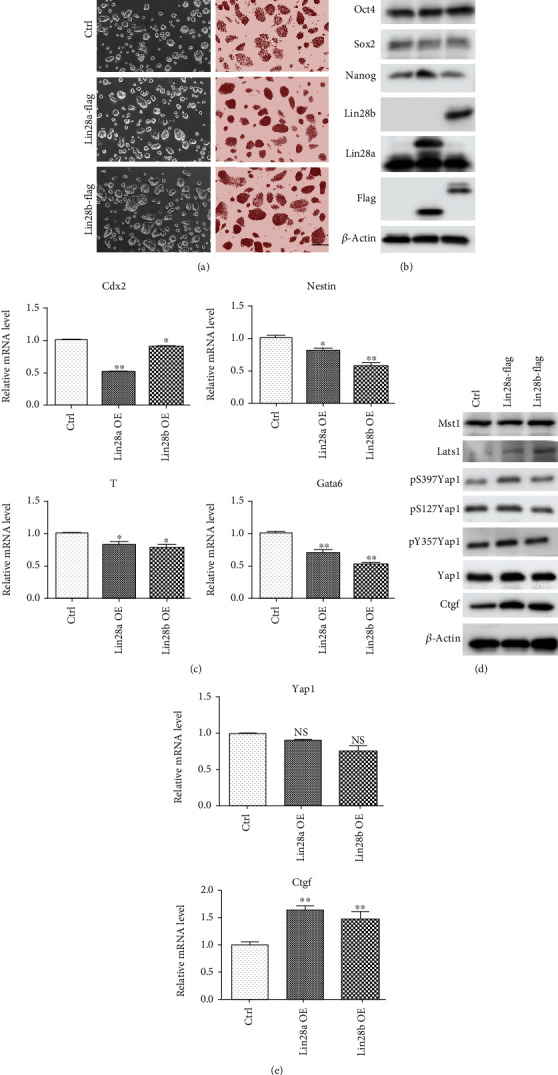
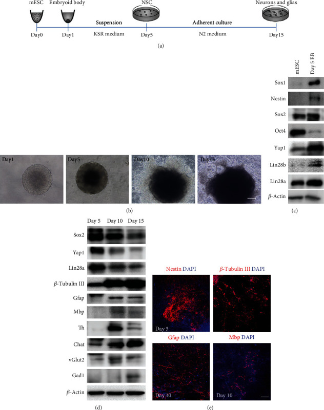
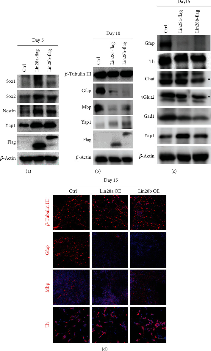
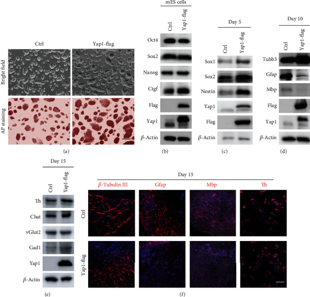
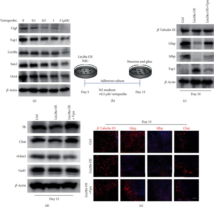
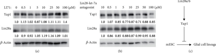
The RNA-binding protein Lin28 regulates neurogliogenesis in mammals, independently of the let-7 microRNA. However, the detailed regulatory mechanism remains obscured. Here, we established Lin28a or Lin28b overexpression mouse embryonic stem cells (ESCs) and found that these cells expressed similar levels of the core pluripotent factors, such as Oct4 and Sox2, and increased Yap1 but decreased lineage-specific markers compared to the control ESCs. Further differentiation of these ESCs to neuronal and glial lineage cells revealed that Lin28a/b overexpression did not affect the expression of neuronal marker βIII-tubulin, but dramatically inhibited the glial lineage markers, such as Gfap and Mbp. Interestingly, overexpression of Yap1 in mouse ESCs phenocopied Lin28a/b overexpression ESCs by showing defect in glial cell differentiation. Inhibition of Yap1/Tead-mediated transcription with verteporfin partially rescued the differentiation defect of Lin28a/b overexpression ESCs. Mechanistically, we demonstrated that Lin28 can directly bind to Yap1 mRNA, and the induction of Yap1 by Lin28a in mESCs is independent of Let7. Taken together, our results unravel a novel Lin28-Yap1 regulatory axis during mESC to glial lineage cell differentiation, which may shed light on glial cell generation in vitro.


Conflict of interest statement

The authors declare no conflicts of interest.

Figures

Figure 1
Figure 1
Constitutive expression of Lin28a/b in mouse ESC induced Yap1 expression and reduced lineage-specific gene expression. (a) Phase-contrast microscopy and AP staining of Ctrl and Lin28a/b constitutively expressed (Lin28a/b OE) mouse ESCs grown under 2i + LIF medium. Scale bar, 200 μm. (b) Western blot analyses of total proteins from Ctrl and Lin28a/b OE mouse ESCs using the indicated antibodies. Oct4, Sox2, and Nanog are pluripotent stem cell markers. (c) Quantitative real-time PCR to examine the mRNA level of lineage-specific gene expression in Ctrl and Lin28a/b OE mouse ESCs, trophectoderm gene Cdx2, ectoderm gene Nestin, mesoderm gene T, and endoderm gene Gata6. Actin was analyzed as an internal control. The data are shown as the mean ± S.D (n = 3). Statistically significant differences were indicated (, P < 0.05 and ∗∗, P < 0.01). (d) Western blot analyses of total proteins from Ctrl and Lin28a/b OE mouse ESCs using the indicated antibodies. (e) Quantitative real-time PCR to examine the mRNA level of Yap1 and its downstream target gene Ctgf expression in Ctrl and Lin28a/b OE mouse ESCs. The data are shown as the mean ± S.D (n = 3).
Figure 2
Figure 2
Establishment of the in vitro differentiation protocol from mouse ESCs to neuronal and glial lineage cells. (a) A schematic drawing of the direct differentiation assay from mouse ESCs to neuronal and glial lineage cells. ESCs were cultured under feeder-free condition with 2i + LIF medium for 1 day and then disassociated to single cells and quickly aggregated in differentiation medium for one day. After 5 days of suspension culture in KSR medium, the aggregates were subjected to adhesion culture for another 10 or 15 days in N2 medium. (b) Phase-contrast microscopy of mouse ESC differentiated cells on days 1, 5, 10, and 15. Scale bar, 200 μm. (c) Western blot analyses of total proteins from mESC differentiated cells on day 0 and 5 using the indicated antibodies. Mouse ESC pluripotent markers: Oct4 and Sox2, neural stem cell markers: Sox1, Nestin, and Sox2. (d) Western blot analyses of total proteins from mouse ESC differentiated cells on days 5, 10, and 15 using the indicated antibodies. Neuronal marker: β-tubulin III, glial markers: Gfap and Mbp, neuronal subtype markers: Th, vGlut2, Gad1, and Chat. (e) Immunofluorescence staining of the neural stem cell marker Nestin on day 5, neuronal marker β-tubulin III and glial markers Gfap and Mbp on day 10. Cell nuclear was stained with DAPI. Scale bar, 200 μm.
Figure 3
Figure 3
Constitutive overexpression of Lin28a/b inhibited the differentiation of mouse ESCs to glial lineage cells. (a) Western blot analyses of total proteins from Ctrl and Lin28a/b OE mouse ESC differentiated cells on day 5 using the indicated neural stem cell markers: Sox1, Nestin, and Sox2. (b) Western blot analyses of total proteins from Ctrl and Lin28a/b OE mouse ESC differentiated cells on day 10 using the indicated neuronal marker: β-tubulin III, glial markers: Gfap and Mbp. (c) Western blot analyses of total proteins from Ctrl and Lin28a/b OE mouse ESC differentiated cells on day 15 using the indicated neuronal subtype markers: Th, vGlut2, Gad1, and Chat. (d) Immunofluorescence staining of the Ctrl and Lin28a/b OE mouse ESC differentiated cells on day 15 using the neuronal marker β-tubulin III and glial markers Gfap and Mbp, and neuronal subtype markers Th. Cell nuclear was stained with DAPI. Scale bar, 200 μm.
Figure 4
Figure 4
Yap1 overexpression in mESCs phenocopied the glial cell lineage defect differentiated from Lin28a/b OE cells. (a) Phase-contrast microscopy and AP staining of Ctrl and Yap1 constitutively expressed (Yap1 OE) mouse ESCs grown under 2i + LIF medium. Scale bar, 200 μm. (b) Western blot analyses of total proteins from Ctrl and Yap1 OE mouse ESCs using the indicated antibodies. Oct4, Sox2, and Nanog are pluripotent stem cell markers. (c) Western blot analyses of total proteins from Ctrl and Yap1 OE mouse ESC differentiated cells on day 5 using the indicated neural stem cell markers: Sox1, Nestin, and Sox2. (d) Western blot analyses of total proteins from Ctrl and Yap1 OE mouse ESC differentiated cells on day 10 using the indicated neuronal marker: β-tubulin III, glial markers: Gfap and Mbp. (e) Western blot analyses of total proteins from Ctrl and Yap1 OE mouse ESC differentiated cells on day 15 using the indicated neuronal subtype markers: Th, vGlut2, Gad1, and Chat. (f) Immunofluorescence staining of the Ctrl and Yap1 OE mouse ESC differentiated cells on day 15 using the neuronal marker β-tubulin III, glial markers Gfap and Mbp, and neuronal subtype markers Th. Cell nuclear was stained with DAPI. Scale bar, 200 μm.
Figure 5
Figure 5
Inhibition of Yap1-Tead interaction using verteporfin partially rescued the glial cell lineage differentiation defect of Lin28a/b OE mouse ESCs. (a) Western blot analyses of total proteins from Ctrl mouse ESCs treated with different concentrations of Yap1-Tead interaction inhibitor (verteporfin) using the indicated antibodies. (b) A schematic drawing of the differentiation assay from neural stem cells (day 5) to neuronal and glial lineage cells (day 15) combining with verteporfin. (c) Western blot analyses of total proteins from Ctrl and Lin28a OE mouse ESC differentiated cells (treated with DMSO and Verteporfin, respectively) on day 10 using the indicated antibodies. (d) Western blot analyses of total proteins from Ctrl and Lin28a OE mouse ESC differentiated cells (treated with DMSO and verteporfin, respectively) on day 15 using the indicated antibodies. (e) Immunofluorescence staining of the Ctrl and Lin28a OE mouse ESC differentiated cells (treated with DMSO and verteporfin, respectively) on day 15 using the neuronal marker β-tubulin III, glial markers Gfap and Mbp, and neuronal subtype markers chat. Cell nuclear was stained with DAPI. Scale bar, 200 μm.
Figure 6
Figure 6
Induction of Yap1 by Lin28a/b in mouse ESCs was independent of Let7 pathway. (a) Western blot analyses of total proteins from Lin28a OE mouse ESCs treated with different concentrations of Lin28-let7 inhibitor (LI71) using the indicated antibodies. (b) Western blot analyses of total proteins from Lin28a OE mouse ESCs treated with different concentrations of Lin28-let7a antagonist using the indicated antibodies. (c) Schematic of the Lin28a/b-Yap1 pathway activity regulating mouse ESC differentiation to glial lineage cells.

Similar articles

Cited by

References

    1. Moss E. G., Lee R. C., Ambros V. The cold shock domain protein LIN-28 controls developmental timing in C. elegans and is regulated by the lin-4 RNA. Cell. 1997;88(5):637–646. doi: 10.1016/S0092-8674(00)81906-6. - DOI - PubMed
    1. Moss E. G., Tang L. Conservation of the heterochronic regulator Lin-28, its developmental expression and microRNA complementary sites. Developmental Biology. 2003;258(2):432–442. doi: 10.1016/S0012-1606(03)00126-X. - DOI - PubMed
    1. Shyh-Chang N., Daley G. Q. Lin28: primal regulator of growth and metabolism in stem cells. Cell Stem Cell. 2013;12(4):395–406. doi: 10.1016/j.stem.2013.03.005. - DOI - PMC - PubMed
    1. Ye Z., Su Z., Xie S., et al. Yap-lin28a axis targets let7-Wnt pathway to restore progenitors for initiating regeneration. Elife. 2020;9 doi: 10.7554/eLife.55771. - DOI - PMC - PubMed
    1. Xiong H., Shen J., Chen Z., et al. H19/let‑7/Lin28 ceRNA network mediates autophagy inhibiting epithelial‑mesenchymal transition in breast cancer. International Journal of Oncology. 2020;56(3):794–806. doi: 10.3892/ijo.2020.4967. - DOI - PubMed