Intracellular pH regulation: characterization and functional investigation of H+ transporters in Stylophora pistillata
- PMID: 33685406
- PMCID: PMC7941709
- DOI: 10.1186/s12860-021-00353-x
Intracellular pH regulation: characterization and functional investigation of H+ transporters in Stylophora pistillata
Abstract
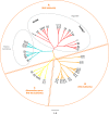
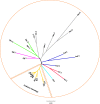
Background: Reef-building corals regularly experience changes in intra- and extracellular H+ concentrations ([H+]) due to physiological and environmental processes. Stringent control of [H+] is required to maintain the homeostatic acid-base balance in coral cells and is achieved through the regulation of intracellular pH (pHi). This task is especially challenging for reef-building corals that share an endosymbiotic relationship with photosynthetic dinoflagellates (family Symbiodinaceae), which significantly affect the pHi of coral cells. Despite their importance, the pH regulatory proteins involved in the homeostatic acid-base balance have been scarcely investigated in corals. Here, we report in the coral Stylophora pistillata a full characterization of the genomic structure, domain topology and phylogeny of three major H+ transporter families that are known to play a role in the intracellular pH regulation of animal cells; we investigated their tissue-specific expression patterns and assessed the effect of seawater acidification on their expression levels.
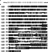
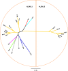
Results: We identified members of the Na+/H+ exchanger (SLC9), vacuolar-type electrogenic H+-ATP hydrolase (V-ATPase) and voltage-gated proton channel (HvCN) families in the genome and transcriptome of S. pistillata. In addition, we identified a novel member of the HvCN gene family in the cnidarian subclass Hexacorallia that has not been previously described in any species. We also identified key residues that contribute to H+ transporter substrate specificity, protein function and regulation. Last, we demonstrated that some of these proteins have different tissue expression patterns, and most are unaffected by exposure to seawater acidification.
Conclusions: In this study, we provide the first characterization of H+ transporters that might contribute to the homeostatic acid-base balance in coral cells. This work will enrich the knowledge of the basic aspects of coral biology and has important implications for our understanding of how corals regulate their intracellular environment.
Keywords: Gene expression; H+ transport; Ocean acidification; Reef-building corals; pH regulation.
Conflict of interest statement
The authors declare that they have no competing interests.
Figures

Similar articles
-
Effects of light and darkness on pH regulation in three coral species exposed to seawater acidification.Sci Rep. 2019 Feb 18;9(1):2201. doi: 10.1038/s41598-018-38168-0. Sci Rep. 2019. PMID: 30778093 Free PMC article.
-
Live tissue imaging shows reef corals elevate pH under their calcifying tissue relative to seawater.PLoS One. 2011;6(5):e20013. doi: 10.1371/journal.pone.0020013. Epub 2011 May 27. PLoS One. 2011. PMID: 21637757 Free PMC article.
-
Impact of seawater acidification on pH at the tissue-skeleton interface and calcification in reef corals.Proc Natl Acad Sci U S A. 2013 Jan 29;110(5):1634-9. doi: 10.1073/pnas.1216153110. Epub 2012 Dec 31. Proc Natl Acad Sci U S A. 2013. PMID: 23277567 Free PMC article.
-
How corals made rocks through the ages.Glob Chang Biol. 2020 Jan;26(1):31-53. doi: 10.1111/gcb.14912. Epub 2019 Dec 14. Glob Chang Biol. 2020. PMID: 31696576 Free PMC article. Review.
-
Environmental impacts of dredging and other sediment disturbances on corals: a review.Mar Pollut Bull. 2012 Sep;64(9):1737-65. doi: 10.1016/j.marpolbul.2012.05.008. Epub 2012 Jun 7. Mar Pollut Bull. 2012. PMID: 22682583 Review.
Cited by
-
Immunolocalization of Metabolite Transporter Proteins in a Model Cnidarian-Dinoflagellate Symbiosis.Appl Environ Microbiol. 2022 Jun 28;88(12):e0041222. doi: 10.1128/aem.00412-22. Epub 2022 Jun 9. Appl Environ Microbiol. 2022. PMID: 35678605 Free PMC article.
-
Post-injury pain and behaviour: a control theory perspective.Nat Rev Neurosci. 2023 Jun;24(6):378-392. doi: 10.1038/s41583-023-00699-5. Epub 2023 May 10. Nat Rev Neurosci. 2023. PMID: 37165018 Free PMC article. Review.
-
Discovery and characterization of Hv1-type proton channels in reef-building corals.Elife. 2021 Aug 6;10:e69248. doi: 10.7554/eLife.69248. Elife. 2021. PMID: 34355697 Free PMC article.
-
Spatial variability of and effect of light on the cœlenteron pH of a reef coral.Commun Biol. 2024 Feb 29;7(1):246. doi: 10.1038/s42003-024-05938-8. Commun Biol. 2024. PMID: 38424314 Free PMC article.
-
Investigating calcification-related candidates in a non-symbiotic scleractinian coral, Tubastraea spp.Sci Rep. 2022 Aug 6;12(1):13515. doi: 10.1038/s41598-022-17022-4. Sci Rep. 2022. PMID: 35933557 Free PMC article.
References
-
- Davies PS. Effect of daylight variations on the energy budgets of shallow-water corals. Mar Biol. 1991;108:137–144. doi: 10.1007/BF01313481. - DOI
-
- Tambutté S, Tambutté E, Zoccola D, Allemand D. Organic Matrix and Biomineralization of Scleractinian Corals. In Handbook of Biomineralization: Biological Aspects and Structure Formation (pp.243 - 259), E. Bauerlein (Ed.).
MeSH terms
LinkOut - more resources
Full Text Sources
Other Literature Sources

