. 2021 Feb 17:11:604470.
doi: 10.3389/fimmu.2020.604470. eCollection 2020.

A Novel Experimental Approach for In Vivo Analyses of the Salivary Gland Microvasculature

Affiliations

A Novel Experimental Approach for In Vivo Analyses of the Salivary Gland Microvasculature

Bernd Uhl et al. Front Immunol. .

Abstract

Microvascular dysfunction plays a fundamental role in the pathogenesis of salivary gland disorders. Restoring and preserving microvascular integrity might therefore represent a promising strategy for the treatment of these pathologies. The mechanisms underlying microvascular dysfunction in salivary glands, however, are still obscure, partly due to the unavailability of adequate in vivo models. Here, we present a novel experimental approach that allows comprehensive in vivo analyses of the salivary gland microvasculature in mice. For this purpose, we employed different microscopy techniques including multi-photon in vivo microscopy to quantitatively analyze interactions of distinct immune cell subsets in the submandibular gland microvasculature required for their infiltration into the surrounding parenchyma and their effects on microvascular function. Confocal microscopy and multi-channel flow cytometry in tissue sections/homogenates complemented these real-time analyses by determining the molecular phenotype of the participating cells. To this end, we identified key adhesion and signaling molecules that regulate the subset- and tissue-specific trafficking of leukocytes into inflamed glands and control the associated microvascular leakage. Hence, we established an experimental approach that allows in vivo analyses of microvascular processes in healthy and diseased salivary glands. This enables us to delineate distinct pathogenetic factors as novel therapeutic targets in salivary gland diseases.

Keywords: immunology; in vivo imaging; inflammation; leukocyte trafficking; microcirculation; microvascular permeability; salivary gland.


Conflict of interest statement

The authors declare that the research was conducted in the absence of any commercial or financial relationships that could be construed as a potential conflict of interest.

Figures

Figure 1
Figure 1
Trafficking of leukocytes into the inflamed submandibular gland. (A) The gating strategy for the differentiation of neutrophils, classical monocytes (cMOs)/monocyte-derived macrophages (MDMs) and non-classical monocytes (ncMOs)/tissue-resident macrophages (TRMs), CD4+ and CD8+ T lymphocytes, as well as B lymphocytes in homogenates of murine submandibular glands in multi-channel flow cytometry analyses. (B) Quantitative data on the recruitment of these leukocyte subsets into submandibular glands upon superfusion with recombinant mouse TNF (1 µg/ml; 5 h) or saline (mean ± SEM for n = 6; *p < .05 vs. saline; n.s., non significant).
Figure 2
Figure 2
Mechanisms of leukocyte trafficking into the inflamed submandibular gland. (A) Representative microscopy images (z-projections) of immunohistochemically stained tissue sections of control (superfusion with saline) and inflamed (superfusion with TNF) submandibular glands illustrating the expression of CD62P/P-selectin, CD54/ICAM-1, and CD106/VCAM-1 (green) in CD31/PECAM-1 (red)-positive postcapillary venules (scale bar: 10 µm). (B) Quantitative data for endothelial expression of CD62P/P-selectin, CD54/ICAM-1, and CD106/VCAM-1 of submandibular glands superfused with TNF or saline (mean ± SEM for n = 4; *p < .05 vs. saline; n.s., not significant). (C) Quantitative data for TNF-induced recruitment of neutrophils and classical monocytes (cMOs)/monocyte-derived macrophages (MDMs) to submandibular glands in animals treated with blocking monoclonal anti-CD62P/P-selectin or anti-CD54/ICAM-1 antibodies or isotype control antibodies (mean ± SEM for n = 6; #p < .05 vs. isotype control).
Figure 3
Figure 3
Experimental setup for in vivo microscopy analyses of the submandibular gland microvasculature. (A) Schematic illustration of the microscopy setup for in vivo analysis of the microcirculation in the mouse submandibular gland. (B) Representative image of the microscopy stage for in vivo imaging of the microcirculation in the mouse submandibular gland. (C) Representative image of the operation situs of the anterior neck region after careful dissection of the submandibular gland. ([1] dipping objective; [2] glass-window for microscopy; [3] mounting of stage; [4] submandibular gland (synonyms in veterinary anatomy: mandibular gland or submaxillary gland); [5] fixation ring for skin sutures; n.s., non significant.
Figure 4
Figure 4
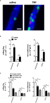
Microvascular leukocyte-endothelial cell interactions in the inflamed submandibular gland. (A) Representative multi-channel epifluorescence in vivo microscopy images illustrating postcapillary venules in control (superfusion with saline) and inflamed (superfusion with TNF) submandibular glands (scale bar: 20 µm; intravascular FITC dextran in blue, Ly-6G/Ly6C+ cells (GR-1) in green, and CD115+ cells in red). (B) Quantitative data for intravascular rolling flux and firm adherence of neutrophils and classical monocytes (cMOs) in postcapillary venules of submandibular glands superfused with TNF or saline (mean ± SEM for n = 4; *p < .05 vs. saline). (C) Quantitative data for TNF-induced intravascular rolling flux and firm adherence of neutrophils and cMOs in postcapillary venules of submandibular glands in animals treated with blocking monoclonal anti-CD62P/P-selectin antibodies or anti-CD54/ICAM-1 antibodies as compared to isotype control antibody-treated animals (mean ± SEM for n = 4; #p < .05 vs. isotype control).
Figure 5
Figure 5
Intravascular interactions between leukocytes in the inflamed submandibular gland. (A) Representative multi-channel epifluorescence in vivo microscopy images illustrating interactions between neutrophils (N) and classical monocytes (cMO) or non-classical monocytes (ncMO) in postcapillary venules of TNF-stimulated submandibular glands (scale bar: 10 µm; intravascular FITC dextran in blue, Ly-6G/Ly6C+ cells (GR-1) in green, and CD115+ cells in red). (B, C) Quantitative data for short (≤ 5s) and long (> 5s) interactions of neutrophils and classical monocytes (cMOs) (B) or of neutrophils and non-classical monocytes (ncMOs) (C) in postcapillary venules of submandibular glands upon superfusion of TNF or saline (mean ± SEM for n = 4; *p < .05 vs. saline). (D) Quantitative data for TNF-elicited recruitment of neutrophils and classical monocytes (cMOs)/monocyte-derived macrophages (MDMs) to submandibular glands in animals treated with neutrophil-depleting anti-Ly6G or isotype control antibodies (mean ± SEM for n = 6; #p < .05 vs. isotype control).
Figure 6
Figure 6
Role of neutrophils for microvascular hyperpermeability in the inflamed submandibular gland. (A) Representative multi-photon in vivo microscopy images illustrating sections of the microvasculature in control (superfusion with saline) and inflamed (superfusion with TNF) submandibular glands treated with isotype control or neutrophil-depleting anti-Ly6G antibodies (scale bar: 50 µm; FITC dextran in green). Quantitative data is shown in (B); mean ± SEM for n = 6; *p < .05 vs. isotype control + saline; #p < .05 vs. isotype control + TNF; n.s., non significant).

Similar articles

Cited by

References

    1. Wilson KF, Meier JD, Ward PD. Salivary gland disorders. Am Fam Phys (2014) 89:882–8. - PubMed
    1. Miletich I. Introduction to salivary glands: structure, function and embryonic development. Front Oral Biol (2010) 14:1–20. - PubMed
    1. Cavanaugh VJ, Deng Y, Birkenbach MP, Slater JS, Campbell AE. Vigorous innate and virus-specific cytotoxic T-lymphocyte responses to murine cytomegalovirus in the submaxillary salivary gland. J Virol (2003) 77:1703–17. - PMC - PubMed
    1. Fukumura D, Duda DG, Munn LL, Jain RK. Tumor microvasculature and microenvironment: novel insights through intravital imaging in pre-clinical models. Microcirculation (2010) 17:206–25. - PMC - PubMed
    1. Zanatta E, Colombo C, D’amico G, D’humieres T, Dal Lin C, Tona F. Inflammation and Coronary Microvascular Dysfunction in Autoimmune Rheumatic Diseases. Int J Mol Sci (2019) 20:5563. - PMC - PubMed

Publication types

MeSH terms

LinkOut - more resources