. 2022 May;13(5):360-378.
doi: 10.1007/s13238-021-00822-1. Epub 2021 Mar 6.

The nucleocapsid protein of rice stripe virus in cell nuclei of vector insect regulates viral replication

Affiliations

The nucleocapsid protein of rice stripe virus in cell nuclei of vector insect regulates viral replication

Wan Zhao et al. Protein Cell. 2022 May.

Erratum in

Abstract

Rice stripe virus (RSV) transmitted by the small brown planthopper causes severe rice yield losses in Asian countries. Although viral nuclear entry promotes viral replication in host cells, whether this phenomenon occurs in vector cells remains unknown. Therefore, in this study, we systematically evaluated the presence and roles of RSV in the nuclei of vector insect cells. We observed that the nucleocapsid protein (NP) and viral genomic RNAs were partially transported into vector cell nuclei by utilizing the importin α nuclear transport system. When blocking NP nuclear localization, cytoplasmic RSV accumulation significantly increased. In the vector cell nuclei, NP bound the transcription factor YY1 and affected its positive regulation to FAIM. Subsequently, decreased FAIM expression triggered an antiviral caspase-dependent apoptotic reaction. Our results reveal that viral nuclear entry induces completely different immune effects in vector and host cells, providing new insights into the balance between viral load and the immunity pressure in vector insects.

Keywords: YY1; importin α; nuclear localization; nucleocapsid protein; rice stripe virus.


Conflict of interest statement

The authors declare no competing interests.

Figures
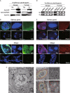
Figure 1
Figure 1
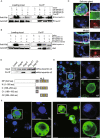
RSV ribonucleoprotein particles localize in the nuclei of planthopper cells. (A) Western blot results show the nuclear location of nucleocapsid protein (NP) in the nuclear (Nc) and cytoplasmic (Cy) extracts from viruliferous planthoppers bearing Jiangsu or Yunnan RSV isolates using a monoclonal anti-NP antibody. Reference proteins for nuclear and cytoplasmic proteins were histone H3 and GAPDH, respectively, which were observed using anti-H3 and anti-GAPDH monoclonal antibodies, respectively. (B) Immunohistochemistry showing the NP nuclear location in the salivary gland and midgut cells. The green signal is from an Alexa Fluor 488-labeled anti-NP monoclonal antibody. The blue signal is the nucleus (Nc) stained with Hoechst. The boxed region is enlarged and shown in two different panels on the right side. The samples without the treatment of anti-NP antibody are shown as negative controls. Scale bars: 10 μm. (C) Colloidal gold immunoelectron microscopy showing the NP nuclear location in the midgut cells. The left panel is the viruliferous midgut section without treatment of a monoclonal anti-NP antibody. Nc, nucleus; NM, nuclear membrane; CM, cell membrane. In the right panels, the cells and nuclei are outlined by red-dotted and yellow-dotted lines, respectively. NP was labeled with 10 nm-gold particles (red strangles). The boxed region is enlarged and shown in the panel on the right side. Scale bars: 1 μm. (D) RSV genomic RNA segments were amplified from nuclear (Nc) and cytoplasmic (Cy) extracts from viruliferous planthoppers bearing the Jiangsu or Yunnan RSV isolates by reverse transcription-PCR. (E) In situ fluorescence hybridization to assess the viral genomic RNA3 location in the salivary gland and midgut cells. The red signal is from the digoxigenin (DIG)-labeled RNA3 probe, and the blue signal is the nucleus (Nc) stained with Hoechst. The boxed region is enlarged and shown in two different panels on the right side. The samples without the treatment of RNA3 probe are shown as negative controls. Scale bars: 10 μm
Figure 2
Figure 2
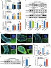
RSV nucleocapsid protein interacts with importin α proteins via. the nuclear localization signal. (A) Recombinantly expressed RSV nucleocapsid protein (NP) with a Flag-tag binds three recombinantly expressed His-importin α proteins in the co-immunoprecipitation (Co-IP) and Western blot assay. The expression products from the pET28a vector were used as a negative control. (B) Three recombinantly expressed His-importin α proteins pulled down the NP from viruliferous planthoppers in the Co-IP and Western blot assay. The expression products from the pET28a vector were used as a negative control. (C) Colocalization of importin α3 and NP examined in the salivary gland and midgut cells via immunohistochemistry analysis. The green signal is from an Alexa Fluor 488-labeled anti-NP monoclonal antibody. The red signal is from an Alexa Fluor 594-labeled anti-importin α3 (IMPα3) polyclonal antibody. The boxed region is enlarged and shown in three different panels on the right side. The samples without the treatment of primary antibodies are shown as negative controls. (D) Co-IP and Western blot assay for the interaction between the recombinantly expressed Flag-N, Flag-C, Flag-C1, or Flag-C2 fragments of NP and His-importin α2. Motifs A, B, C and D comprise the putative nuclear localization signals (NLS). The overlapping amino acid residues shared by the motifs are marked in yellow. The expression products from the pET28a vector were used as a negative control. (E) Immunohistochemistry analysis of the subcellular location of expressed NP in S2 cells. The green signal is from an Alexa Fluor 488-labeled anti-NP monoclonal antibody. The boxed region is enlarged and shown in two different panels on the right side. (F) Subcellular location of expressed GFP and the GFP-C2 fragment in S2 cells was observed. The boxed region is enlarged and shown in two different panels on the right side. Scale bars in 2C, 2E, and 2F: 10 μm. The blue signal is the nuclei (Nc) stained with Hoechst
Figure 3
Figure 3
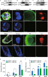
Inhibition of NP nuclear entry significantly promotes RSV accumulation in planthoppers. (A) Relative RNA levels of RSV NP and genomic RNA3 7 d after the injection of dsRNA of importin α1 (dsIMPα1), importin α2 (dsIMPα2), importin α3 (dsIMPα3), or GFP (dsGFP), as measured by quantitative real-time PCR (qPCR). The RNA levels of NP and RNA3, and the transcript levels of importin α genes are normalized to that of EF2. (B) The nuclear accumulation of NP in the nuclear protein extracts of viruliferous planthoppers after the injection of dsIMPα1, dsIMPα2, dsIMPα3 or dsGFP for 7 d, as assessed by Western blot assay. NP was detected using a monoclonal anti-NP antibody. The reference protein for the nuclear proteins was histone H3, which was detected using a monoclonal anti-H3 antibody. Four independent replicates are shown for each group. The relative optical densities of NP to that of H3 were calculated. (C) Immunohistochemistry analysis of the subcellular location of NP in the midgut cells after the injection of dsIMPα1, dsIMPα2, dsIMPα3, a mixture of dsRNA of the three importin α genes (ds3IMPα), or dsGFP for 7 d. NP was detected using a monoclonal anti-NP antibody. The blue signal is the nuclei (Nc) stained with Hoechst. The boxed region is enlarged and shown in two different panels on the right side. Scale bars: 20 μm. (D) The relative transcript levels of importin α1, α2 and α3 after the injection of ds3IMPα or dsGFP for 7 d measured by qPCR. (E) Nuclear accumulation of NP in the nuclear protein extracts of viruliferous planthoppers after the injection of ds3IMPα or dsGFP for 7 d, as assessed via Western blot assay. Four independent replicates are shown for each group. The relative optical densities of NP to that of H3 are calculated. (F) Relative RNA levels of RSV NP and genomic RNA3 after injection of ds3IMPα or dsGFP for 7 d measured by qPCR. ns, no significant difference. **P < 0.01. ***P < 0.001
Figure 4
Figure 4
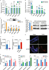
Binding of NP to YY1 in the nuclei leads to an antiviral reaction in planthoppers. (A) Recombinantly expressed His-YY1 binds recombinantly expressed Flag-NP in the co-immunoprecipitation (Co-IP) and Western blot assay. The expression products from the pET28a vector were used as a negative control. (B) NP was pulled down from viruliferous planthoppers in the Co-IP and Western blot assay using an anti-human YY1 polyclonal antibody. An IgG antibody was used as a negative control. (C) YY1 was pulled down from viruliferous planthoppers in the Co-IP and Western blot assay using an anti-NP monoclonal antibody. An IgG antibody was used as a negative control. (D) Subcellular colocalization of NP and YY1 in the salivary gland and midgut cells of viruliferous planthoppers as determined by immunohistochemistry analysis. The green signal is from an Alexa Fluor 488-labeled anti-NP monoclonal antibody. The red signal is from an Alexa Fluor 594-labeled anti-human YY1 polyclonal antibody. The blue signal is the nuclei stained with Hoechst. The boxed region is enlarged and shown in three different panels on the right side. The samples without the treatment of primary antibodies are shown as negative controls. Scale bars: 20 μm. (E) Relative RNA levels of RSV NP and genomic RNA3 and YY1 in the planthoppers at 3 d after injection of dsRNA of YY1 (dsYY1) and RSV crude preparations measured by quantitative real-time PCR (qPCR). The control group was injected with dsRNA of GFP (dsGFP) and RSV crude preparations. The RNA levels of NP, RNA3, and YY1 are normalized to that of EF2. (F) The relative RNA levels of RSV NP and genomic RNA3 and YY1 in the planthoppers at 7 d after injection of dsRNA of YY1 (dsYY1) and RSV crude preparations measured by qPCR. ns, no significant difference. *P < 0.05. **P < 0.01. ***P < 0.001
Figure 5
Figure 5
YY1 positively regulates the expression of FAIM to modulate cell apoptosis. (A) Relative transcript levels of the potential target genes of YY1 in nonviruliferous planthoppers (N) at 7 d after injection of dsRNA of YY1 (dsYY1) or GFP (dsGFP) measured by quantitative real-time PCR (qPCR). The transcript levels of these genes were normalized to that of EF2. (B) The relative transcript levels of the potential target genes of YY1 in the planthoppers at 3 d after injection of dsYY1 and RSV crude preparations measured by qPCR. The control group was injected with dsGFP and RSV crude preparations. (C) Relative enrichment of promoter regions of the putative targets of YY1 measured by chromatin immunoprecipitation (ChIP) combined with qPCR. The anti-human YY1 polyclonal antibody efficiently captured the YY1 from nonviruliferous planthoppers in the ChIP as shown by the Western blot. IgG was used instead of the anti-YY1 antibody in the immunoprecipitation step as the negative control. (D) Relative enrichment of FAIM promoter binding with YY1 to that of IgG immunoprecipitation group (fold enrichment) in viruliferous (V) and nonviruliferous (N) planthoppers measured by ChIP-qPCR. The comparable amounts of YY1 precipitated from viruliferous and nonviruliferous insects were quantified using the Western blot assay with the anti-human YY1 polyclonal antibody. Eight biological replicates were prepared for each group. (E) The relative transcript levels of FAIM to that of EF2 in nonviruliferous and viruliferous planthoppers. N, nonviruliferous insects. V, viruliferous insects. (F) The relative transcript levels of FAIM to that of EF2 in viruliferous insects at 7 d after injection of dsRNA mixture of the three importin α genes (ds3IMPα) or GFP (dsGFP). (G) Apoptotic response in the midgut cells of nonviruliferous planthoppers at 7 d after injection of dsRNA of FAIM (dsFAIM) or dsGFP assayed with the TUNEL assay. Scale bars: 20 μm. (H) Caspase activities in planthoppers measured with the human caspase 1, 3 and 8 Activity Assay kits. The activities were calculated as µmol/L of the product p-nitroaniline (pNA). For nonviruliferous planthoppers (N), dsFAIM or dsYY1 was injected. For viruliferous planthoppers (V), ds3IMPα was injected. The control group was injected with dsGFP. The caspase activities were measured at 7 d after injection. ns, no significant difference. *P < 0.05. **P < 0.01. ***P < 0.001
Figure 6
Figure 6
NP nuclear entry activates cell apoptosis to inhibit viral replication in planthoppers. (A) Apoptotic response in the midgut cells of nonviruliferous and viruliferous planthoppers as assessed by immunohistochemistry assays. N, nonviruliferous insects. V, viruliferous insects. The green signal is from an Alexa Fluor 488-labeled anti-NP monoclonal antibody. The red signal is from TUNEL label. The blue signal is the nuclei stained with Hoechst. (B) The relative RNA levels of RSV NP and genomic RNA3 in the midguts of viruliferous planthoppers at 6 d after injection of the pan-caspase inhibitor measured by quantitative real-time PCR. DMSO was injected in the control group. The RNA levels of NP and RNA3 are normalized to that of EF2. **P < 0.01. (C) Immunohistochemistry for the midguts of viruliferous planthoppers at 6 d after injection of the pan-caspase inhibitor. DMSO was injected in the control group. (D) Immunohistochemistry for the midguts of viruliferous planthoppers at 7 d after injection of dsRNA mixture of the three importin α genes (ds3IMPα). dsRNA of GFP (dsGFP) was injected in the control group. Scale bars in 6A, 6C, and 6D: 20 μm
Figure 7
Figure 7
Model of RSV nuclear entry to inhibit virus replication in vector insect cells. The nucleocapsid protein (NP) and genomic RNAs of RSV are transported to the nuclei of vector insects through binding to three importin α proteins, which are part of the nuclear transport system of vector cells. In nuclei, NP interacts with the transcription factor YY1 and inhibits the transcriptional regulation of YY1 to FAIM. The reduced expression of FAIM activates a caspase-dependent apoptotic response, which inhibits viral replication in the cytoplasm

Similar articles

Cited by

References

    1. Ashour J, Laurent-Rolle M, Shi PY, García-Sastre A. NS5 of dengue virus mediates STAT2 binding and degradation. J Virol. 2009;83:5408–5418. doi: 10.1128/JVI.02188-08. - DOI - PMC - PubMed
    1. Audsley MD, Jans DA, Moseley GW. Roles of nuclear trafficking in infection by cytoplasmic negative-strand RNA viruses: paramyxoviruses and beyond. J Gen Virol. 2016;97:2463–2481. doi: 10.1099/jgv.0.000575. - DOI - PubMed
    1. Austen M, Luscher B, Luscher-Firzlaff JM. Characterization of the transcriptional regulator YY1. The bipartite transactivation domain is independent of interaction with the TATA box-binding protein, transcription factor IIB, TAFII55, or cAMP-responsive element-binding protein (CPB)-binding protein. J Biol Chem. 1997;272:1709–1717. doi: 10.1074/jbc.272.3.1709. - DOI - PubMed
    1. Bhuvanakantham R, Chong MK, Ng ML. Specific interaction of capsid protein and importin-alpha/beta influences West Nile virus production. Biochem Biophys Res Commun. 2009;389:63–69. doi: 10.1016/j.bbrc.2009.08.108. - DOI - PubMed
    1. Bień K, Sokołowska J, Bąska P, Nowak Z, Stankiewicz W, Krzyzowska M. Fas/FasL pathway participates in regulation of antiviral and inflammatory response during mousepox infection of lungs. Mediators Inflamm. 2015;2015:281613. doi: 10.1155/2015/281613. - DOI - PMC - PubMed

Publication types

Substances