

Genetic Mechanisms Underlying Cortical Evolution in Mammals
- PMID: 33659245
- PMCID: PMC7917222
- DOI: 10.3389/fcell.2021.591017
Genetic Mechanisms Underlying Cortical Evolution in Mammals
Abstract
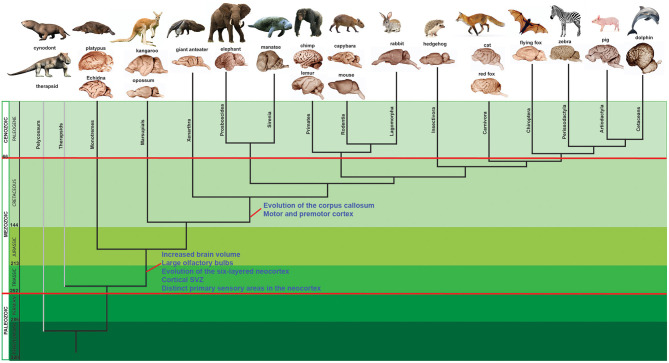
The remarkable sensory, motor, and cognitive abilities of mammals mainly depend on the neocortex. Thus, the emergence of the six-layered neocortex in reptilian ancestors of mammals constitutes a fundamental evolutionary landmark. The mammalian cortex is a columnar epithelium of densely packed cells organized in layers where neurons are generated mainly in the subventricular zone in successive waves throughout development. Newborn cells move away from their site of neurogenesis through radial or tangential migration to reach their specific destination closer to the pial surface of the same or different cortical area. Interestingly, the genetic programs underlying neocortical development diversified in different mammalian lineages. In this work, I will review several recent studies that characterized how distinct transcriptional programs relate to the development and functional organization of the neocortex across diverse mammalian lineages. In some primates such as the anthropoids, the neocortex became extremely large, especially in humans where it comprises around 80% of the brain. It has been hypothesized that the massive expansion of the cortical surface and elaboration of its connections in the human lineage, has enabled our unique cognitive capacities including abstract thinking, long-term planning, verbal language and elaborated tool making capabilities. I will also analyze the lineage-specific genetic changes that could have led to the modification of key neurodevelopmental events, including regulation of cell number, neuronal migration, and differentiation into specific phenotypes, in order to shed light on the evolutionary mechanisms underlying the diversity of mammalian brains including the human brain.
Keywords: brain; cetacea; cortex; elephant; human; human accelerated region; primates; synapsids.
Copyright © 2021 Franchini.
Conflict of interest statement
The author declares that the research was conducted in the absence of any commercial or financial relationships that could be construed as a potential conflict of interest.
Figures


Similar articles
-
The evolution of brains from early mammals to humans.Wiley Interdiscip Rev Cogn Sci. 2013 Jan;4(1):33-45. doi: 10.1002/wcs.1206. Epub 2012 Nov 8. Wiley Interdiscip Rev Cogn Sci. 2013. PMID: 23529256 Free PMC article. Review.
-
Evolution and development of the mammalian cerebral cortex.Brain Behav Evol. 2014;83(2):126-39. doi: 10.1159/000357753. Epub 2014 Apr 24. Brain Behav Evol. 2014. PMID: 24776993 Free PMC article.
-
Comparative aspects of cortical neurogenesis in vertebrates.J Anat. 2007 Aug;211(2):164-76. doi: 10.1111/j.1469-7580.2007.00769.x. Epub 2007 Jul 17. J Anat. 2007. PMID: 17634059 Free PMC article. Review.
-
Phylogenetic variation in cortical layer II immature neuron reservoir of mammals.Elife. 2020 Jul 21;9:e55456. doi: 10.7554/eLife.55456. Elife. 2020. PMID: 32690132 Free PMC article.
-
Laminar and columnar auditory cortex in avian brain.Proc Natl Acad Sci U S A. 2010 Jul 13;107(28):12676-81. doi: 10.1073/pnas.1006645107. Epub 2010 Jun 28. Proc Natl Acad Sci U S A. 2010. PMID: 20616034 Free PMC article.
Cited by
-
Opportunities to Discuss Diversity-Related Topics in Neuroscience Courses.J Undergrad Neurosci Educ. 2022 Oct 1;20(3):A361-A375. doi: 10.59390/AOIN4016. eCollection 2022 Spring. J Undergrad Neurosci Educ. 2022. PMID: 39036724 Free PMC article.
-
Evaluation of advances in cortical development using model systems.Dev Neurobiol. 2022 Jul;82(5):408-427. doi: 10.1002/dneu.22879. Epub 2022 May 29. Dev Neurobiol. 2022. PMID: 35644985 Free PMC article. Review.
-
Research models of neurodevelopmental disorders: The right model in the right place.Front Neurosci. 2022 Oct 20;16:1031075. doi: 10.3389/fnins.2022.1031075. eCollection 2022. Front Neurosci. 2022. PMID: 36340790 Free PMC article. Review.
-
Mercury Toxicity and Neurogenesis in the Mammalian Brain.Int J Mol Sci. 2021 Jul 14;22(14):7520. doi: 10.3390/ijms22147520. Int J Mol Sci. 2021. PMID: 34299140 Free PMC article. Review.
-
Editorial: A view of cell migration dynamics at the single-cell level.Front Cell Dev Biol. 2023 Dec 12;11:1348039. doi: 10.3389/fcell.2023.1348039. eCollection 2023. Front Cell Dev Biol. 2023. PMID: 38149048 Free PMC article. No abstract available.
References
Publication types
LinkOut - more resources
Full Text Sources
Other Literature Sources