. 2021 Mar 3;7(10):eabe8671.
doi: 10.1126/sciadv.abe8671. Print 2021 Mar.

Anti-EGFR VHH-armed death receptor ligand-engineered allogeneic stem cells have therapeutic efficacy in diverse brain metastatic breast cancers

Affiliations

Anti-EGFR VHH-armed death receptor ligand-engineered allogeneic stem cells have therapeutic efficacy in diverse brain metastatic breast cancers

Yohei Kitamura et al. Sci Adv. .

Abstract

Basal-like breast cancer (BLBC) shows brain metastatic (BM) capability and overexpresses EGFR and death-receptors 4/5 (DR4/5); however, the anatomical location of BM prohibits efficient drug-delivery to these targetable markers. In this study, we developed BLBC-BM mouse models featuring different patterns of BMs and explored the versatility of estem cell (SC)-mediated bi-functional EGFR and DR4/5-targeted treatment in these models. Most BLBC lines demonstrated a high sensitivity to EGFR and DR4/5 bi-targeting therapeutic protein, EVDRL [anti-EGFR VHH (EV) fused to DR ligand (DRL)]. Functional analyses using inhibitors and CRISPR-Cas9 knockouts revealed that the EV domain facilitated in augmenting DR4/5-DRL binding and enhancing DRL-induced apoptosis. EVDRL secreting stem cells alleviated tumor-burden and significantly increased survival in mouse models of residual-tumor after macrometastasis resection, perivascular niche micrometastasis, and leptomeningeal metastasis. This study reports mechanism based simultaneous targeting of EGFR and DR4/5 in BLBC and defines a new treatment paradigm for treatment of BM.


Figures

Fig. 1
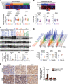
Fig. 1. EGFR and DR4/5 are up-regulated in BLBC-BM.
(A) Top: Heatmap of mRNA levels of EGFR, DR5, and DR4 in patient samples of four subtypes (BL, basal-like; HE, HER2-enriched; LA, luminal A; LB, luminal B) of BC from TCGA database (n = 526). Bottom: Comparison of EGFR, DR5, and DR4 mRNA levels between subtypes. (B) Top: Heatmap of mRNA levels of EGFR, DR5, and DR4 in cell lines of BLBC or non-BLBC from TCGA database (n = 52). Bottom: Comparison of EGFR, DR5, and DR4 mRNA levels between subtypes. (C) Top: Western blot (WB) of EGFR, DR5, and DR4 in 18 BC cell lines (PE, pleural effusion; RPT, repeat; loading control–adjusted ratios are provided under blots). Bottom: Relative expression of EGFR, DR5, and DR4 in BLBC and non-BLBC. (D) Top: Cell surface protein levels of EGFR, DR5, and DR4 analyzed by flow cytometry in 18 BC cell lines. Bottom: Comparison of cell surface expression of EGFR, DR5, and DR4 in BLBC and non-BLBC. PE-A, phycoerythrin-area. (E) Left: Representative micrograph of immunohistochemistry of EGFR, DR5, and DR4 in primary tumors and BM of TNBC. Scale bars, 100 μm. Right: Quantifications of immunohistochemical staining densities by ImageJ (primary, n = 57; BM, n = 13).
Fig. 2
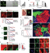
Fig. 2. Developing and characterizing clinically relevant mouse models for BLBC-BM.
(A) Schematic representation of three clinical scenarios of BM. (B) Left: Schematic of micrometastasis model. Right: BLI signal curves of each mouse after ICA injection of BMET02-FmC and representative pictures. (C) Top: Chronological representative bright-field (BF) and fluorescence photographs of brain samples from ICA-injected BMET02-FmC–bearing mice. Scale bar, 10 mm. Bottom: Photomicrograph of coronal sections of the samples. Scale bar, 1 mm. (D) Chronological photomicrographs of immunohistochemistry of CD31 in brain sections that had ICA injection of BMET02-FmC. Scale bars, 100 μm. Critical moment of extravasation of cancer cells was observed on day 2 (inset of d2). (E) Left: Schematic of leptomeningeal metastasis model. Right: BLI signal curve of IT-injected BMET02-FmC–bearing mice (n = 2) and representative photographs. (F) Center: Representative photograph of brain and spine samples from mice 23 days after IT injection of BMET02-FmC. Scale bar, 10 mm. Surrounding: Representative microphotograph of fluorescence and hematoxylin and eosin (H&E) staining of the brain and spine samples. Scale bars, 100 μm. (G) Schematic of macrometastasis resection model. (H) Left: Representative intraoperative BF and fluorescence photographs of brain of pre- and postresection of BMET02-FmC tumor. Scale bars, 1 mm. Right: Representative pictures of BLI. (I) Representative photomicrograph of brain sections of pre- and postresection of tumor. Scale bars, 1 mm. Photo credit: Yohei Kitamura, Brigham and Women’s Hospital. DAPI, 4′,6-diamidino-2-phenylindole.
Fig. 3
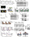
Fig. 3. BLBC is sensitive to EVDRL, and EV domain of EVDRL enhances apoptosis-inducing effect of DRL depending on tumor cell surface EGFR expression.
(A) Representative microphotograph of low and high (the insets) magnifications of H&E and immunohistochemistry of EGFR and DR4/5 in patient samples of TNBC- BM. Scale bars, 100 μm (main images) and 10 μm (insets). (B) Schematic showing the construction of anti-EGFR VHH-DRL (EVDRL) and anti-EGFR scFv-DRL (ESDRL) proteins. (C) Cell viability of BLBC-BM lines after 72-hour treatment with control media (Ctrl), ESDRL, or EVDRL. (n = 3, technical replicates). (D) WB showing cleavage of caspases and poly(ADP-ribose) polymerase (PARP) in BLBC-BM lines after 8-hour treatment with Ctrl, ESDRL, or EVDRL (n = 3, technical replicates). (Loading control–adjusted ratios are provided under blots; only cleaved part was quantified). (E) Cell viability of 18 BC cell lines after 72-hour treatment with different concentrations of Ctrl, EV, DRL, or EVDRL (n = 3, technical replicates). (F) Correlation between cell surface DR4/5 expression and growth inhibition effect of DRL at the time point of 24 hours. (G) Correlation between cell surface EGFR expression and growth inhibition ratio between DRL and EVDRL at the time point of 24 hours. (H) Correlation between cell surface DR5 and EGFR expression and the growth inhibition efficacy of EVDRL. (I) WB showing phosphorylation of EGFR and its downstream elements in BLBC-BM lines with EGF treatment after pretreatment with various concentrations of EVDRL (n = 3, technical replicates). (J) WB showing cleavage of caspases and PARP in BLBC-BM lines after 8-, 16-, and 24-hour treatment with Ctrl or EVDRL (n = 3, technical replicates). (K) Caspase-Glo 3/7 assay of BLBC-BM lines after 8-hour treatment with Ctrl or EVDRL (n = 3, technical replicates).
Fig. 4
Fig. 4. EV domain of EVDRL enhances the apoptosis-inducing efficacy of DRL.
(A and B) Cell viability of BLBC-BM lines after 72-hour treatment (A) and WB of cleavage of caspases and PARP in BLBC-BM lines after 8-hour treatment (B) with control media (Ctrl), EV, DRL, EV + DRL, and EVDRL (n = 3, technical replicates). (C) Confocal images of unstimulated BMET02 cells stably expressing EGFR-YFP and transiently transfected with DR5-CFP. (D) Real-time FRET (sensitized emission) imaging in NIH-3T3 cells coexpressing DR5-CFP and EGFR-YFP. Ratio images depicting the bottom focal plane of the cell show FRET before (left image) and 30 min after treatment EVDRL (right image). (E) Schematic of EGFR inhibitors used for blocking experiments of EVDRL. (F and G) Cell viability (F) and WB showing cleavage of caspases and PARP (G) of BLBC-BM lines after 24-hour treatments with DRL and EVDRL after pretreatment with various concentrations of cetuximab (n = 3, technical replicates). (H) Coimmunoprecipitation (Co-IP) and WB analyses showing EGFR-EVDRL-DR4/5 complex formation in the presence of EVDRL and the attenuation of the complex by cetuximab in BMET02 (n = 2, technical replicates). (I) Cell viability of BLBC-BM lines treated by EVDRL for 24 hours with or without pretreatment by 1 μM erlotinib (n = 3, technical replicates). ns, not significant. (J) Flow cytometry showing reduction of cell surface expression of DR4/5 in BMET02 lines with CRISPR-Cas9 knockout (KO) of DR4, DR5, and DR4/5. (K) Cell viability of BMET02-DR4/5 KO lines after 72-hour treatment with DRL and EVDRL (n = 3, technical replicates). (L) Left: Co-IP and WB analyses showing levels of DRL bound to DR4/5 in BLBC-BM lines after 8-hour treatment with separated EV plus DRL (S) or combined EVDRL (C). Right: Quantification of levels of DRL bound to DR4/5 (n = 3). (M) Schematic showing functional difference between DRL (left) and EVDRL (right).
Fig. 5
Fig. 5. EVDRL-secreting stem cells have antitumor effects against BLBC in vitro and in vivo.
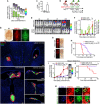
(A) Left: Photomicrograph of EVDRL-secreting hMSC. Scale bar, 100 μm. Right: Concentration of EVDRL in culture media of hMSC-EVDRL quantified by enzyme-linked immunosorbent assay (ELISA) (n = 2, technical replicates). (B) Top: Photomicrographs of BMET02-FmC cocultured with hMSC-GFP/DRL/EVDRL for 72 hours. Scale bars, 100 μm. Bottom: Cell viability of BMET02-FmC after 72-hour coculture with increasing percentages of hMSC-GFP, hMSC-DRL, or hMSC-EVDRL (n = 3, technical replicates). (C) Photomicrographs of different engineered stem cells (left) (scale bars, 100 μm) and cell viability of BMET02FmC cocultured with increasing percentages (0 to 100) of the stem cells (right) (n = 3, technical replicates). (D) Left: Photomicrograph of BMET02-FmC cocultured with sECM-encapsulated hMSC-GFP, hMSC-DRL, or hMSC-EVDRL. Scale bar, 1 mm. Right: Relative number of BMET02-FmC cells 72-hour following coculture with sECM-encapsulated hMSC-GFP, hMSC-DRL, or hMSC-EVDRL (n = 3, technical replicates). (E) Left: Experimental outline for testing efficacy of sECM-encapsulated hMSC-EVDRL in BMET02-FmC–bearing mice. Right: BLI signals before and after resection (n = 20). (F) Intraoperative photographs of light and fluorescence of mice implanted sECM-hMSC into the resection cavity of BMET02-FmC tumor. Scale bars, 1 mm. (G) Representative photomicrographs of brain section from mice 2 and 4 days after resection of BMET02-FmC tumor and implantation of sECM-hMSC. Scale bars, 100 μm. (H) Estimate of relative tumor volume after resection in treatment groups based on Fluc signal intensity of BMET02-FmC (hMSC-GFP, n = 6; hMSC-DRL, n = 7; hMSC-EVDRL, n = 7). (I) Kaplan-Meier survival curves of the mice with median survival (days) indicated in the legend. (J) Immunohistochemistry of cleaved caspase-3 of brain sections from treated and control mice. Scale bars, 100 μm. Photo credit: Yohei Kitamura, Brigham and Women’s Hospital
Fig. 6
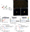
Fig. 6. EVDRL-secreting stem cells show antitumor efficacy for micrometastasis of BLBC.
(A) Experimental outline for testing efficacy of ICA injection of mNSC-EVDRL-TK in mice that had ICA injection of BMET02-FmC 7 days before. (B) Top: Representative photomicrograph of whole brain section from ICA-injected BMET02-FmC–bearing mice 2 days after ICA injection of mNSC. Scale bar, 1 mm. Bottom: Representative photomicrograph of immunohistochemistry of CD31 of the brain section. Scale bars, 100 μm. (C) BLI signal curves and photographs of mice treated with mNSC-GFP/EVDRL-TK +/−GCV (mNSC-GFP, n = 6; mNSC-EVDRL-TK −GCV, n = 7; mNSC-EVDRL-TK +GCV, n = 7). (D) Kaplan-Meier curves of macrometastasis-free survival. The presence of macrometastasis was judged from the substantial BLI signal around 1 × 10⁴ photons/min. Median macrometastasis-free survivals (days) are indicated in the legend. (E) Kaplan-Meier curves of overall survival of mice with median overall survival (days) indicated in the legend.
Fig. 7
Fig. 7. EVDRL-secreting stem cells have antitumor effects in leptomeningeal metastasis of BLBC.
(A) BLI signal and photographs of IT-injected hMSC-GFP-Fluc (GFl)–bearing mice (n = 3). (B) Top: Representative photograph of whole-brain sample of an IT-established, BMET02-FmC leptomeningeal metastasis–bearing mice, 2 days after IT injection of hMSC. Scale bar, 10 mm. Bottom: Representative photomicrographs of brain and spine sections from the mice. Scale bars, 100 μm. (C) Concentration of EVDRL in CSF from mice before and 2 days after IT injection of hMSC-EVDRL quantified by ELISA (n = 3). (D) Experimental outline for testing efficacy of IT injections of hMSC-EVDRL in mice that had IT injection of BMET02-FmC 7 days before. (E) Representative BLI pictures of IT-injected BMET02-FmC–bearing mice treated with hMSC-GRl or hMSC-EVDRL. (F) Fluc signal curves of BMET02-FmC treated with hMSC-GRl or hMSC-EVDRL and Rluc signals of injected hMSC-GRl (hMSC-GRl, n = 7; hMSC-EVDRL, n = 10). (G) Representative photographs of whole-brain sample of IT-injected BMET02-FmC–bearing mice treated with hMSC-GRl or hMSC-EVDRL for 7 days. Scale bars, 10 mm. (H) Kaplan-Meier curves of overall survival of mice. Median survivals (days) are indicated in the legend. (I) Fluc signal curves and representative BLI images of mice bearing MDA231-BrM2-FmC tumors treated with hMSC-GRl or hMSC-EVDRL (hMSC-GRl, n = 6; hMSC-EVDRL, n = 5). (J) Kaplan-Meier curves of overall survival of mice with median survival (days) indicated in the legend. (K) Immunohistochemistry of cleaved caspase-3 in brain sections from IT-injected BMET02-FmC–bearing mice treated with hMSC-GRl or hMSC-EVDRL. Scale bars, 100 μm.

Similar articles

Cited by

References

    1. Tabouret E., Chinot O., Metellus P., Tallet A., Viens P., Gonçalves A., Recent trends in epidemiology of brain metastases: An overview. Anticancer Res 32, 4655–4662 (2012). - PubMed
    1. Lin N. U., Bellon J. R., Winer E. P., CNS metastases in breast cancer. J. Clin. Oncol. 22, 3608–3617 (2004). - PubMed
    1. Siegel R. L., Miller K. D., Jemal A., Cancer statistics, 2018. CA Cancer J. Clin. 68, 7–30 (2018). - PubMed
    1. Mack F., Baumert B. G., Schäfer N., Hattingen E., Scheffler B., Herrlinger U., Glas M., Therapy of leptomeningeal metastasis in solid tumors. Cancer Treat. Rev. 43, 83–91 (2016). - PubMed
    1. Prat A., Adamo B., Cheang M. C. U., Anders C. K., Carey L. A., Perou C. M., Molecular characterization of basal-like and non-basal-like triple-negative breast cancer. Oncologist 18, 123–133 (2013). - PMC - PubMed

Publication types