

Isolation and Culture of Single Myofiber and Immunostaining of Satellite Cells from Adult C57BL/6J Mice
- PMID: 33654822
- PMCID: PMC7854163
- DOI: 10.21769/BioProtoc.3313
Isolation and Culture of Single Myofiber and Immunostaining of Satellite Cells from Adult C57BL/6J Mice
Abstract
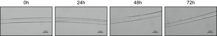
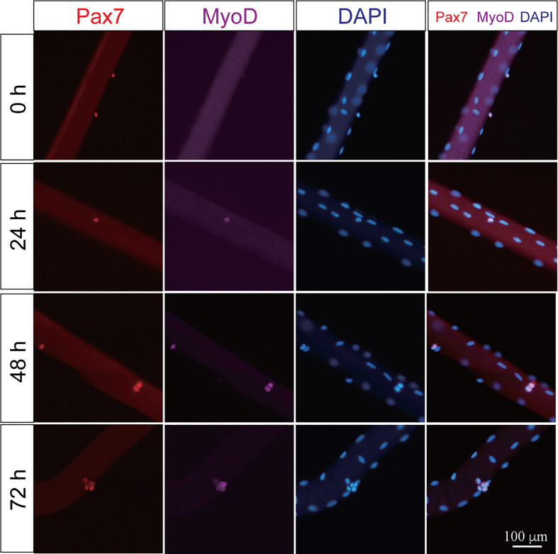
Myofiber isolation followed with ex vivo culture could recapitulate and visualize satellite cells (SCs) activation, proliferation, and differentiation. This approach could be taken to understand the physiology of satellite cells and the molecular mechanism of regulatory factors, in terms of the involvement of intrinsic factors over SCs quiescence, activation, proliferation and differentiation. Single myofiber culture has several advantages that the traditional approach such as FASC and cryosection could not compete with. For example, myofiber isolation and culture could be used to observe SCs activation, proliferation and differentiation at a continuous manner within their physiological "niche" environment while FACS or cryosection could only capture single time-point upon external stimulation to activate satellite cells by BaCl2, Cardiotoxin or ischemia. Furthermore, in vitro transfection with siRNA or overexpression vector could be performed under ex vivo culture to understand the detailed molecular function of a specific gene on SCs physiology. With these advantages, the physiological state of SCs could be analyzed at multiple designated time-points by immunofluorescence staining. In this protocol, we provide an efficient and practical protocol to isolate single myofiber from EDL muscle, followed with ex vivo culture and immunostaining.
Keywords: Immunofluorescence staining; Muscle satellite cells; Myofiber isolation and culture.
Copyright © 2019 The Authors; exclusive licensee Bio-protocol LLC.
Conflict of interest statement
Competing interestsThe authors declare that there is no conflict of financial or research interest.
Figures
Similar articles
-
Isolation and culture of individual myofibers and their satellite cells from adult skeletal muscle.J Vis Exp. 2013 Mar 22;(73):e50074. doi: 10.3791/50074. J Vis Exp. 2013. PMID: 23542587 Free PMC article.
-
Single EDL Myofiber Isolation for Analyses of Quiescent and Activated Muscle Stem Cells.Methods Mol Biol. 2018;1686:149-159. doi: 10.1007/978-1-4939-7371-2_11. Methods Mol Biol. 2018. PMID: 29030819
-
Targeting the Expression of Long Noncoding RNAs in Murine Satellite Cells from Single Myofibers.Bio Protoc. 2021 Nov 5;11(21):e4209. doi: 10.21769/BioProtoc.4209. eCollection 2021 Nov 5. Bio Protoc. 2021. PMID: 34859124 Free PMC article.
-
Plasticity of the Muscle Stem Cell Microenvironment.Adv Exp Med Biol. 2017;1041:141-169. doi: 10.1007/978-3-319-69194-7_8. Adv Exp Med Biol. 2017. PMID: 29204832 Free PMC article. Review.
-
Blood vessels and the satellite cell niche.Curr Top Dev Biol. 2011;96:121-38. doi: 10.1016/B978-0-12-385940-2.00005-X. Curr Top Dev Biol. 2011. PMID: 21621069 Review.
Cited by
-
Retinoic acid and RARγ maintain satellite cell quiescence through regulation of translation initiation.Cell Death Dis. 2022 Sep 29;13(9):838. doi: 10.1038/s41419-022-05284-9. Cell Death Dis. 2022. PMID: 36175396 Free PMC article.
-
Dynamic changes in butyrate levels regulate satellite cell homeostasis by preventing spontaneous activation during aging.Sci China Life Sci. 2024 Apr;67(4):745-764. doi: 10.1007/s11427-023-2400-3. Epub 2023 Dec 20. Sci China Life Sci. 2024. PMID: 38157106
-
Transferrin receptor 1 ablation in satellite cells impedes skeletal muscle regeneration through activation of ferroptosis.J Cachexia Sarcopenia Muscle. 2021 Jun;12(3):746-768. doi: 10.1002/jcsm.12700. Epub 2021 May 6. J Cachexia Sarcopenia Muscle. 2021. PMID: 33955709 Free PMC article.
References
-
- Bischoff R.(1986). Proliferation of muscle satellite cells on intact myofibers in culture. Dev Biol 115(1): 129-139. - PubMed
-
- Collins C. A., Olsen I., Zammit P. S., Heslop L., Petrie A., Partridge T. A. and Morgan J. E.(2005). Stem cell function, self-renewal, and behavioral heterogeneity of cells from the adult muscle satellite cell niche. Cell 122(2): 289-301. - PubMed
LinkOut - more resources
Full Text Sources
Other Literature Sources
