. 2021 Mar 2:10:e51581.
doi: 10.7554/eLife.51581.

New light shed on the early evolution of limb-bone growth plate and bone marrow

Affiliations

New light shed on the early evolution of limb-bone growth plate and bone marrow

Jordi Estefa et al. Elife. .

Abstract

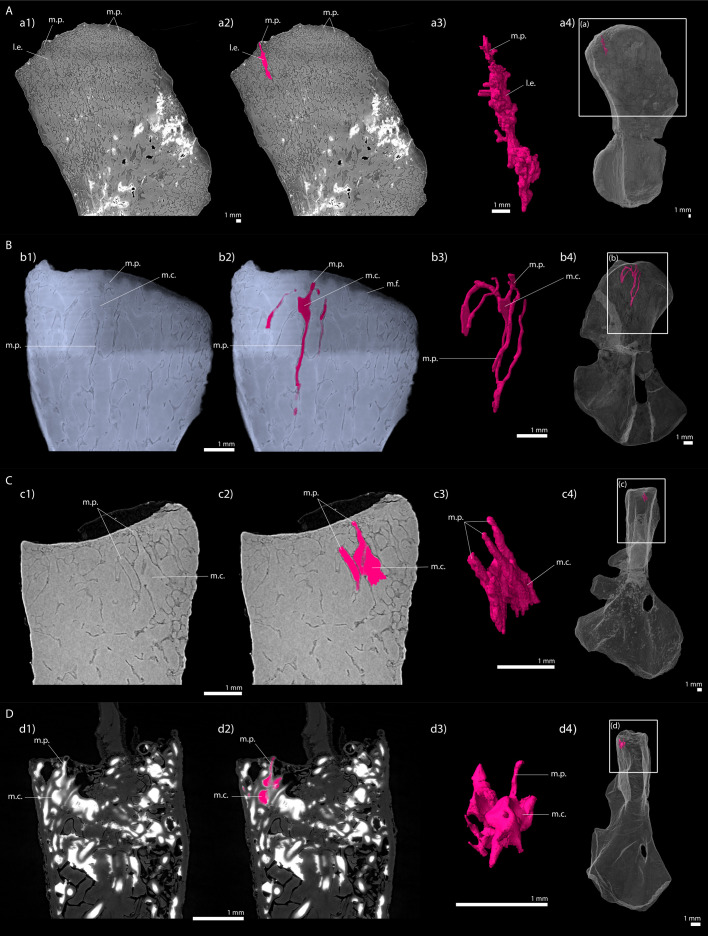
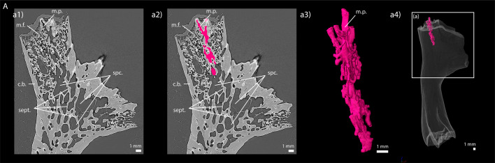
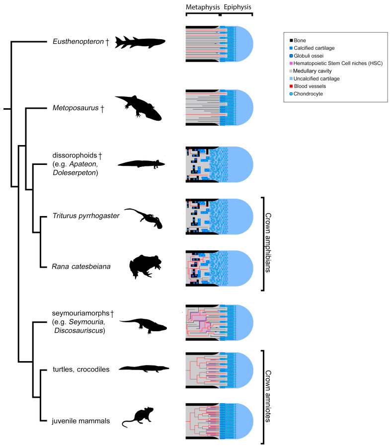
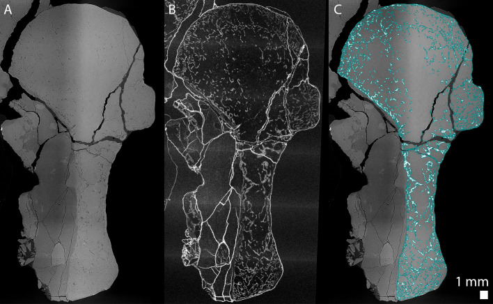
The production of blood cells (haematopoiesis) occurs in the limb bones of most tetrapods but is absent in the fin bones of ray-finned fish. When did long bones start producing blood cells? Recent hypotheses suggested that haematopoiesis migrated into long bones prior to the water-to-land transition and protected newly-produced blood cells from harsher environmental conditions. However, little fossil evidence to support these hypotheses has been provided so far. Observations of the humeral microarchitecture of stem-tetrapods, batrachians, and amniotes were performed using classical sectioning and three-dimensional synchrotron virtual histology. They show that Permian tetrapods seem to be among the first to exhibit a centralised marrow organisation, which allows haematopoiesis as in extant amniotes. Not only does our study demonstrate that long-bone haematopoiesis was probably not an exaptation to the water-to-land transition but it sheds light on the early evolution of limb-bone development and the sequence of bone-marrow functional acquisitions.

Keywords: amphibians; evolutionary biology; haematopoiesis; propagation phase-contrast synchrotron microtomography; stem amniotes; tetrapod terrestrialisation; three-dimensional virtual palaeohistology.

Plain language summary

For many aquatic creatures, the red blood cells that rush through their bodies are created in organs such as the liver or the kidney. In most land vertebrates however, blood-cell production occurs in the bone marrow. There, the process is shielded from the ultraviolet light or starker temperature changes experienced out of the water. It is possible that this difference evolved long before the first animal with a backbone crawled out of the aquatic environment and faced new, harsher conditions: yet very little fossil evidence exists to support this idea. A definitive answer demands a close examination of fossils from the water-to-land transition including lobe-finned fish and early limbed vertebrates. To support the production of red blood cells, their fin and limb bones would have needed an internal cavity that can house a specific niche that opens onto a complex network of blood vessels. To investigate this question, Estefa et al. harnessed the powerful x-ray beam produced by the European Synchrotron Radiation Facility and imaged the fin and limb bones from fossil lobe-finned fish and early limbed vertebrates. The resulting three-dimensional structures revealed spongy long bones with closed internal cavities where the bone marrow cells were probably entrapped. These could not have housed the blood vessels needed to create an environment that produces red blood cells. In fact, the earliest four-legged land animals Estefa et al. found with an open marrow cavity lived 60 million years after vertebrates had first emerged from the aquatic environment, suggesting that blood cells only began to be created in bone marrow after the water-to-land transition. Future work could help to pinpoint exactly when the change in blood cell production occurred, helping researchers to identify the environmental and biological factors that drove this change.


Conflict of interest statement

JE, PT, AC, JK, GN, CB, SS No competing interests declared

Figures

Figure 1.
Figure 1.. Schematic drawing of the long-bone epiphyses of extant amniotes (A) and amphibians (B).
Four conditions are figured here. They are separated by yellow dashed lines: A1, condition in crocodiles (interpreted from Haines, 1938); A2, condition in mammals at an early developmental stage before the appearance of the secondary ossification centre (Anderson and Shapiro, 2010; Tanaka, 1976); B1, condition in Triturus (Cynops) pyrrhogaster (Quilhac et al., 2014; Tanaka, 1976); B2, condition in Rana catesbeiana (Francillon, 1981; Tanaka, 1976). Abbreviations: c., cortex; Dia., diaphysis; e., endosteal bone; Epi., epiphysis; h.c., hypertrophied chondrocytes; Meta., metaphysis; m.p., marrow process; s., sinusoids; sept., septum; trab., trabeculae.
Figure 2.
Figure 2.. Juvenile specimen of Apateon caducus, GPIM-N 1297.
(A) Skeleton. (a) Right limb. (B) Epiphyseal and metaphyseal histology of the proximal end of the humerus. (C) Epiphyseal and metaphyseal histology of the proximal end of the radius (c2-3) and ulna (c1). Abbreviations: c.b., cortical bone; c.c., cluster of chondrocytes; c.f., calcification front; c.m., cartilage matrix; dia., diaphysis; e.b., erosion bay; e.l., erosion lacunae; g.o., globuli ossei; H., humerus; l.r., Liesegang’s rings; meta., metaphysis; m.f., mineralisation front; o.n., ossification notch; Prox., proximal end; R. and U., radius and ulna; t., trabeculae.
Figure 3.
Figure 3.. Adult specimen of Apateon caducus, GPIM-N 1572.
(A) Skeleton. (a) Right limb. (B) Epiphyseal and metaphyseal histology of the proximal end of the radius (b1) and ulna (b2). Abbreviations: c.b., cortical bone; c.m., cartilage matrix; dia., diaphysis; e.b., erosion bay; e.l., erosion lacunae; g.o., globuli ossei; H., humerus; l.r., Liesegang’s rings; meta., metaphysis; Prox., proximal end; R. and U., radius and ulna.
Figure 4.
Figure 4.. Adult specimen of Apateon pedestris, SMNS 54981.
(A) Skeleton. (a) Right limb. (B) Epiphyseal and metaphyseal histology of the distal end of the humerus. (C) Epiphyseal and metaphyseal histology of the proximal end of the radius (c2) and ulna (c1). Abbreviations: c.b., cortical bone; c.c.t., calcified-cartilage trabecula; Dist., distal end; e.b., erosion bay; g.o., globuli ossei; H., humerus; l.r., Liesegang’s rings; meta., metaphysis; m.f., mineralisation front; m.t., mineralised trabecula; Prox., proximal end; R. and U., radius and ulna.
Figure 5.
Figure 5.. Adult specimen of Apateon pedestris, SMNS 54988.
(A) Skeleton. (a) Right limb. (B) Epiphyseal and metaphyseal histology of the distal end of the humerus. (C) Epiphyseal and metaphyseal histology of the proximal end of the radius and ulna. Abbreviations: c.b., cortical bone; c.m., cartilage matrix; dia., diaphysis; Dist., distal end; e.b., erosion bay; e.l., erosion lacunae; g.o., globuli ossei; H., humerus; meta., metaphysis; Prox., proximal end; R. and U., radius and ulna.
Figure 6.
Figure 6.. Left humerus of a (sub-)adult specimen of Metoposaurus sp., MUZ PGI OS-220/171 imaged using PPC-SRµCT.
(A) Frontal view. (a1) Longitudinal virtual thin section (40 µm thick) and (a2) longitudinal virtual thin section of the segmented model of the bone (50 µm thick). The longitudinally-oriented trabeculae are highlighted in purple (white arrows), while the transversally-oriented trabeculae appear in green. (B) Ventral view. (a) Longitudinal virtual thin section of the proximal metaphysis (40 µm thick), (b) transverse virtual thin section made in the metaphysis and (c) longitudinal thin section made in the distal metaphysis. Abbreviations: c.b., cortical bone; dia., diaphysis; Dist., distal end; l.r., Liesegang’s rings; meta., metaphysis; m.f., mineralisation front; m.p., marrow process; Prox., proximal end; s., sediment; t., trabeculae.
Figure 7.
Figure 7.. Left humerus of a juvenile specimen of Seymouria sanjuanensis, MNG 7747 imaged using PPC-SRµCT.
(A) Frontal view. (a1) Longitudinal virtual thin section (40 µm thick), the darker part is an artefact in the original data due to electron reinjection in the synchrotron storage ring (refilling) during the scan and (a2) longitudinal virtual thin section of the segmented model of the bone (250 µm thick). The longitudinally-oriented trabeculae (pointed by horizontal arrows) are highlighted in purple, while the transversally-oriented trabeculae appear in green. Note that, due to the shape of the metaphysis, the trabeculae exhibit an overall fan-like configuration which progressively tilts to 90 degrees at the location of the deltopectoral crest (Asterisk). For that reason, the longitudinal trabeculae appear green and the transverse trabeculae appear purple at this location. (B) Ventral view. (a) Longitudinal virtual thin section in the proximal metaphysis (40 µm thick), (b) transverse virtual thin section in the metaphysis, the large ring artefact results from the synchrotron electron refilling visible in a1, (c) detail of (b) showing marrow processes and cavities in transverse section. Abbreviations: c., cavity; dia., diaphysis; Dist., distal end; meta., metaphysis; m.f., mineralisation front; m.p., marrow process; Prox., proximal end; s., sediment; t., trabeculae.
Figure 8.
Figure 8.. Right humerus of an adult Seymouria sanjuanensis, CM 28597 imaged using PPC-SRµCT.
(A) Frontal view. (a1) Longitudinal virtual thin section (40 µm thick) and (a2) longitudinal section of the segmented model of the bone (450 µm thick). The longitudinally-oriented trabeculae (pointed by horizontal arrows) are highlighted in purple, while the transversally-oriented trabeculae appear in green. Note that, due to the shape of the metaphysis, the trabeculae exhibit an overall fan-like configuration which progressively tilts to 90 degrees at the location of the deltopectoral crest (Asterisk). For that reason, the longitudinal trabeculae appear green and the transverse trabeculae appear purple at this location. (B) Dorsal view. (a) Longitudinal virtual thin section of the proximal metaphysis (40 µm thick), (b) transverse virtual thin section in the metaphysis (40 µm thick), (c) detail of (b) showing marrow processes and cavities in transverse section. Abbreviations: c., cavity; dia., diaphysis; Dist., distal end; meta., metaphysis; m.f., mineralisation front; m.p., marrow process; Prox., proximal end; s., sediment; t., trabeculae.
Figure 8—figure supplement 1.
Figure 8—figure supplement 1.. Humeral microanatomical architecture of the stem amniote Seymouria sanjuanensis (CM 28597) and the tortoise Centrochelys sulcata.
(A) Radiography of the humerus of S. sanjuanensis in anterior view (on the left) and ventral view (on the right). The red arrows show the presence of a resting surface both in the proximal and distal metaphyses. (B) Virtual 5 mm-thick section made in the humerus of C. sulcata. Both metaphyses are zoomed in (as 2-mm-thick sections) to show the presence of multiple resting surfaces (red arrows). Abbreviations: Prox., proximal end.
Figure 9.
Figure 9.. Right humerus of a subadult Discosauriscus austriacus, SNM Z 15568 imaged using PPC-SRµCT.
Due to processing to convert the scan data into a stack of images, the images have been flipped, thereby resulting in a flipped 3D model. (A) Frontal view. (a1) Longitudinal virtual thin section (40 µm thick) and (a2) longitudinal section of the segmented model of the bone (160 µm thick). The longitudinally-oriented trabeculae are highlighted in purple, while the transversally-oriented trabeculae appear in green. (B) Ventral view. (a) Longitudinal virtual thin section of the proximal metaphysis (40 µm thick) and (b) transverse virtual thin section in the proximal metaphysis (40 µm thick). Abbreviations: c.b., cortical bone; dia., diaphysis; Dist., distal end; meta., metaphysis; m.f., mineralisation front; m.p., marrow process; Prox., proximal end; s., sediment; t., trabeculae.
Figure 10.
Figure 10.. Longitudinal virtual sections and three-dimensional (3D) segmentation from PPC-SRµCT of marrow processes and marrow cavities in the humeral proximal ends of: A, Metoposaurus sp.
(MUZ PGI OS-220/171); B, Seymouria sanjuanensis (MNG 7747); C, S. sanjuanensis (CM 28597); D, Discosauriscus austriacus (SNM Z 15568). (a1, b1, c1, d1) Longitudinal virtual thin section (60 µm thick); (a2, b2, c2, d2) marrow processes and cavities segmented; (a3, b3, c3, d3) 3D models of the segmentations. Note that the marrow cavities have not been completely segmented in 3D to allow the full visualisation of the marrow processes; (a4, b4, c4, d4) respective locations of a3, b3, c3, d3 in the humeri. Abbreviations: l.e., region of local erosion; m.c., marrow cavity; m.f., mineralisation front; m.p., marrow process.
Figure 10—figure supplement 1.
Figure 10—figure supplement 1.. Longitudinal virtual sections and three-dimensional (3D) segmentation from PPC-SRµCT of marrow processes in the humeral proximal end of (A) Andrias sp.
(MNHN-ZA-AC-2005–72, Museum national d’Histoire naturelle, Paris, France). (a1) Longitudinal virtual thin section (60 µm thick), (a2) marrow processes segmented, (a3) 3D model of the segmentation and (a4) location of a3 in the humerus. The humerus of Andrias has been imaged using the protocol published by Sanchez et al., 2014. Abbreviations: c.b., cortical bone; m.f., mineralisation front; m.p., marrow process; sept., septa; spc., spaces.
Figure 11.
Figure 11.. Evolution of growth-plate patterns and metaphyseal organisations of the long bones of the studied taxa in a phylogenetic context (e.g., Ruta and Coates, 2007; Schoch, 2019).
Hypothesis on haematopoietic activity is herein contextualised. Black silhouettes represent the taxa studied. Crosses (†) have been attributed to fossil taxa.
Figure 12.
Figure 12.. Longitudinal virtual thin sections of the right humerus of an adult Seymouria sanjuanensis (CM 28597).
(A) Tomogram showing low frequency artefacts resulting in an image divided into a darker and brighter part, (B) image processed with a filter for tomographic texture enhancement to remove low frequency artefacts (Cau et al., 2017) and (C) overlap of A and the bone segmentation with the aid of B used to produce Figure 8Aa2. The segmentation is highlighted by the blue line in C.

Comment in

Similar articles

Cited by

References

    1. Ahlberg PE. Follow the footprints and mind the gaps: a new look at the origin of tetrapods. Earth and Environmental Science Transactions of the Royal Society of Edinburgh. 2018;109:115–137. doi: 10.1017/S1755691018000695. - DOI
    1. Akiyoshi H, Inoue AM. Comparative histological study of hepatic architecture in the three orders amphibian livers. Comparative Hepatology. 2012;11:2. doi: 10.1186/1476-5926-11-2. - DOI - PMC - PubMed
    1. Amemiya CT, Saha NR, Zapata A. Evolution and development of immunological structures in the lamprey. Current Opinion in Immunology. 2007;19:535–541. doi: 10.1016/j.coi.2007.08.003. - DOI - PMC - PubMed
    1. Amizuka N. Histology of epiphyseal cartilage calcification and endochondral ossification. Frontiers in Bioscience. 2012;E4:2085–2100. doi: 10.2741/e526. - DOI - PubMed
    1. Anderson JS. Incorporating ontogeny into the matrix: a phylogenetic evaluation of developmental evidence for the origin of modern amphibians. In: Anderson J. S, Sues H. -D, editors. Major Transitions in Vertebrate Evolution. Bloomington: Indiana University Press; 2007. pp. 182–227.

Publication types

Grants and funding

The funders had no role in study design, data collection and interpretation, or the decision to submit the work for publication.

LinkOut - more resources