. 2021 Jul 7;29(7):2268-2280.
doi: 10.1016/j.ymthe.2021.02.025. Epub 2021 Feb 27.

Targeting choroidal vascular dysfunction via inhibition of circRNA-FoxO1 for prevention and management of myopic pathology

Affiliations

Targeting choroidal vascular dysfunction via inhibition of circRNA-FoxO1 for prevention and management of myopic pathology

Dan Li et al. Mol Ther. .

Abstract

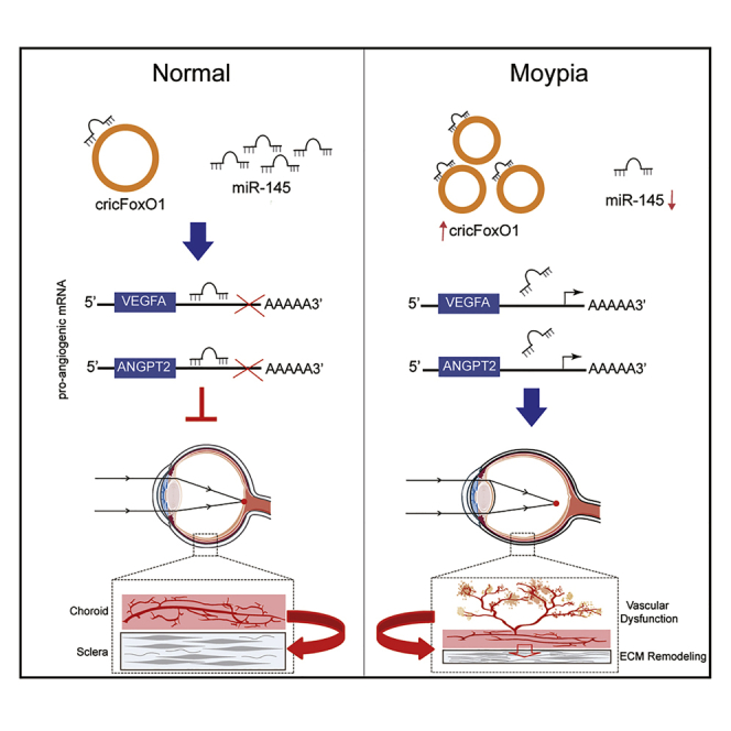
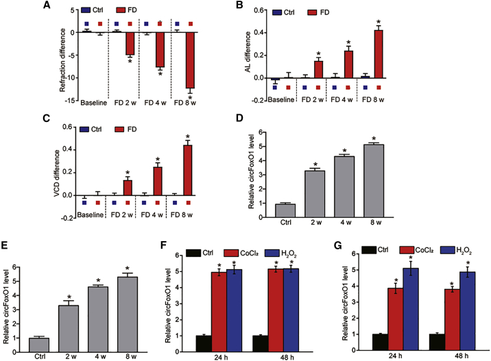
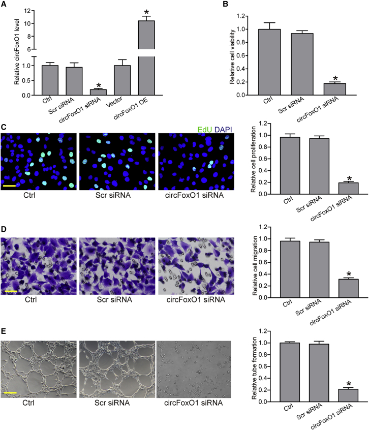
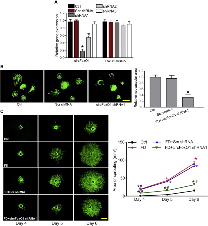
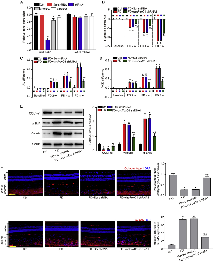
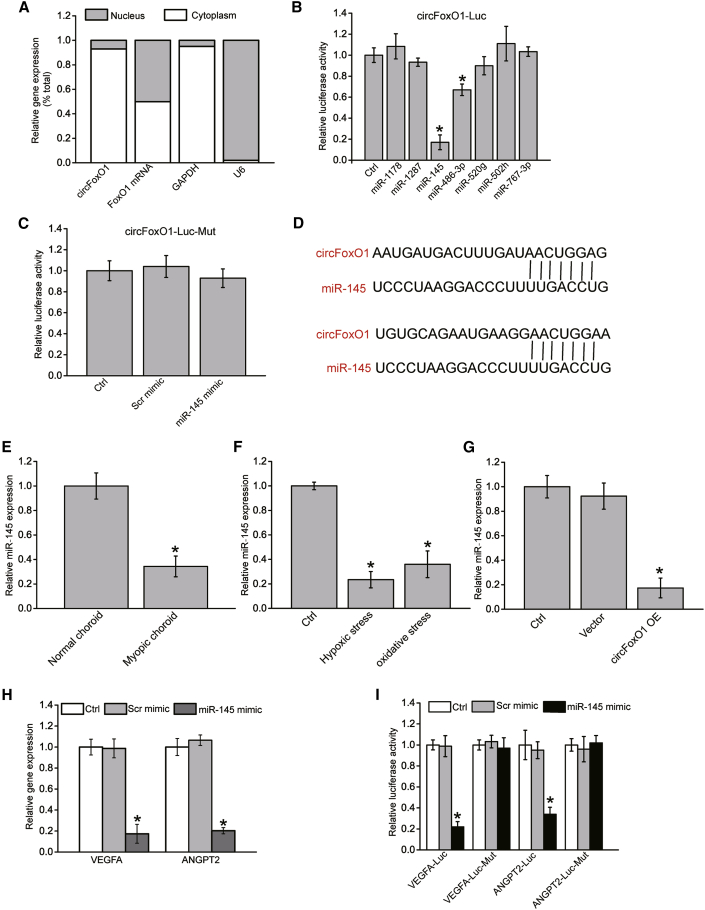
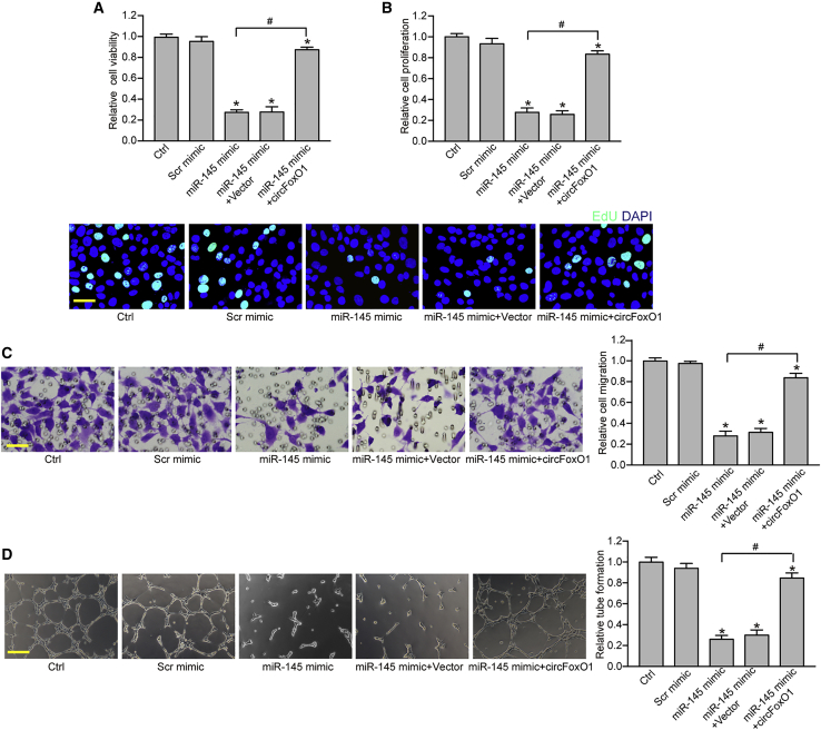
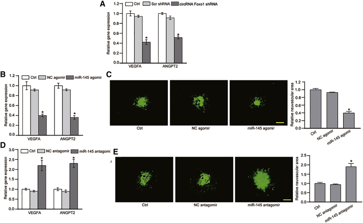
Myopia has become a global public health problem due to high prevalence. Although the etiological factors of myopia have been gradually recognized, the underlying mechanism remains largely elusive. Choroidal vascular dysfunction is recognized as a critical vision-threatening complication in myopia. Circular RNAs (circRNAs) are shown as the critical regulators in many biological processes and human diseases. In this study, we investigated the role of circRNAs in choroidal vascular dysfunction in myopia. The level of circFoxO1 was significantly upregulated in myopic choroid. circFoxO1 silencing suppressed choroidal endothelial cell viability, proliferation, migration, and tube formation in vitro and alleviated choroidal vascular dysfunction in vivo and ex vivo. circFoxO1 silencing retarded the progression of myopia as shown by reduced extracellular matrix remodeling and improved refractive error and axial elongation. Mechanistically, circFoxO1 acted as the sponge of miR-145 to sequester and inhibit miR-145 activity, thereby inducing VEGFA or ANGPT2 expression. miR-145 could mimic the effects of circFoxO1 silencing on choroidal endothelial phenotypes. Collectively, intervention of choroidal vascular dysfunction via regulating circFoxO1 level is a potential strategy for the prevention and management of myopia.

Keywords: choroidal vascular dysfunction; circular RNA; microRNA sponge; myopia.


Conflict of interest statement

Declaration of interests The authors declare no competing interests.

Figures

None
Graphical abstract
Figure 1
Figure 1
Myopia leads to an increased level of circFoxO1 during choroidal vascular dysfunction (A–C) The guinea pigs underwent monocular form deprivation (FD) using the translucent eye shield. Three ocular biometric parameters, including refractive state (A), axial length (AL; B), and vitreous chamber depth (VCD; C), were measured to verify the successful establishment of a myopia model (n = 6 retinas per group; Mann-Whitney U, Bonferroni test). (D) qRT-PCR assays were conducted to detect the expression of circFoxO1 in the choroidal samples of guinea pigs after 0 (control [Ctrl]), 2, 4, or 8 weeks of FD treatment (n = 6 retinas per group; Kruskal-Wallis test, Bonferroni test). (E) qRT-PCR assays were conducted to detect the expression of circFoxO1 in the choroid of C57BL/6 mice after 0 (Ctrl), 2, 4, or 8 weeks of FD treatment (n = 6 retinas per group; Kruskal-Wallis test, Bonferroni test). (F and G) circFoxO1 expression was detected by qRT-PCRs in RF/6A cells or primarily isolated choroidal endothelial cells (ECs) cultured in the medium containing normal medium (Ctrl), CoCl2 (200 μM), or H2O2 (100 μM) for 24 and 48 h (n = 4; ∗p < 0.05 versus Ctrl group, one-way ANOVA, Bonferroni test).
Figure 2
Figure 2
circFoxO1 regulates choroidal EC function in vitro (A) RF/6A cells were transfected with scrambled (Scr) siRNA, circFoxO1 siRNA, pcDNA 3.1 vector, pcDNA 3.1-circFoxO1 (circFoxO1 OE), or were left untreated (Ctrl) for 48 h. qRT-PCR assays were conducted to detect the levels of circFoxO1 (n = 4; ∗p < 0.05 versus Ctrl group, one-way ANOVA, Bonferroni test). (B–E) RF/6A cells were transfected with Scr siRNA, circFoxO1 siRNA, or were left untreated (Ctrl) for 48 h. Cell viability was detected using an MTT assay (B, n = 4; ∗p < 0.05 versus Ctrl group, one-way ANOVA, Bonferroni test). Cell proliferation was detected using an EdU detection kit to analyze the incorporation of EdU during DNA synthesis (C, n = 4; ∗p < 0.05 versus Ctrl group, one-way ANOVA, Bonferroni test). Scale bar, 20 μm. Cell migration was determined using the transwell assay, and the cells that migrated through the transwell were quantified (D, n = 4; ∗p < 0.05 versus Ctrl group, one-way ANOVA, Bonferroni test). Scale bar, 20 μm. RF/6A cells were seeded on the Matrigel matrix. The tube-like structures were observed 6 h after cell seeding. The average length of tube formation for each field was statistically analyzed (E, n = 4; ∗p < 0.05 versus Ctrl group, one-way ANOVA, Bonferroni test). Scale bar, 100 μm.
Figure 3
Figure 3
circFoxO1 regulates choroidal vascular dysfunction in vivo and ex vivo (A) C57BL/6 mice (males, 8 weeks old) received an intravitreal injection of Scr shRNA, circFoxO1 shRNA1–3, or were left untreated (Ctrl) for 2 weeks. qRT-PCR assays were conducted to detect the levels of circFoxO1 and FoxO1 mRNA in choroid (n = 6; ∗p < 0.05 versus Ctrl group, Kruskal-Wallis test, Bonferroni test). (B) C57BL/6 mice received an intravitreal injection of Scr shRNA, circFoxO1 shRNA1, or were left untreated (Ctrl). Two weeks after laser injury, IB4 labeling was conducted to label the neovascular area in flat-mounted choroidal tissues. White circles denote the neovascular area (n = 6; ∗p < 0.05 versus Ctrl group, Kruskal-Wallis test, Bonferroni test). Scale bar, 100 μm. (C) Choroidal sprouting assays were conducted to determine the angiogenic potency of choroidal explants. CD31 staining was conducted to label choroidal sprouting. Representative images of choroidal sprouting and quantification results are shown (n = 6; ∗p < 0.05 versus Ctrl group; #p < 0.05 FD+Scr shRNA group versus FD+circFoxO1 shRNA1 group; Kruskal-Wallis test, Bonferroni test). Scale bar, 500 μm.
Figure 4
Figure 4
circFoxO1 silencing retards the progression of myopia in vivo (A) The eyes of guinea pigs received an intravitreal injection of Scr shRNA, circFoxO1 shRNA1–3, or were left untreated (Ctrl) for 2 months. qRT-PCR assays were conducted to detect the levels of circFoxO1 (n = 6; ∗p < 0.05 versus Ctrl group, Kruskal-Wallis test, Bonferroni test). (B–D) Refraction difference (B), AL (C), and VCD (D) in guinea pigs after FD treatment was measured to determine the role of circFoxO1 in myopia. (E and F) The expression of a fibroblast marker (COL1α1) and myofibroblast markers (α-SMA and vinculin) in the sclera after circFoxO1 silencing were detected by western blot or immunofluorescence staining (n = 6; ∗p < 0.05 versus Ctrl group; #p < 0.05 FD+Scr shRNA group versus FD+circFoxO1 shRNA1 group; Kruskal-Wallis test, Bonferroni test). Scale bars, 100 μm.
Figure 5
Figure 5
circFoxO1 acts as a miRNA sponge in choroidal ECs (A) The expression of nucleus Ctrl transcript (U6), cytoplasmic Ctrl transcript (GAPDH), FoxO1 mRNA, and circFoxO1 was detected by qRT-PCRs in the nucleus and cytoplasmic fractions of RF/6A cells (n = 4). (B) The entire sequence of circFoxO1 was cloned into the pRL-TK luciferase reporter to construct the LUC-circFoxO1 vector. RF/6A cells were co-transfected LUC-circFoxO1 with different miRNA mimics. Luciferase activity was detected using a dual-luciferase assay 48 h after transfection (n = 4; ∗p < 0.05 versus Ctrl group, one-way ANOVA, Bonferroni test). (C) RF/6A cells were co-transfected LUC-circFoxO1-Mut (without miR-145 binding site) with miR-145 mimic, Scr mimics, or were left untreated (Ctrl). Luciferase activity was detected using a dual- luciferase assay at 48 h post-transfection (n = 4; ∗p < 0.05 versus Ctrl group, one-way ANOVA, Bonferroni test). (D) The putative binding sites of miR-145 on circFoxO1 transcript are shown. (E) qRT-PCR assays were conducted to detect the expression level of miR-145 in the choroidal tissues of C57BL/6 mice after 8-week FD induction (n = 6; ∗p < 0.05 versus normal choroid, Mann-Whitney U test, Bonferroni test). (F) RF/6A cells were incubated with normal culture medium, H2O2 (200 μM, oxidative stress), or CoCl2 (100 μM, hypoxic stress) for 24 h. qRT-PCR assays were conducted to detect miR-145 expression (n = 4; ∗p < 0.05 versus Ctrl group, one-way ANOVA, Bonferroni test). (G) RF/6A cells were transfected with pcDNA3.1 (vector), pcDNA3.1-circFoxO1, or were left untreated (Ctrl) for 24 h. qRT-PCR assays were conducted to detect miR-145 expression levels (n = 4; ∗p < 0.05 versus Ctrl group, one-way ANOVA, Bonferroni test). (H) RF/6A cells were transfected with Scr mimic, miR-145 mimic, or were left untreated (Ctrl) for 24 h. qRT-PCR assays were conducted to detect VEGFA and ANGPT2 expression (n = 4; ∗p < 0.05 versus Ctrl group, one-way ANOVA, Bonferroni test). (I) RF/6A cells were co-transfected VEGFA-Luc, VEGFA-Luc mutant (Mut), ANGPT2-Luc, or ANGPT2-Luc Mut with Scr mimic, miR-145 mimic, or were left untreated (Ctrl) for 24 h. Luciferase activity was detected at 24 h post-transfection (n = 4; ∗p < 0.05 versus Ctrl group, one-way ANOVA, Bonferroni test).
Figure 6
Figure 6
The circFoxO1/miR-145/VEGFA or ANGPT2 axis regulates choroidal EC function in vitro (A) RF/6A cells were treated as shown. Cell viability was detected by an MTT assay (n = 4; ∗p < 0.05 versus Ctrl group, one-way ANOVA, Bonferroni test). (B) Cell proliferation was detected using EdU detection kits to analyze the incorporation of EdU during DNA synthesis (n = 4; ∗p < 0.05 versus Ctrl group, one-way ANOVA, Bonferroni test). A representative image along with the quantification result is shown. Scale bar, 20 μm. (C) Transwell assay and quantification analysis was conducted to determine the migration of RF/6A cells (n = 4; ∗p < 0.05 versus Ctrl group, one-way ANOVA, Bonferroni test). Representative images along with the quantification results are shown. Scale bar, 20 μm. (D) RF/6A cells were seeded on the Matrigel matrix. The tube-like structures were observed at 6 h after cell seeding. The average length of tube formation for each field was statistically analyzed (n = 4; ∗p < 0.05 versus Ctrl group, one-way ANOVA, Bonferroni test). Scale bar, 100 μm. ∗p < 0.05 versus Ctrl group; #p < 0.05 indicates significant difference between the marked group.
Figure 7
Figure 7
The circFoxO1/miR-145/VEGFA or ANGPT2 axis regulates choroidal vascular dysfunction in vivo (A) C57BL/6 mice (males, 8 weeks old) received an intravitreal injection of Scr shRNA, circFoxO1 shRNA, or were left untreated (Ctrl) for 2 weeks. qRT-PCR assays were conducted to detect the levels of VEGFA and ANGPT2 in choroid. (B and C) C57BL/6 mice received an intravitreal injection of negative Ctrl (NC) agomir, miR-145 agomir, or were left untreated (Ctrl). Two weeks after laser injury, qRT-PCR assays were conducted to detect the levels of VEGFA and ANGPT2 in choroid. IB4 labeling was conducted to detect neovascular area in flat-mounted choroidal tissues. (D and E) C57BL/6 mice received an intravitreal injection of NC antagomir, miR-145 antagomir, or were left untreated (Ctrl). Two weeks after laser injuryqRT-PCR assays were conducted to detect the levels of VEGFA and ANGPT2 in choroid. IB4 labeling was conducted to detect neovascular area in flat-mounted choroidal tissues. n = 6; ∗p < 0.05 versus Ctrl group, Kruskal-Wallis test, Bonferroni test. Scale bars, 100 μm.

Similar articles

Cited by

References

    1. Morgan I.G., Ohno-Matsui K., Saw S.M. Myopia. Lancet. 2012;379:1739–1748. - PubMed
    1. Bressler N.M. Reducing the progression of myopia. JAMA. 2020;324:558–559. - PubMed
    1. Morgan I.G., French A.N., Ashby R.S., Guo X., Ding X., He M., Rose K.A. The epidemics of myopia: aetiology and prevention. Prog. Retin. Eye Res. 2018;62:134–149. - PubMed
    1. Li Y., Zhang Y., Li P., Mi G., Tu J., Sun L., Webster T.J., Shen Y. Ion-paired pirenzepine-loaded micelles as an ophthalmic delivery system for the treatment of myopia. Nanomedicine (Lond.) 2017;13:2079–2089. - PubMed
    1. Barsam A., Allan B.D. Excimer laser refractive surgery versus phakic intraocular lenses for the correction of moderate to high myopia. Cochrane Database Syst. Rev. 2014;(6):CD007679. - PMC - PubMed

Publication types

MeSH terms