Expression of TIGIT in splenic and circulatory T cells from mice acutely infected with Toxoplasma gondii
- PMID: 33629951
- PMCID: PMC7906093
- DOI: 10.1051/parasite/2021010
Expression of TIGIT in splenic and circulatory T cells from mice acutely infected with Toxoplasma gondii
Abstract
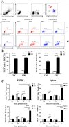
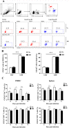
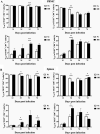
The surface protein TIGIT (T cell immunoglobulin and immunoreceptor tyrosine-based inhibitory motif (ITIM) domain) has been characterized as an important regulator of cell-mediated immune responses in various infections. However, TIGIT expression in immune cells of mice infected with Toxoplasma gondii has not been investigated. Here, we detected TIGIT expression and related phenotypes by flow cytometry and real-time PCR in splenic and circulatory T cells of mice infected with the T. gondii RH strain. We found that the expression of TIGIT on the surface of CD4+ T cells and CD8+ T cells from the spleen and peripheral blood mononuclear cells decreased in the early stage, but increased significantly in the late stage of acute T. gondii infection in mice. Importantly, TIGIT expression was positively correlated with lesions in the murine spleen. In addition, T. gondii-specific TIGIT+TCM cells in the spleen were activated and transformed into TIGIT+ TEM cells. Hematoxylin and eosin staining of spleen sections and real-time PCR showed that the severity of splenic lesions was positively correlated with the T. gondii load. This study demonstrates that acute T. gondii infection can regulate the expression of TIGIT in T cells and affect immune cell function.
Title: Expression de TIGIT dans les cellules T spléniques et circulatoires de souris lourdement infectées par Toxoplasma gondii.
Abstract: La protéine de surface TIGIT a été caractérisée comme un régulateur important des réponses immunitaires à médiation cellulaire dans diverses infections. Cependant, l’expression de TIGIT dans les cellules immunitaires de souris infectées par Toxoplasma gondii n’a pas été étudiée. Ici, nous avons détecté l’expression de TIGIT et les phénotypes associés par cytométrie en flux et PCR en temps réel dans les cellules T spléniques et circulatoires de souris infectées par la souche RH de T. gondii. Nous avons constaté que l’expression de TIGIT à la surface des cellules T CD4 + et des cellules T CD8 + de la rate et des cellules mononucléées du sang périphérique diminuait au stade précoce, mais augmentait de manière significative au stade avancé de l’infection aiguë à T. gondii chez la souris. Surtout, l’expression de TIGIT était positivement corrélée avec les lésions de la rate de la souris. De plus, des cellules TIGIT+TCM spécifiques de T. gondii dans la rate ont été activées et transformées en cellules TEM. La coloration à l’hématoxyline et à l’éosine (H&E) des coupes de rate et la PCR en temps réel ont montré que la gravité des lésions spléniques était positivement corrélée à la charge en T. gondii. Cette étude démontre qu’une infection aiguë par T. gondii peut réguler à la hausse l’expression de TIGIT dans les cellules T et affecter la fonction des cellules immunitaires.
Keywords: CD226; T cells; TIGIT; Toxoplasma gondii.
© S. Wang et al., published by EDP Sciences, 2021.
Figures


Similar articles
-
Dynamic Expressions of TIGIT on Splenic T Cells and TIGIT-Mediated Splenic T Cell Dysfunction of Mice With Chronic Toxoplasma gondii Infection.Front Microbiol. 2021 Aug 5;12:700892. doi: 10.3389/fmicb.2021.700892. eCollection 2021. Front Microbiol. 2021. PMID: 34421855 Free PMC article.
-
Limited Impact of the Inhibitory Receptor TIGIT on NK and T Cell Responses during Toxoplasma gondii Infection.Immunohorizons. 2021 Jun 4;5(6):384-394. doi: 10.4049/immunohorizons.2100007. Immunohorizons. 2021. PMID: 34088852
-
Dynamic changes in TIGIT expression on the T-cell surface and TIGIT-mediated T-cell dysfunction in the brains of mice with chronic Toxoplasma gondii infection.Acta Trop. 2023 May;241:106871. doi: 10.1016/j.actatropica.2023.106871. Epub 2023 Mar 1. Acta Trop. 2023. PMID: 36863503
-
[Progress of CD8+ T cell-mediated immune response to Toxoplasma gondii infection].Zhongguo Ji Sheng Chong Xue Yu Ji Sheng Chong Bing Za Zhi. 2014 Apr;32(2):143-7. Zhongguo Ji Sheng Chong Xue Yu Ji Sheng Chong Bing Za Zhi. 2014. PMID: 25065216 Review. Chinese.
-
CD8+ T Cell Responses to Toxoplasma gondii: Lessons from a Successful Parasite.Trends Parasitol. 2019 Nov;35(11):887-898. doi: 10.1016/j.pt.2019.08.005. Epub 2019 Oct 7. Trends Parasitol. 2019. PMID: 31601477 Review.
Cited by
-
Dynamic Expressions of TIGIT on Splenic T Cells and TIGIT-Mediated Splenic T Cell Dysfunction of Mice With Chronic Toxoplasma gondii Infection.Front Microbiol. 2021 Aug 5;12:700892. doi: 10.3389/fmicb.2021.700892. eCollection 2021. Front Microbiol. 2021. PMID: 34421855 Free PMC article.
References
-
- Ackermann C, Smits M, Woost R, Eberhard JM, Peine S, Kummer S, Marget M, Kuntzen T, Kwok WW, Lohse AW, Jacobs T, Boettler T, Schulze Zur Wiesch J. 2019. HCV-specific CD4+ T cells of patients with acute and chronic HCV infection display high expression of TIGIT and other co-inhibitory molecules. Scientific Reports, 9(1), 10624. - PMC - PubMed
-
- Bottino C, Castriconi R, Pende D, Rivera P, Nanni M, Carnemolla B, Cantoni C, Grassi J, Marcenaro S, Reymond N, Vitale M, Moretta L, Lopez M, Moretta A. 2003. Identification of PVR (CD155) and Nectin-2 (CD112) as cell surface ligands for the human DNAM-1 (CD226) activating molecule. Journal of Experimental Medicine, 198(4), 557–567. - PMC - PubMed
MeSH terms
Substances
LinkOut - more resources
Full Text Sources
Other Literature Sources
Medical
Research Materials
