Review
. 2021 Jun 20:90:709-737.
doi: 10.1146/annurev-biochem-081820-092427. Epub 2021 Feb 19.

Mechanisms for Regulating and Organizing Receptor Signaling by Endocytosis

Affiliations
Review

Mechanisms for Regulating and Organizing Receptor Signaling by Endocytosis

Mark von Zastrow et al. Annu Rev Biochem. .

Abstract

Intricate relationships between endocytosis and cellular signaling, first recognized nearly 40 years ago through the study of tyrosine kinase growth factor receptors, are now known to exist for multiple receptor classes and to affect myriad physiological and developmental processes. This review summarizes our present understanding of how endocytosis orchestrates cellular signaling networks, with an emphasis on mechanistic underpinnings and focusing on two receptor classes-tyrosine kinase and G protein-coupled receptors-that have been investigated in particular detail. Together, these examples provide a useful survey of the current consensus, uncertainties, and controversies in this rapidly advancing area of cell biology.

Keywords: G protein–coupled receptors; downregulation; endocytosis; endosomes; receptor tyrosine kinases; signaling.


Figures
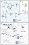
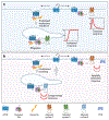
Figure 1
Figure 1
Pathways of endocytosis and postendocytic sorting of RTKs and GPCRs. (a) Simplified scheme for regulated endocytosis and postendocytic sorting of RTKs. Ligand binding results in activation of the receptor kinase; tyrosine phosphorylation of the C-terminal tail (red P); binding of Cbl, either directly to the receptor pTyr or through Grb2; and RTK ubiquitination. RTK is recruited into CCPs either via an interaction between ubiquitin moieties and adaptors in CCPs (which bind to AP-2 or clathrin), direct binding of internalization motifs (LL motif is shown) to AP-2, or an indirect interaction between AP-2 and additional adaptor proteins. RTKs internalized through CIE and CME accumulate in the same early/sorting endosomes containing EEA1 and Rab5. Ubiquitinated RTK is sorted to ILVs in MVEs via interactions with ESCRTs, whereas nonubiquitinated RTK is capable of recycling back to the plasma membrane via a fast route involving Rab4 or a slower route involving additional recycling compartments containing Rab11 or Rab25. (b) Trans-endocytosis of the Ephrin-B:EphB complex. Binding of Ephrin-B expressed in a donor cell to EphB expressed in the acceptor cell results in CME of the ligand together with the surrounding plasma membrane of the donor cell into the acceptor cell. Postendocytic sorting pathways of EphRs are similar to those shown in panel a. (c) Simplified scheme for regulated endocytosis and postendocytic sorting of GPCRs. Endocytosis is regulated through agonist-dependent Ser/Thr phosphorylation of the GPCR tail (red P), which promotes recruitment of β-arrestin from the cytoplasm and a conformational change in β-arrestin that exposes latent endocytic motifs. Agonists can selectively regulate endocytosis through an allosteric selection process mediated by distinct GRK recruitment modes that encode different phosphorylation patterns into a modular Ser/Thr substrate sequence (or barcode) in the receptor tail. Internalized receptors traffic to lysosomes by engaging ESCRTs, either through receptor ubiquitination or alternative protein connectivity not requiring receptor ubiquitination, as described in the text. A number of GPCRs are sorted by sequence-directed recycling; a PDZ motif–directed mechanism that is engaged by β2AR is depicted. There is considerable diversity among GPCRs in their overall postendocytic itinerary and kinetics of transit. Abbreviations: β2AR, β-2 adrenergic receptor; ASRT, actin–SNX27–retromer tubule; CCP, clathrin-coated pit; CIE, clathrin-independent endocytosis; CME, clathrin-mediated endocytosis; ESCRT, endosomal sorting complex required for transport; FEME, fast endophilin-mediated endocytosis; GPCR, G protein–coupled receptor; GRK, GPCR kinase; ILV, intraluminal vesicle; MVE, multivesicular endosome; P, phosphate; PIP2, phosphatidylinositol 4,5-phosphate; RTK, receptor tyrosine kinase; TGN, trans-Golgi network; Ub, ubiquitin. Figure adapted from images created with BioRender.com.
Figure 2
Figure 2
Downregulation of RTK and GPCR signaling by endocytic trafficking to lysosomes and examples of regulatory mechanisms controlling signal attenuation. Receptor ligands, proteins, and posttranslational modifications are shown that augment or impair the endocytosis and postendocytic targeting for lysosomal degradation of GPCRs (left) and RTKs (right) by retaining receptors at the cell surface and modulating receptors or the core trafficking machinery. Downregulation of signaling by an RTK can also be minimized by its overexpression and through heterodimerization with an internalization- or degradation-impaired RTK. Abbreviations: DUB, deubiquitination enzyme; FCHSD2, FCH and double SH3 domain-containing protein; GPCR, G protein–coupled receptor; GRK, GPCR kinase; pTyr, phosphorylated tyrosine; RCP, Rab-coupling protein; RTK, receptor tyrosine kinase; SORLA, sortilin-related receptor 1; Ub, ubiquitin. Figure adapted from images created with BioRender.com.
Figure 3
Figure 3
Endocytosis disrupts or facilitates the proximity of RTKs and their downstream signaling effectors. (a) Examples of the spatial separation of RTKs from their effectors during endocytosis are shown, such as separation of the plasma membrane Ca2+ channel and RTK–enzymatic complexes (with PI3K and PLCγ1 or Grb2–SOS) from their substrates (PIP2 or Ras) and disruption of cooperative RTK–integrin signaling (b) Example of endosomal signaling 1. Akt is activated in endosomes through binding to APPL1/2 and/or PI3,4P22. Liver-specific PI3K–C2γ binds to activated (GTP-loaded) Rab5 and converts PI4P to PI3,4P2. Therefore, activation of Akt results in the inhibitory phosphorylation of GSK3β in APPL- and Rab5-containing endosomes. (c) Example of endosomal signaling 2. Rac1 is located in endosomes and activated by its GEF, VAV2, translocated to endosomes in a complex with an activated RTK or TIAM1, and recruited to GTP–Rab5, whose activity is increased by RTK signaling. Abbreviations: ECM, extracellular matrix; GEF, guanine exchange factor; GTP, guanosine triphosphate; PI3K, phosphatidylinositol 3-kinases; PI3,4P2, phosphatidylinositol 3,4-bisphosphate; PIP2, phosphatidylinositol 4,5-phosphate; PIP3, phosphatidylinositol 3,4,5-trisphosphate; PLC, phospholipase C; RTK, receptor tyrosine kinase. Figure adapted from images created with BioRender.com.
Figure 4
Figure 4
Current models for how endocytosis affects GPCR–G protein signaling via cAMP. (a) Example of temporal control using endocytosis to produce a sustained cAMP response. GPCRs remain phosphorylated and bound to β-arrestin after endocytosis. The GPCR-β-arrestin complex assembles with Gs in an alternate signaling complex (megaplex) on the endosome-limiting membrane. This prolongs GPCR residence in endosomes and increases the efficiency of Gs activation, producing a sustained cAMP response that persists even after agonist removal from the extracellular milieu. In contrast, cAMP production from the plasma membrane is transient due to desensitization mediated by receptor phosphorylation, binding to β-arrestin, and endocytic removal from the plasma membrane. (b) Example of spatial control using endocytosis to change proximity to adenylyl cyclase isoforms and phosphodiesterases. GPCRs are dephosphorylated and dissociated from β-arrestin during or shortly after arrival in the endosome-limiting membrane. This enables a second phase of Gs activation and cAMP production in endosomes that is transient due to receptor recycling. Iterative cycling that occurs with prolonged agonist exposure drives repeated bursts of cAMP production from the plasma membrane and endosomes. cAMP produced from endosomes can signal over a longer range due to reduced local proximity to PDEs, and cAMP production from endosomes differs in its control by other pathways due to isoform-specific localization and trafficking of adenylyl cyclase. Abbreviations: cAMP, cyclic adenosine monophosphate; GPCR, G protein–coupled receptor; P, phosphate; PDE, phosphodiesterase. Figure adapted from images created with BioRender.com.

Similar articles

Cited by

References

    1. Goh LK, Sorkin A. 2013. Endocytosis of receptor tyrosine kinases. Cold Spring Harb. Perspect. Biol. 5(5):a017459. - PMC - PubMed
    1. Sorkin A, von Zastrow M. 2009. Endocytosis and signalling: intertwining molecular networks. Nat. Rev. Mol. Cell Biol. 10(9):609–22 - PMC - PubMed
    1. Sigismund S, Confalonieri S, Ciliberto A, Polo S, Scita G, Di Fiore PP. 2012. Endocytosis and signaling: Cell logistics shape the eukaryotic cell plan. Physiol. Rev. 92(1):273–366 - PMC - PubMed
    1. Bergeron JJM, Di Guglielmo GM, Dahan S, Dominguez M, Posner BI. 2016. Spatial and temporal regulation of receptor tyrosine kinase activation and intracellular signal transduction. Annu. Rev. Biochem. 85:573–97 - PubMed
    1. Irannejad R, Tsvetanova NG, Lobingier BT, von Zastrow M. 2015. Effects of endocytosis on receptor-mediated signaling. Curr. Opin. Cell Biol. 35:137–43 - PMC - PubMed

Publication types

Substances

LinkOut - more resources