

Case of early-onset Alzheimer's disease with atypical manifestation
- PMID: 33585790
- PMCID: PMC7845665
- DOI: 10.1136/gpsych-2020-100283
Case of early-onset Alzheimer's disease with atypical manifestation
Abstract
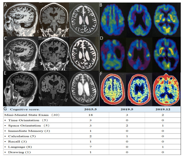
Short-term memory decline is the typical clinical manifestation of Alzheimer's disease (AD). However, early-onset AD usually has atypical symptoms and may get misdiagnosed. In the present case study, we reported a patient who experienced symptoms of memory loss with progressive non-fluent aphasia accompanied by gradual social withdrawal. He did not meet the diagnostic criteria of AD based on the clinical manifestation and brain MRI. However, his cerebrospinal fluid examination showed a decreased level of beta-amyloid 42, and increased total tau and phosphorylated tau. Massive amyloid β-protein deposition by 11C-Pittsburgh positron emission tomography confirmed the diagnosis of frontal variant AD. This case indicated that early-onset AD may have progressive non-fluent aphasia as the core manifestation. The combination of individual and precision diagnosis would be beneficial for similar cases.
Keywords: cognition disorders; diagnosis; dual (psychiatry).
© Author(s) (or their employer(s)) 2021. Re-use permitted under CC BY-NC. No commercial re-use. See rights and permissions. Published by BMJ.
Conflict of interest statement
Competing interests: None declared.
Figures
Similar articles
-
Typical and atypical appearance of early-onset Alzheimer's disease: A clinical, neuroimaging and neuropathological study.Neuropathology. 2017 Apr;37(2):150-173. doi: 10.1111/neup.12364. Epub 2017 Jan 17. Neuropathology. 2017. PMID: 28093855
-
Subtypes of progressive aphasia: application of the International Consensus Criteria and validation using β-amyloid imaging.Brain. 2011 Oct;134(Pt 10):3030-43. doi: 10.1093/brain/awr216. Epub 2011 Sep 9. Brain. 2011. PMID: 21908392
-
Assessment of cerebrospinal fluid (CSF) beta-amyloid (1-42), phosphorylated tau (ptau-181) and total Tau protein in patients with Alzheimer's disease (AD) and other dementia at Siriraj Hospital, Thailand.J Med Assoc Thai. 2011 Feb;94 Suppl 1:S77-83. J Med Assoc Thai. 2011. PMID: 21721431
-
Frontal variant of Alzheimer's disease with asymmetric presentation mimicking frontotemporal dementia: Case report and literature review.Brain Behav. 2020 Mar;10(3):e01548. doi: 10.1002/brb3.1548. Epub 2020 Jan 27. Brain Behav. 2020. PMID: 31989779 Free PMC article. Review.
-
Cerebrospinal Fluid Biomarkers for Early and Differential Alzheimer's Disease Diagnosis.J Alzheimers Dis. 2018;62(3):1199-1209. doi: 10.3233/JAD-170680. J Alzheimers Dis. 2018. PMID: 29562530 Free PMC article. Review.
References
Publication types
LinkOut - more resources
Full Text Sources
Other Literature Sources