. 2021 Feb 11;4(4):e202000873.
doi: 10.26508/lsa.202000873. Print 2021 Apr.

A conserved PI(4,5)P2-binding domain is critical for immune regulatory function of DOCK8

Affiliations

A conserved PI(4,5)P2-binding domain is critical for immune regulatory function of DOCK8

Tetsuya Sakurai et al. Life Sci Alliance. .

Abstract

DOCK8 is a Cdc42-specific guanine-nucleotide exchange factor that is essential for development and functions of various subsets of leukocytes in innate and acquired immune responses. Although DOCK8 plays a critical role in spatial control of Cdc42 activity during interstitial leukocyte migration, the mechanism remains unclear. We show that the DOCK homology region (DHR)-1 domain of DOCK8 binds specifically to phosphatidylinositol 4,5-bisphosphate (PI(4,5)P2) and is required for its recruitment to the plasma membrane. Structural and biochemical analyses reveal that DOCK8 DHR-1 domain consists of a C2 domain-like core with loops creating the upper surface pocket, where three basic residues are located for stereospecific recognition of phosphoinositides. Substitution of the two basic residues, K576 and R581, with alanine abolished PI(4,5)P2 binding in vitro, ablated the ability of DOCK8 to activate Cdc42 and support leukocyte migration in three-dimensional collagen gels. Dendritic cells carrying the mutation exhibited defective interstitial migration in vivo. Thus, our study uncovers a critical role of DOCK8 in coupling PI(4,5)P2 signaling with Cdc42 activation for immune regulation.


Conflict of interest statement

The authors declare that they have no conflict of interest.

Figures

Figure 1.
Figure 1.. DOCK8 binds to PI(4,5)P2 specifically through the DHR-1 domain.
(A) Immunoblots showing the phosphoinositide-binding specificity of DOCK8. Lysates of BW5147αβ cells expressing HA-tagged wild-type (WT) DOCK8 were used as an input for incubation with lipid vesicles containing each phosphoinositide at indicated concentrations (%). Lipid-associated fractions were analyzed by immunoblotting with anti-HA antibody. Positions of the size marker were shown on the right. (B) Schematic diagram of DOCK8 constructs used in the experiments. (C) Immunoblots showing no detectable binding to PI(4,5)P2 of a DOCK8 mutant in which the DHR-1 domain is deleted (ΔDHR-1). Lysates of BW5147αβ cells expressing HA-tagged WT DOCK8 (top) or DOCK8-ΔDHR-1 (bottom) were used as an input for lipid-binding assays. (D) Immunoblots showing PI(4,5)P2 binding of recombinant GST-fusion DOCK8 DHR-1 domain (top) and GST-fusion PLCδ1 PH domain (bottom).
Figure 2.
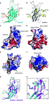
Figure 2.. Crystal structure of the DOCK8 DHR-1 domain.
(A) Ribbon diagram of the DOCK8 DHR-1 domain. The β-sandwich core is colored in cyan; β2-β3 loop and β7-β8 insertion are in orange. Three loops on the upper surface are labeled with numbers (L1 through L3). (B) Ribbon diagram of the DOCK1 DHR-1 domain (PDB ID: 3L4C; gray and light-yellow). (C, D, E, F) Surface charge representation of the DOCK8 DHR-1 domain (C, E) and the DOCK1 DHR-1 domain (D, F). Blue and red represent positive and negative electrostatic potential, respectively. The views in (C, D) are the same as those in (A, B); their top views are shown in (E, F), respectively. The upper surface region, β-groove, and basic pocket are indicated. (G) Ribbon diagram of the DOCK8 DHR-1 domain in the presence of 0.84 mM diC8-PI(4,5)P2 (purple) superposed with the one in the absence of PI(4,5)P2 shown in (A) (cyan). (H) Close-up view of the upper surface pocket of the DOCK8 DHR-1 domain. The boxed region in (G) is slightly tilted for the frontal view of the L1 loop region. Residues R570, K576, and R581 are highlighted by a stick model to show the different conformations.
Figure S1.
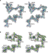
Figure S1.. Electron density map around the L1 loop of DOCK8 DHR-1.
(A, B) Stereo views of the 2Fo-Fc electron density map (contoured at 0.6 σ) around L1 loop of DOCK8 DHR-1 in the absence (A), and presence (B) of 0.84 mM diC8-PI(4,5)P2 are shown.
Figure 3.
Figure 3.. Identification of critical residues in the DOCK8 DHR-1 domain essential for PI(4,5)P2 binding.
(A) Immunoblots showing the effect of point mutations on PI(4,5)P2 binding of GST-fusion DOCK8 DHR-1 domain. Mutant proteins carry alanine substitution at the indicated residue. A doubly mutant carrying K576A and R581A is designated as “KARA.” (B, C) Measurement of PI(4,5)P2 binding to DOCK8 DHR-1 by isothermal titration calorimetry. Conditions: 0.062 mM DOCK8 DHR-1 protein titrated with 2 μl aliquotes of 1 mM diC8-PI(4,5)P2 in 20 mM Tris (pH 8.0) and 16 mM NaCl at 25°C. (B) Representative titration plots for each DOCK8 DHR-1 (n = 3). Data were best fitted to acquire the stoichiometry and thermodynamic parameters. (C) Summary of the experiments. Kd: dissociation constant; ΔH: enthalpy change; TΔS: temperature (K) x entropy change; N: stoichiometry. Data were expressed as means ± SD (n = 3). (D) Immunoblots showing no detectable binding of DOCK8 KARA mutant to PI(4,5)P2. Lysates of BW5147αβ cells expressing HA-tagged WT DOCK8 (top) or DOCK8 KARA (bottom) were used as an input for lipid-binding assays.
Figure S2.
Figure S2.. Measurement of PI(3,4,5)P3 binding to DOCK8 DHR-1 by isothermal titration calorimetry.
Conditions: 0.062 mM DOCK8 DHR-1 titrated with 2 μl aliquotes of 1 mM diC8-PI(3,4,5)P3 in 20 mM Tris (pH 8.0), 16 mM NaCl at 25°C. (A) Representative titration plots for WT DOCK8 DHR-1 and KARA mutant (n = 5). (B) Data were best fitted to acquire the stoichiometry and thermodynamic parameters.
Figure 4.
Figure 4.. Model for PI(4,5)P2 binding to the DOCK8 DHR-1 domain.
(A) Images of diC8-PI(4,5)P2 docked into the upper surface pocket of the DOCK8 DHR-1 domain. PI(4,5)P2 are highlighted by a stick model. DOCK8 DHR-1 is shown in ribbon diagram (green). (B) Close-up view of the upper surface pocket in (A). Residues predicted to form the pocket and/or bind phospholipid are shown. (C) Surface charge representation of DOCK8 DHR-1 in (A) showing the electrostatic surface of the phospholipid-binding pocket. (D) Close-up view of the binding pocket in (C).
Figure S3.
Figure S3.. Sequence alignment of the L1-loop regions of the DHR-1 domains.
Shown are the L1-loop regions of the DOCK-C subfamily: vertebrate DOCK8 (human/mouse/bird/reptile/frog/fish), mouse DOCK6 and DOCK7, and Drosophila Zizimin–related (Zir), and the DOCK-A subfamily: mouse DOCK1 and DOCK2, and Drosophila myoblast city (Mbc), aligned by the Clustal Omega software. Numbers on the top indicate the amino acid position in mouse DOCK8. Conserved basic residues are marked by red boxes. The secondary structure is indicated.
Figure S4.
Figure S4.. Docking model for PI(4,5)P2 binding to DOCK8 DHR-1 R570S mutant.
Close-up view of the upper surface pocket of DOCK8 DHR-1 R570S mutant (in ribbon diagram) docked with diC8-PI(4,5)P2 (shown in yello stick). S570 is capable of forming a hydrogen bond with the phosphate at one position of the inositol ring.
Figure 5.
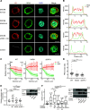
Figure 5.. DHR-1 domain is required for plasma membrane (PM) targeting of DOCK8 and its ability to activate Cdc42 and facilitate 3D cell migration in collagen gels.
(A) Confocal images showing the cellular localization of HA-tagged WT and mutant DOCK8. BW5147αβ cells stably expressing indicated proteins were analyzed by immunofluorescence using anti-HA antibody (red). Alexa Fluor 488–conjugated WGA (green) and DAPI (4′,6-diamidino-2-phenylindole, blue) were used to stain the cell surface membrane and nucleus, respectively. Scale bar: 10 μm. (B) Line scanned intensity profiles for HA and WGA fluorescence in the respective cells depicted in (A). Fluorescence intensity of HA and WGA stainings was scanned along the dotted lines in (A). The x-axis indicates arbitrary position on the line. (C) Quantification of PM localization of WT and mutant DOCK8. The intensity profiles for HA (red) and WGA (green) fluorescence from mupltiple cells were averaged (n = 48/24, 44/22, and 34/18 regions/cells for DOCK8 WT, KARA, and ΔDHR-1, respectively). Data are means ± SD. Positions of the peak intensity of WGA fluorescence were defined as the PM, and the fluorescence intensity at PM was set as 1.0 for normalization of HA and WGA fluorescence in individual cells. Distance from PM was plotted on x-axis. (D) Plasma membrane to cytoplasmic ratio of HA fluorescence for WT and mutant DOCK8 protein. The ratio of HA fluorescence intensity at the PM and cytoplasm (Cyto) in (C) was plotted for individual cells. **P < 0.0001 by a two-tailed unpaired Mann-Whitney test. (E) Fluorescence resonance energy transfer (FRET)–based measurement of Cdc42 activity in living cells. COS-7 cells were co-transfected with a FRET-based biosensor (Raichu-Cdc42), and the pBJ-neo or the respective DOCK8 constructs. FRET imaging was performed during 26–32 h after transfection. Relative emission ratio (YFP/CFP) of the whole cell area was calculated (n = 10, 20, 17, and 14 cells from three independent experiments for control, WT, KARA, and ΔDHR-1, respectively). P-values by a two-tailed unpaired t test. Right panel: Immunoblots showing the expression of transfected DOCK8 and Raichu-Cdc42 (probed with anti-HA and anti-GFP antibodies, respectively). (F) 3D migration in collagen gels of BW5147αβ cells expressing HA-tagged DOCK2 and FLAG-tagged WT or mutant DOCK8 (n = 232–286 cells per group from three independent experiments). Two independent clones were analyzed for KARA mutation. Each box plot indicates the median (the line in the middle), 25th and 75th percentiles (box ends), and 10th and 90th percentiles (whiskers). The number on each column indicates the average speed in μm/min. **P < 0.0001 by a two-tailed unpaired Mann–Whitney test. Right panel: immunoblots showing the expression level of DOCK2 and DOCK8 in the cells. Actin blot is shown as a loading control; the positions for the size markers on the right.
Figure S5.
Figure S5.. Subcellular distribution of DOCK8 in BW5147α−β− cells.
(A) Immunoblots showing the subcellular distribution of WT and mutant DOCK8. Cytoplasmic “C” and membrane “M” fractions were prepared from BW5147αβ cells expressing HA-tagged DOCK8 WT, KARA, or ΔDHR-1 and analyzed by immunoblotting. Input “I” (total cell lysate), C, and M fractions were loaded at 1:2:4 ratio for visualization. LAT (the linker for activation of T cells; transmembrane protein) and Cdc42 (mainly cytoplasmic) were also probed to assess the integrity of the fractions. (B) Plot showing the membrane to cytoplasmic ratio for each DOCK8. Data from densitometric analyses of immunoblots from three independent experiments.
Figure 6.
Figure 6.. KARA mutation in DOCK8 significantly attenuates 3D migration of mDCs.
(A) Immunoblot analysis of the expression level of DOCK8 protein in DCs derived from DOCK8WT/−, DOCK8KARA/−, and DOCK8−/− mice. Actin blot is shown as a loading control. The positions for the size markers were indicated on the right. (B) Impaired migration of DOCK8KARA/− and DOCK8−/− mature DCs (mDCs) in 3D collagen gels. Migration of LPS-stimulated DOCK8WT/−, DOCK8KARA/−, and DOCK8−/− mDCs toward CCL21 source was recorded for 120 min by time-lapse video microscopy. Representative tracks of individual mDCs. (C) The migration speed, directionality, and forward migration index were compared among DOCK8WT/−, DOCK8KARA/−, and DOCK8−/− mDCs (n = 132 (109), 127 (75), and 100 (41), respectively, from three independent experiments). For directionality and forward migration index, the cells that had migrated at 0.3 μm/min or faster were analyzed (cell numbers in the parentheses). Each box plot indicates the median (the line in the middle), 25th and 75th percentiles (box ends), and 10th and 90th percentiles (whiskers). P-values by a two-tailed unpaired Mann–Whitney test. (D) In vivo migration efficiency of DOCK8WT/−, DOCK8KARA/−, and DOCK8−/− mDCs. DCs in a pair were mixed at 1:1 ratio, injected into footpads of C57BL/6 mice and recovered from the popliteal LNs after 48 h. Data are means ± SD for six pairs of DOCK8WT/− and DOCK8KARA/− DCs with data for two pairs of DOCK8WT/− and DOCK8−/− DCs. P-value by a two-tailed unpaired Mann–Whitney test.
Figure S6.
Figure S6.. Normal expression of CCR7 in DOCK8WT/−, DOCK8KARA/−, and DOCK8−/− DCs.
(A) CCR7 mRNA expression was quantified by real time qPCR from three independent DC preparations. Data are shown in fold increase relative to the level of DOCK8WT/− DCs (means ± SD). Neutophils were analyzed as a negative control. (B) Cell surface presentation of CCR7 was analyzed by flow cytometry with DCs from three independent preparations. (C) Representative flow cytometry plots. DCs were stained with phosphatidylethanolamine–conjugated anti-CCR7 or isotype-matched control antibody.
Figure 7.
Figure 7.. Schematic illustrating critical roles of the DOCK family proteins in linking phosphoinositide signaling to specific Rho-Wiskott-Aldrich syndrome protein family pairs.
Through the DHR-1 domain, DOCK8 is localized to a PI(4,5)P2–enriched compartment of the plasma membrane, where, upon encounter with GDP-Cdc42, the DHR-2 domain catalyzes the nucleotide exchange reaction of Cdc42 (the current work marked in a box). GTP-loaded Cdc42 and PI(4,5)P2 serve as the coincidence detection signals for activation of Wiskott-Aldrich syndrome protein, which stimulates Arp2/3 complex–mediated actin polymerization for reorganization of the actin cytoskeleton. In a similar, but distinct way, the DOCK-A subfamily member (e.g., DOCK2), which makes a signaling complex with engulfment and cell motility (ELMO) protein through the SH3 domain (Hanawa-Suetsugu et al, 2012; Chang et al, 2020), is recruited to the leading edge of migrating cells through the PI(3,4,5)P3–binding DHR-1 domain in response to chemoattractant signals. The DHR-2 domain of the DOCK-A subfamily activates Rac, which in turn acts in synergy with PI(3,4,5)P3 to activate WAVE complex. See text for details.

Similar articles

Cited by

References

    1. Adams PD, Afonine PV, Bunkoczi G, Chen VB, Davis IW, Echols N, Headd JJ, Hung LW, Kapral GJ, Grosse-Kunstleve RW, et al. (2010) PHENIX: A comprehensive Python-based system for macromolecular structure solution. Acta Crystallogr D Biol Crystallogr 66: 213–221. 10.1107/s0907444909052925 - DOI - PMC - PubMed
    1. Aoki K, Matsuda M (2009) Visualization of small GTPase activity with fluorescence resonance energy transfer-based biosensors. Nat Protoc 4: 1623–1631. 10.1038/nprot.2009.175 - DOI - PubMed
    1. Biggs CM, Keles S, Chatila TA (2017) DOCK8 deficiency: Insights into pathophysiology, clinical features and management. Clin Immunol 181: 75–82. 10.1016/j.clim.2017.06.003 - DOI - PMC - PubMed
    1. Bouma G, Burns S, Thrasher AJ (2007) Impaired T-cell priming in vivo resulting from dysfunction of WASp-deficient dendritic cells. Blood 110: 4278–4284. 10.1182/blood-2007-06-096875 - DOI - PubMed
    1. Butty AC, Perrinjaquet N, Petit A, Jaquenoud M, Segall JE, Hofmann K, Zwahlen C, Peter M (2002) A positive feedback loop stabilizes the guanine-nucleotide exchange factor Cdc24 at sites of polarization. EMBO J 21: 1565–1576. 10.1093/emboj/21.7.1565 - DOI - PMC - PubMed

Publication types

MeSH terms

Substances

LinkOut - more resources