Chronic stress increases the tyrosine phosphorylation in female reproductive organs: An experimental study
- PMID: 33554006
- PMCID: PMC7851478
- DOI: 10.18502/ijrm.v19i1.8183
Chronic stress increases the tyrosine phosphorylation in female reproductive organs: An experimental study
Abstract
Background: Changes in tyrosine-phosphorylated (TyrPho) protein expressions have demonstrated stress in males. In females, chronic stress (CS) is a major cause of infertility, especially anovulation. However, the tyrosine phosphorylation in the female reproductive system under stress conditions has never been reported.
Objective: To investigate the alteration of TyrPho protein expression in ovary, oviduct, and uterus of CS rats.
Materials and methods: In this experimental study, 16 female Sprague-Dawley rats (5 wk: 220-250 gr) were divided into control and CS groups (n = 8/group). Every day, the CS animals were immobilized within a restraint cage and individually forced to swim in cold water for 60 consecutive days. Following the stress induction, the ovary, oviduct, and uterus of all rats were observed for their morphologies. The total protein profiles of all tissues were revealed by sodium dodecyl sulphate polyacrylamide gel electrophoresis (SDS-PAGE) before detecting TyrPho proteins using western blot. Intensity analysis was used to compare the expression of proteins between groups.
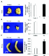
Results: The results showed that the morphology and weights of ovary and oviduct in the CS group were not different from control. In contrast, the CS significantly increased the uterine weight as compared to control. Moreover, the expressions of TyrPho proteins in the ovary (72, 43, and 28 kDas), oviduct (170, 55, and 43 kDas), and uterus (55, 54, and 43 kDas) were increased in CS group as compared to those of control.
Conclusion: The increased expressions of TyrPho proteins in ovary, oviduct, and uterus could be potential markers used to explain some machanisms of female infertility caused from chronic stress.
Keywords: Phosphorylation.; Uterus; Ovary; Oviduct.
Copyright © 2021 Bunsueb et al.
Conflict of interest statement
The authors declare no conflict of interest in the present study.
Figures


Similar articles
-
Localization (and profiles) of tyrosinephosphorylated proteins in female reproductive organs of adult rats.Clin Exp Reprod Med. 2020 Sep;47(3):180-185. doi: 10.5653/cerm.2020.03573. Epub 2020 Sep 1. Clin Exp Reprod Med. 2020. PMID: 32911588 Free PMC article.
-
Chronic stress affects tyrosine phosphorylated protein expression and secretion of male rat epididymis.Andrologia. 2021 Apr;53(3):e13981. doi: 10.1111/and.13981. Epub 2021 Jan 20. Andrologia. 2021. PMID: 33469986
-
Effect of chronic stress on expression and secretion of seminal vesicle proteins in adult rats.Andrologia. 2021 Feb;53(1):e13800. doi: 10.1111/and.13800. Epub 2020 Aug 20. Andrologia. 2021. PMID: 32816406
-
NTP Research Report on the CLARITY-BPA Core Study: A Perinatal and Chronic Extended-Dose-Range Study of Bisphenol A in Rats: Research Report 9 [Internet].Research Triangle Park (NC): National Toxicology Program; 2018 Sep. Research Triangle Park (NC): National Toxicology Program; 2018 Sep. PMID: 31305969 Free Books & Documents. Review.
-
NTP Developmental and Reproductive Toxicity Technical Report on the Modified One-Generation Study of Bisphenol AF (CASRN 1478-61-1) Administered in Feed to Sprague Dawley (Hsd:Sprague Dawley® SD®) Rats with Prenatal, Reproductive Performance, and Subchronic Assessments in F1 Offspring: DART Report 08 [Internet].Research Triangle Park (NC): National Toxicology Program; 2022 Sep. Research Triangle Park (NC): National Toxicology Program; 2022 Sep. PMID: 36383702 Free Books & Documents. Review.
References
-
- Vander Borghtb M, Wyns Ch. Fertility and infertility: Definition and epidemiology. Clin Biochem 2018; 62: 2–10. - PubMed
-
- Barbieri RL. Female infertility. In: Strauss J, Barbieri R. Yen and Jaffe's reproductive endocrinology. 8 Ed. Netherland: Elsevier; 2019. 556–581.
-
- Pandey AK, Gupta A, Tiwari M, Prasad Sh, Pandey AN, Yadav PK, et al. Impact of stress on female reproductive health disorders: Possible beneficial effects of shatavari (Asparagus racemosus). Biomed Pharmacother 2018; 103: 46–49. - PubMed
-
- Farkas J, Rigó A, Demetrovics Z. Psychological aspects of the polycystic ovary syndrome. Gynecol Endocrinol 2013; 30: 95–99. - PubMed
LinkOut - more resources
Full Text Sources
Other Literature Sources
