Review
. 2021 Jan 28;26(3):692.
doi: 10.3390/molecules26030692.

Discussions of Fluorescence in Selenium Chemistry: Recently Reported Probes, Particles, and a Clearer Biological Knowledge

Affiliations
Review

Discussions of Fluorescence in Selenium Chemistry: Recently Reported Probes, Particles, and a Clearer Biological Knowledge

Ariq Abdillah et al. Molecules. .

Abstract

In this review from literature appearing over about the past 5 years, we focus on selected selenide reports and related chemistry; we aimed for a digestible, relevant, review intended to be usefully interconnected within the realm of fluorescence and selenium chemistry. Tellurium is mentioned where relevant. Topics include selenium in physics and surfaces, nanoscience, sensing and fluorescence, quantum dots and nanoparticles, Au and oxide nanoparticles quantum dot based, coatings and catalyst poisons, thin film, and aspects of solar energy conversion. Chemosensing is covered, whether small molecule or nanoparticle based, relating to metal ion analytes, H2S, as well as analyte sulfane (biothiols-including glutathione). We cover recent reports of probing and fluorescence when they deal with redox biology aspects. Selenium in therapeutics, medicinal chemistry and skeleton cores is covered. Selenium serves as a constituent for some small molecule sensors and probes. Typically, the selenium is part of the reactive, or active site of the probe; in other cases, it is featured as the analyte, either as a reduced or oxidized form of selenium. Free radicals and ROS are also mentioned; aggregation strategies are treated in some places. Also, the relationship between reduced selenium and oxidized selenium is developed.

Keywords: fluorescence; luminescence; particle; phosphorescence; probe; selenium; tellurium.


Conflict of interest statement

The authors declare no conflict of interest.

Figures

Figure 1
Figure 1
Uracil derivatization with selenophene by Nuthanakanti et al.
Figure 2
Figure 2
Structure of the Sec probe reported by Zhang et al. and its transformation.
Figure 3
Figure 3
Detection mechanism of Mito–diNO2 for Sec by Han et al.
Figure 4
Figure 4
Reactions and selenium compound synthesized electrochemically by Li et al.
Figure 5
Figure 5
Structures of the acridine from the report by Chen et al.
Figure 6
Figure 6
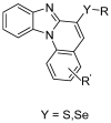
Molecular structures of the novel benzimidazo[1,2–a]quinoline derivatives reported by Da Silva et al.
Figure 7
Figure 7
The chemical structure of the amphiphilic copolymer and the water–soluble copolymer studied in the context of nucleophilic attack and fluorescence changes by Yu et al.
Figure 8
Figure 8
The diselenide system prepared by Tang et al. and its self–stacking form when introduced into water.
Figure 9
Figure 9
The organotriazoles studied by Soares et al.
Figure 10
Figure 10
The compound and its aggregation tendency in water was described by Wu et al. The position of the selenium is specific and discussed.
Figure 11
Figure 11
Chemical structure and reactivity of the rhodamine–containing selenide (RhSe) system designed for Pd2+ binding and recognition by Soares–Paulino et al.
Figure 12
Figure 12
The first TrxR diselenide–based probe reported by Mafireyi, et al.
Figure 13
Figure 13
The recent novel diselenide probe reported by Madibone et al.
Figure 14
Figure 14
Image of SNARFSSPy and SNARFSeSPy by Zhang et al.
Figure 15
Figure 15
Structures of TPP–Se–CDs by Huang et al. and MitoTracker Green and MitoSox Red.
Figure 16
Figure 16
The structure and reactivity of the reversible/reserrable Se–Ph containing species reported by Zang et al.
Figure 17
Figure 17
The selenium-containing probe by Gonçalves et al. that engages in π–π stacking and binds to mercury.
Figure 18
Figure 18
The structure of chemosensor 1–(4–ethyl–2–oxo–2H–chromen–7–yl)–3–phenyl–selenourea (L) which, as synthesized by Casula et al. demonstrated, the detection of S2− and CN.
Figure 19
Figure 19
Near–infrared (NIR) chemosensor based on azo–BODIPY and diphenylselenol (BD–di–SeH) probes by Gao et al. for sulfane sulfur analyte detection.
Figure 20
Figure 20
Coum–Se chemosensor for HOCl probe reported by Xie et al.
Figure 21
Figure 21
The structure of 5–bromo–p–2–(phenylselanyl)–thiophene–substituted BODIPY and the interfacial angle by Kim et al.
Figure 22
Figure 22
FRET probe CmNp–Sec by Zhao et al.
Figure 23
Figure 23
Structures from the report by Suarez et al. describe Selenosulfide gem–Dimethyl Ester conjugation for the goal of improving future H2S chemosensing.
Figure 24
Figure 24
The polyhedral and oligomeric silsequioxane based compound POSSSe serving as a chemosensor, by Liu et al.
Figure 25
Figure 25
The intriguing dibenzobarrelene structures DbbSeO and MbbSeO used to detect H2S reversibly/resetably that involves a ka/selenide redox system by Annaka et al.
Figure 26
Figure 26
One–step activation of Fast–TRFS reported by Li et al.
Figure 27
Figure 27
Chemical structure of the tandem fluorophore-substituted phenyl selenide moiety able to undergo oxidation at the Se center by Desmukh et al.
Figure 28
Figure 28
Rhodamine–based phenyl telluride–substituted system used for mercuric detection by Soares–Paulino et al.
Figure 29
Figure 29
A tellurium–rhodamine B (TRB)–based probe that was studied for Hg2+ detection by Soares–Paulino et al.
Figure 30
Figure 30
The structures of DAAQ and the proposed mechanism of the probe for differentiating Cys, Hcy, and GSH by Tian et al.
Figure 31
Figure 31
Selective detection of GSH using a coumarin in which the SePh group and CHO group are both serving as adjacent reactive groups.
Figure 32
Figure 32
Structure of 7–hydroxy–4–methyl–5–(phenylselanyl)isobenzofuran–1,3–dione probe (Probe–1) reported by Mulay et al.
Figure 33
Figure 33
Proposed mechanism of the probe for detection of hypochlorite by Kim et al.
Figure 34
Figure 34
The structure and utility of the CyDiSe probe used in analyte detection by Chen et al.
Figure 35
Figure 35
Structures of SC1 and SC2 by Su et al. and their reaction with Ag+ and Na2S.
Figure 36
Figure 36
Structure of PA5 and a schematic of detection mechanism (chemical) reported by Gaur et al.
Figure 37
Figure 37
Structures of BODIPY based probes for the detection of OCl by Mulay et al.
Figure 38
Figure 38
Compound 7f squarine compound by Martin et al.
Figure 39
Figure 39
Structure of fluorophore based on hemicyanine synthesized by Han et al.
Figure 40
Figure 40
Probe design strategy for sulfane sulfur detection by Gao et al.
Figure 41
Figure 41
BODIPY system containing an aliphatic Se and protonated amino group synthesized by Xu and Xian.
Figure 42
Figure 42
Structure of CQDs–SL synthesized by Devi et al.
Figure 43
Figure 43
The structure of ABDO synthesized by Guan et al. and the binding of selenite analyte to achieve recognition with this probe.
Figure 44
Figure 44
Structure of HBTSe and its reaction towards H2Se by Xin et al.
Figure 45
Figure 45
Detection of selenol compound with gold nanoparticles and Nile Blue by Guo et al.
Figure 46
Figure 46
Molecular detection of selenol with gold nanoparticles and Cy–5,5 by Hu et al.
Figure 47
Figure 47
Structure of Hcy–H2Se and its action of undergoing reaction with and detecting H2Se by Kong et al.
Figure 48
Figure 48
Structure and intended reactivity for chemosensing probe NO2L studied for selenocysteine detection by Areti et al.
Figure 49
Figure 49
The structure of probe 1 analyzed for selenocysteine based on the 2,4 dinitrophenyl motif by Li et al.
Figure 50
Figure 50
Selenocysteine detection using Mito–Cy–Sec, a cyanine based dye by Luo et al.
Figure 51
Figure 51
Bioluminescent probes initiated from the 2,4–dinitritro phenyl– group cleavage upon encountering selenocysteine by Zhang et al.
Figure 52
Figure 52
Reaction of NapEb by Ungati et al. with H2O2 and its reversible/resettable reaction with GSH.
Figure 53
Figure 53
The structure and activity of the chemosensing probe that frees the Se from the N,N–chelation by Tian et al. The nature of the heterocycle changes dramatically; the native color of the 1,8–naphthalamide is restored.
Figure 54
Figure 54
Detection mechanism of Sel–p1 and Sel–p2 by Dai et al. towards selenols.
Figure 55
Figure 55
H2Se probe NIR–H2Se by Kong et al. involving the benzene selenodiazole unit.
Figure 56
Figure 56
The investigation of GAT as a selective Se(IV) fluorescent probe by Liu et al.
Figure 57
Figure 57
The reaction of the O–hNRSel fluorescence probe by Zhang et al. and its selective reaction with selenocysteine.
Figure 58
Figure 58
Structure of BPP, a simple sulfilimine model system was described by Luan et al.
Figure 59
Figure 59
Structure of NIR–H2Se synthesized by Gao et al. and its reaction with H2Se. Structure of dihydroethidium and rhodamine 110 are also depicted.
Figure 60
Figure 60
Structure of DCFA–DA by Zwolak et al.
Figure 61
Figure 61
Selenium nanoparticles are formed by reducing them with ascorbic acid with the help of beta–lactoglobulin stabilizer. Investigations of development, physicochemical characterization and cytotoxicity of selenium nanoparticles stabilized by beta–lactoglobulin by Zhang et al.
Figure 62
Figure 62
Cu2+ detection by functionalized CdSe reported by Zhao et al.
Figure 63
Figure 63
Oxidation of tellurium by superoxide and its reversible reaction with glutathione in carbon dots by Zhang et al.
Figure 64
Figure 64
Structure of QDs/Se@Ru(A) by He et al. that is used to antagonize glioblastoma.
Figure 65
Figure 65
Structure of QD–Ab for targeting tumor cell synthesized by Vibin et al.
Figure 66
Figure 66
Structure of m–CdSe and its action for the detection of melamine by Singh et al.
Figure 67
Figure 67
Hg2+–based detection from ZnO nanorod doped with selenium and coated with 3–mercaptopropionic acid (3–MPA) by Rao et al.
Figure 68
Figure 68
The probing of selenite based on the nanodevice structure and strategy discussed by Yu et al.
Figure 69
Figure 69
Structure of SY1080 synthesized by Zhang et al.
Figure 70
Figure 70
Structure of Probe 3 synthesized by Sun et al.
Figure 71
Figure 71
The aggregation-induced fluorescence achieved with a recent probe by Qin et al.
Figure 72
Figure 72
The detection method of H2Se using CdTe quantum dots reported by Huang et al.
Figure 73
Figure 73
Structure of the GSH- selective probe HGc reported by Wang.
Figure 74
Figure 74
Cleavage by peroxynitrite by Fan, Wong, Wu et al.
Figure 75
Figure 75
Hydrazone cleavage upon peroxynitrite addition.

Similar articles

Cited by

References

    1. Hoover G.C., Seferos D.S. Photoactivity and optical applications of organic materials containing selenium and tellurium. Chem. Sci. 2019;10:9182–9188. doi: 10.1039/C9SC04279B. - DOI - PMC - PubMed
    1. Seçkin H.Y., Kalkan G., Bütün I., Akbaş A., Baş Y., Karakuş N., Benli I. Analysis of Manganese Superoxide Dismutase and Glutathione Peroxidase 1 Gene Polymorphisms in Vitiligo. Biochem. Genet. 2016;54:438–447. doi: 10.1007/s10528-016-9730-4. - DOI - PubMed
    1. Wu D., Chen L., Kwon N., Yoon J. Fluorescent Probes Containing Selenium as a Guest or Host. Chem. 2016;1:674–698. doi: 10.1016/j.chempr.2016.10.005. - DOI
    1. Xiao Y., Xu S., Zhao S., Liu K., Lu Z., Hou Z. Protective effects of selenium against zearalenone-induced apoptosis in chicken spleen lymphocyte via an endoplasmic reticulum stress signaling pathway. Cell Stress Chaperon. 2019;24:77–89. doi: 10.1007/s12192-018-0943-9. - DOI - PMC - PubMed
    1. Hussain R., Luo K. Geochemical valuation and intake of F, As, and Se in coal wastes contaminated areas and their potential impacts on local inhabitants, Shaanxi China. Environ. Geochem. Health. 2018;40:2667–2683. doi: 10.1007/s10653-018-0131-y. - DOI - PubMed

LinkOut - more resources