. 2021 Jan 29;17(1):e1009275.
doi: 10.1371/journal.ppat.1009275. eCollection 2021 Jan.

Ebola virus triggers receptor tyrosine kinase-dependent signaling to promote the delivery of viral particles to entry-conducive intracellular compartments

Affiliations

Ebola virus triggers receptor tyrosine kinase-dependent signaling to promote the delivery of viral particles to entry-conducive intracellular compartments

Corina M Stewart et al. PLoS Pathog. .

Abstract

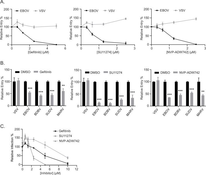
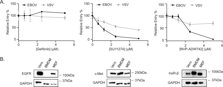
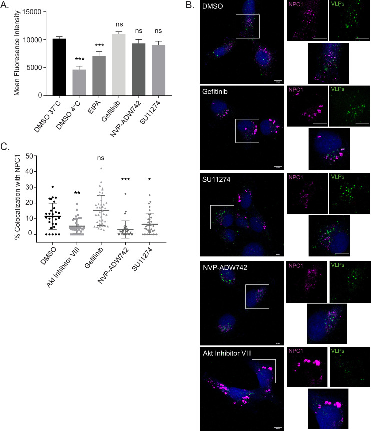
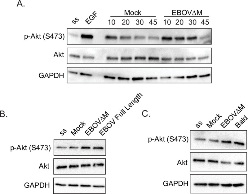
Filoviruses, such as the Ebola virus (EBOV) and Marburg virus (MARV), are causative agents of sporadic outbreaks of hemorrhagic fevers in humans. To infect cells, filoviruses are internalized via macropinocytosis and traffic through the endosomal pathway where host cathepsin-dependent cleavage of the viral glycoproteins occurs. Subsequently, the cleaved viral glycoprotein interacts with the late endosome/lysosome resident host protein, Niemann-Pick C1 (NPC1). This interaction is hypothesized to trigger viral and host membrane fusion, which results in the delivery of the viral genome into the cytoplasm and subsequent initiation of replication. Some studies suggest that EBOV viral particles activate signaling cascades and host-trafficking factors to promote their localization with host factors that are essential for entry. However, the mechanism through which these activating signals are initiated remains unknown. By screening a kinase inhibitor library, we found that receptor tyrosine kinase inhibitors potently block EBOV and MARV GP-dependent viral entry. Inhibitors of epidermal growth factor receptor (EGFR), tyrosine protein kinase Met (c-Met), and the insulin receptor (InsR)/insulin like growth factor 1 receptor (IGF1R) blocked filoviral GP-mediated entry and prevented growth of replicative EBOV in Vero cells. Furthermore, inhibitors of c-Met and InsR/IGF1R also blocked viral entry in macrophages, the primary targets of EBOV infection. Interestingly, while the c-Met and InsR/IGF1R inhibitors interfered with EBOV trafficking to NPC1, virus delivery to the receptor was not impaired in the presence of the EGFR inhibitor. Instead, we observed that the NPC1 positive compartments were phenotypically altered and rendered incompetent to permit viral entry. Despite their different mechanisms of action, all three RTK inhibitors tested inhibited virus-induced Akt activation, providing a possible explanation for how EBOV may activate signaling pathways during entry. In sum, these studies strongly suggest that receptor tyrosine kinases initiate signaling cascades essential for efficient post-internalization entry steps.


Conflict of interest statement

The authors have declared that no competing interests exist.

Figures

Fig 1
Fig 1. A kinase inhibitor screen to identify signaling pathways required for EBOV and MARV GP-mediated entry.
MLV pseudotypes encoding LacZ and harboring EBOV, MARV, or VSV glycoproteins were used to screen the Selleckchem L1200 kinase inhibitor library at 1μM in Vero cells (A) Volcano plots of the log2 values of the ratios of the means of biological replicates of EBOV (left panel) or MARV (right panel) vs VSV pseudotype transduction relative to the DMSO controls over the–log10 values of the p-value. Dots in red represent hits as determined with at least 2-fold ratio and p<0.05. Results are means of three independent experiments performed in duplicates. (B) Venn diagram of the hits for EBOV and MARV, with 22 total hits for EBOV, 23 total hits for MARV, and 10 hits shared between the two viruses. (C) Heat maps of the log2(normalized transduction efficiency of EBOV or MARV vs. that of VSV) for the total hits for both viruses. (D) Pie charts of the signaling pathways (grouped into the following categories: receptor tyrosine kinase (RTK) inhibitors, Janus kinase/signal transducers and activators of transcription (JAK/STAT) pathway inhibitors, mitogen-activated protein kinase (MAPK) pathway inhibitors, non-receptor tyrosine kinase (NRTK) inhibitors, and phosphatidylinositol-3-kinase (PI3K)/Akt inhibitors, or others) of the inhibitors in the library (top), EBOV hits (middle), and MARV (bottom). The representation in percentage and the number of the RTK inhibitors is indicated.
Fig 2
Fig 2. RTK inhibitors block filovirus entry in Vero cells.
(A) Entry of βlam VLPs harboring the EBOV GP or VSV G in Vero cells treated with vehicle (DMSO, 0.1%) or increasing concentrations of Gefitinib, SU11274, or NVP-ADW742. Entry was detected via flow cytometry after loading cells with βlam substrate (CCF2) and measuring the percentage of inhibitor treated cells with cleaved CCF2. Data are expressed as percentages relative to DMSO-treated cells. (B) Entry of βlam VLPs bearing the GPs of EBOV, SUDV, BDBV, MARV, or VSV G in the presence of 1 μM Gefitinib, 1 μM SU11274, 1 μM NVP-ADW742, or vehicle (DMSO, 0.1%). (C) Infection of Vero cells with replication-competent EBOV expressing GFP at increasing concentrations of the indicated inhibitor. Infection was measured by GFP fluorescence 3 days post-infection and normalized to vehicle-treated cells. Results are expressed as mean ± s.d. of triplicates and are representative of 3 experiments. * p < 0.05, ** p < 0.01, *** p < 0.001.
Fig 3
Fig 3. RTK inhibitors do not inhibit entry mediated by a panel of other viral GPs.
Vero cells were transduced with MLV pseudotypes encoding LacZ and harboring EBOV, LFV, LCMV, Nipah, or Junin glycoproteins in the presence of Gefitinib (1 μM), SU11274 (1 μM), NVP-ADW742 (1 μM) or vehicle (DMSO, 0.1%). Results are expressed as mean ± s.d. of triplicates and are representative of 3 experiments. * p < 0.05, ** p < 0.01, *** p < 0.001.
Fig 4
Fig 4. Effect of RTK inhibitor treatment on EBOV GP-mediated entry in BMDMs.
(A) Entry of βlam VLPs harboring EBOV GP or VSV G in BMDMs treated with vehicle (DMSO, 0.1%) or increasing concentrations of Gefitinib, SU11274, or NVP-ADW742. (B) Vero cells, BMDMs, and MEFs were serum-starved, lysed, and expression of EGFR, Met, InsR-β, and GAPDH was detected by immunoblotting. Results in (A) are expressed as mean ± s.d. of triplicates and are representative of 3 experiments. Results in (B) are representative blots of 3 independent experiments.
Fig 5
Fig 5. RTK inhibitors do not block EBOV VLP internalization, but SU11274 and NVP-ADW742 interfere with trafficking to NPC1+ compartments.
(A) Fluorescent mCherry VLPs harboring EBOV ΔM GP were pre-bound to Vero cells by spinoculation at 4°C, followed by washing and incubation with vehicle (DMSO, 0.1%), EIPA (30 μM), Gefitinib (5 μM), SU11274 (2.5 μM), or NVP-ADW742 (2.5 μM) at 37°C for one hour, or vehicle (DMSO, 0.1%) at 4°C for one hour. Cells were then trypsinized with 0.5% trypsin and mCherry fluorescence analyzed by flow cytometry. (B) Infection of HT1080 cells pre-treated with DMSO (0.1%), Akt Inhibitor VIII (10 μM), Gefitinib (5 μM), SU11274 (2.5 μM), or NVP-ADW742 (2.5 μM) with fluorescent VLPs (Green) harboring the fusion deficient ΔM GPF535R for 3 h. 30 min prior to fixation, CMAC cytoplasmic dye (Blue) was added. Cells were then fixed, permeabilized, and immunostained with rabbit anti-NPC1 and DY650-conjugated antiserum (Magenta). Cells were imaged on an LSM800 confocal microscope (Zeiss). Images are displayed as maximum intensity z-projections, bar = 10 μm. (C) Colocalization between VLPs and NPC1 (of a minimum of 25 cells per condition) were analyzed using Imaris software (Bitplane). Data are representative of 3 independent experiments. * p < 0.05, ** p < 0.01, *** p < 0.001.
Fig 6
Fig 6. Gefitinib interferes with the biology of NPC1+ compartments.
(A) Infection of HT1080 cells pre-treated with vehicle (DMSO, 0.1%) or Gefitinib (5 μM) with fluorescent VLPs (Green) harboring the fusion deficient EBOV GPF535R for 3 h. 30 min prior to fixation, CMAC cytoplasmic dye (Blue) was added. Cells were then fixed, permeabilized, and stained for NPC1 (Magenta). Cells were imaged on an LSM800 confocal microscope (Zeiss). Images are displayed as maximum intensity z-projections, bar = 10 μm. (B) Average volume (μm3) of NPC1+ compartments per cell (of a minimum of 25 cells per condition) was determined by modeling these compartments using Imaris software (Bitplane). (C) HT1080 cells were treated with DMSO (0.1%), Gefitinib (5 μM), Akt Inhibitor VIII (10 μM), or NVP-ADW742 (2.5 μM) for 4 h. Cells were then fixed, permeabilized, and immunostained with rabbit anti-NPC1 and mouse anti-LBPA, followed by DY650-conjugated antiserum (Magenta) or AF555-conjugated antiserum (Green). Following immunostaining, cells were stained with Hoechst (Blue) and imaged on an LSM800 confocal microscope (Zeiss). Images are displayed as maximum intensity z-projections, bar = 10 μm. Data are representative of 3 independent experiments. * p < 0.05, ** p < 0.01, *** p < 0.001.
Fig 7
Fig 7. EBOV ΔM GP, EBOV Full Length GP, and Bald EboV VLPs all stimulate Akt phosphorylation in Vero cells.
Vero cells were serum starved in HBSS for 1h, followed by stimulation with (A) EGF (50 ng/mL), Mock, or βlam EBOV ΔM GP VLPs for 10, 20, 30, or 45 min, (B) Mock, βlam EBOV ΔM GP, or βlam EBOV Full Length GP VLPs for 20 min., or (C) Mock, βlam EBOV ΔM GP VLPs, or Bald EBOV VLPs for 20min. Cells were lysed and phosphorylated Akt (p-Akt—S473), total Akt (Akt), and GAPDH were detected by immunoblot. Abbreviations are serum starved (ss), βlam EBOV ΔM GP VLPs (EBOV), βlam EBOV Full Length GP VLPs (Full Length). Data are representative of 3 independent experiments.
Fig 8
Fig 8. RTK inhibitors block EBOV VLP-induced Akt phosphorylation.
Vero cells were pre-treated with Gefitinib (5 μM), SU11274 (2.5 μM), NVP-ADW742 (2.5 μM) or vehicle (DMSO, 0.1%) in serum-free HBSS for 1h. Cells were then stimulated with (A) EGF (50 ng/mL), IGF (50 ng/mL), or HGF (200 ng/mL) for 10, 20, 30, or 45 min, or (B) with Mock, βlam EBOV ΔM GP VLPs (MOI = 100) for 20 min. Cells were lysed and phosphorylated Akt (p-Akt—S473), total Akt (Akt), and GAPDH were detected by immunoblot. Abbreviations are serum starved (ss), βlam EBOV ΔM GP VLPs (EBOV), Gefitinib (GEF), NVP-ADW742 (NVP), and SU11274 (SU). Data are representative of 3 independent experiments.

Similar articles

Cited by

References

    1. Harrison SC. Viral membrane fusion. Virology. 2015;479–480:498–507. Epub 2015/04/14. 10.1016/j.virol.2015.03.043 - DOI - PMC - PubMed
    1. Carette JE, Raaben M, Wong AC, Herbert AS, Obernosterer G, Mulherkar N, et al. Ebola virus entry requires the cholesterol transporter Niemann-Pick C1. Nature. 2011;477(7364):340–3. Epub 2011/08/26. 10.1038/nature10348 - DOI - PMC - PubMed
    1. Miller EH, Obernosterer G, Raaben M, Herbert AS, Deffieu MS, Krishnan A, et al. Ebola virus entry requires the host-programmed recognition of an intracellular receptor. EMBO J. 2012;31(8):1947–60. Epub 2012/03/08. 10.1038/emboj.2012.53 - DOI - PMC - PubMed
    1. Cote M, Misasi J, Ren T, Bruchez A, Lee K, Filone CM, et al. Small molecule inhibitors reveal Niemann-Pick C1 is essential for Ebola virus infection. Nature. 2011;477(7364):344–8. Epub 2011/08/26. 10.1038/nature10380 - DOI - PMC - PubMed
    1. Jae LT, Raaben M, Herbert AS, Kuehne AI, Wirchnianski AS, Soh TK, et al. Virus entry. Lassa virus entry requires a trigger-induced receptor switch. Science. 2014;344(6191):1506–10. Epub 2014/06/28. 10.1126/science.1252480 - DOI - PMC - PubMed

Publication types

MeSH terms

Substances