Activation of the medial preoptic area (MPOA) ameliorates loss of maternal behavior in a Shank2 mouse model for autism
- PMID: 33491217
- PMCID: PMC7917557
- DOI: 10.15252/embj.2019104267
Activation of the medial preoptic area (MPOA) ameliorates loss of maternal behavior in a Shank2 mouse model for autism
Abstract
Impairments in social relationships and awareness are features observed in autism spectrum disorders (ASDs). However, the underlying mechanisms remain poorly understood. Shank2 is a high-confidence ASD candidate gene and localizes primarily to postsynaptic densities (PSDs) of excitatory synapses in the central nervous system (CNS). We show here that loss of Shank2 in mice leads to a lack of social attachment and bonding behavior towards pubs independent of hormonal, cognitive, or sensitive deficits. Shank2-/- mice display functional changes in nuclei of the social attachment circuit that were most prominent in the medial preoptic area (MPOA) of the hypothalamus. Selective enhancement of MPOA activity by DREADD technology re-established social bonding behavior in Shank2-/- mice, providing evidence that the identified circuit might be crucial for explaining how social deficits in ASD can arise.
Keywords: SHANK3; autism spectrum disorders; bonding; social behavior; synapse.
© 2021 The Authors. Published under the terms of the CC BY NC ND 4.0 license.
Conflict of interest statement
The authors declare that they have no conflict of interest.
Figures

- A
In contrast to the immediate care of Shank2 +/+ dams shortly after delivery, Shank2 −/− dams neglect the offspring. No proper nest building is observed, and pups are lying randomly scattered within the bedding. Right diagram: Average percentage of pups surviving until weaning per pregnancy. None of the litters of Shank2 −/− mice survived after delivery, Mann–Whitney test, ***P < 0.001, Shank2 +/+ n = 12, Shank2 −/− n = 9.
- B–F
(B, a–h) Series of pictures displaying the neglected appearance of pups delivered by Shank2 −/− dams: (B,a, C), the percentage of pups gathered in the nest location is significantly reduced in Shank2 −/− mice. Mann–Whitney test, ***P < 0.001. (B,d, D) Shank2 −/− mothers fail to remove extra‐embryonical tissue after delivery, Mann–Whitney test, *P = 0.036. (B,e,f, E) Shank2 −/− dams showed impaired placentophagia (arrowheads), Mann–Whitney test, ***P < 0.001. (B,g,h, F) Shank2 −/− dams attack their pups inducing injury in the head and body region (arrowheads), Mann–Whitney test, *P = 0.013, Shank2 +/+, n = 12, Shank2 −/− n = 9.

- A
Shank2 −/− dams failed to nurture their pups. Pups nurtured by Shank2 +/+ mice display milk in their stomach (arrowheads, upper left panel), while milk was absent in the stomach of Shank2 −/− pups (arrowhead, lower left panel). Pups of Shank2 +/+ mice gradually gained weight (black circles) whereas no weight gain was observed in pups delivered by Shank2 −/− mothers (blue circles), two‐way mixed ANOVA, effect of genotype: ***P < 0.001, effect of day: P = 0.962, day × genotype interaction: ***P < 0.001, Shank2 +/+ n = 12, Shank2 −/− n = 9.
- B, C
Pups of Shank2 −/− mice (genotype Shank2 +/−) were cross‐fostered by a Shank2 +/+ female, while pups of the wild‐type mouse (genotype Shank2 +/+) were given to a Shank2 −/− mothers. In contrast to pups (+/−) given to WT mothers (black circles), +/+ pups gradually lost weight if cross‐fostered by Shank2 −/− mothers (blue circles). Two‐way mixed ANOVA effect of genotype: *P = 0.041, effect of day: P = 0.134, day × genotype interaction: ***P < 0.001, Shank2 +/+ n = 5, Shank2 −/− n = 6.
- D
Olfactory habituation/dishabituation ability was evaluated in female and male Shank2 +/+ and Shank2 −/− by the cumulative time spent sniffing a sequential series of nonsocial odors (water, almond, banana) and social odors (unfamiliar pup urine; unfamiliar male or female urine) delivered on cotton swabs.
- E
Shank2 −/− female mice showed a clear preference (dishabituation: banana #3 vs. pup odor #1) for pup odor in comparison to Shank2 +/+ mice, two‐way mixed ANOVA, effect of trial: ***P < 0.001, effect of genotype: P = 0.923, trial × genotype interaction: P = 0.892. Additionally, Shank2 −/− female mice displayed normal habituation response (pup odor #1–3) toward pup odor, two‐way mixed ANOVA, effect of trial: **P < 0.006, effect of genotype: P = 0.742, trial × genotype interaction: P = 0.907. Shank2 +/+ n = 8, Shank2 −/− n = 10.
- F
Shank2 −/− male mice showed a clear preference (dishabituation: banana #3 vs. pup odor #1) for pup odor in comparison with Shank2 +/+ mice, two‐way mixed ANOVA, effect of trial: ***P < 0.001, effect of genotype: P = 0.787, trial × genotype interaction: P = 0.769. Additionally, Shank2 −/− male mice displayed normal habituation response (pup odor #1–3) toward pup odor, two‐way mixed ANOVA, effect of trial: ***P < 0.001, effect of genotype: P = 0.553, trial × genotype interaction: P = 0.813, Shank2 +/+ n = 10, Shank2 −/− n = 10.
- G
Schematic illustration of the novel object recognition test. After a 30 min habituation phase, Shank2 +/+ and Shank2 −/− mice were allowed to investigate two identical objects in the open field arena (training session). 10 min later, one of the objects was replaced with a novel object (test session). Lower panels show a representative tracking path for a mouse in each test session.
- H
Shank2 +/+ and Shank2 −/− mice displayed a significant preference for the novel object vs. the familiar one in the test session, two‐way mixed ANOVA, effect of object: ***P < 0.001, effect of genotype: P = 0.167, object × genotype interaction: P = 0.667, Shank2 +/+ n = 9, Shank2 −/− n = 9.
- I
Additionally, no significant difference between Shank2 +/+ and Shank2 −/− was evident in the spontaneous alternation behavior during a Y Maze task, unpaired, two‐tailed Student’s t‐test, P = 0.146. Shank2 +/+ n = 10, Shank2 −/− n = 10.

Carmine‐stained whole‐mount preparations of mammary glands of the fourth inguinal gland isolated from naive and pregnant (1 day before delivery) Shank2 +/+ and Shank2 −/− mice. Images shown are representatives of three mice per genotype at the age of 8 weeks. Scale bars: 2 mm whole mount (left panel); 200 µm in higher magnifications (right panel). Mammary glands of naive Shank2 −/− mice (upper panel) showed no gross abnormalities in ductal morphogenesis in comparison with Shank2 +/+ mice. Additionally, during gestation (lower panel), Shank2 −/− mice developed normal lobuloalveolar structures (arrowheads) intended for milk production. LN, lymph node; P, primary duct; S, secondary branch; T, side branch.
Functional assessment of the mammary glands in Shank2 +/+ and Shank2 −/− dams. Milk transport and milk secretion from the alveoli into the mammary gland ducts were induced by myoepithelial contraction trigged through treatment with oxytocin. Representative images of thoracic gland #2–3, isolated from Shank2 +/+ (upper panel) and Shank2 −/− mice (lower panel) 1 day before delivery. Mammary glands were incubated for 1 min with (1 mg/ml) oxytocin. After 1 min, images were taken for the visualization of milk ejection in the mammary gland ducts. Both genotypes displayed milk ejection after oxytocin exposure.

- A
Experimental setup of the pup retrieval paradigm. After 1‐h pup deprivation, pups were placed in three corners of the home cage that did not contain the nest. The mother retrieved the pups and crouches over them, engaging in maternal care responses (pup grooming, crouching, and nest building).
- B
Shank2 −/− mice showed significantly less pup retrieval both on postnatal day 1 and 2, two‐way mixed ANOVA, effect of genotype: ***P < 0.001, effect of day: P = 0.258, day × genotype interaction: P = 0.258, Shank2 +/+ n = 12, Shank2 −/− n = 9.
- C
Tracking path of a Shank2 +/+ mother and a Shank2 −/− mother during the pup retrieval assay. Left panel: Shank2 +/+ mothers immediately retrieved the pups and started crouching over them in the nest location. Right panel: Shank2 −/− mothers rarely retrieved the provided pups and failed to crouch over them in the nest location. In addition, Shank2 −/− mothers showed no or impaired nest building.
- D–G
Behavioral analysis of Shank2 +/+ and Shank2 −/− dams demonstrated a significant reduction in all major components of maternal behavior: (D) pup grooming, unpaired, two‐tailed Student’s t‐test, ***P < 0.001 (E) crouching, Mann–Whitney test, ***P < 0.001 (F) nest building, Mann–Whitney test, ***P < 0.001 and (G) Maternal interaction, unpaired, two‐tailed Student’s t‐test, ***P < 0.001. Shank2 +/+ n = 12, Shank2 −/− n = 9.
- H
Left panel: Example of a tracking trajectory of a Shank2 −/− dams during the pup retrieval test. Shank2 −/− dams investigated the provided pups (upper panel), which was further evident in the nose‐tracking path of a Shank2 −/− dam (lower panel). No significant difference was detected in the latency to approach the provided pups between Shank2 +/+ and Shank2 −/− mothers, Mann–Whitney test, P = 0.943. Shank2 +/+ n = 12, Shank2 −/− n = 9.
- I
Levels of Oxytocin plasma and tissue concentration in Shank2 +/+ and Shank2 −/− mice. Left panel: No significant difference was detected between Oxytocin plasma concentration comparing Shank2 +/+ with Shank2 −/− mice, unpaired, two‐tailed Student’s t‐test, P = 0.276, Shank2 +/+ n = 5, Shank2 −/− n = 5. Right panel: Additionally, Shank2 −/− mice displayed no significant differences in hypothalamic or pituitary Oxytocin concentrations, unpaired, two‐tailed Student’s t‐test, P = 0.536, Shank2 +/+ n = 6, Shank2 −/− n = 5; two‐tailed Student’s t‐test, P = 0.585, Shank2 +/+ n = 5, Shank2 −/− n = 5.
- J
Left panel: Electron micrographs of the posterior pituitary gland from Shank2 +/+ and Shank2 −/− mice in lower (left panel) and higher magnification. Scale bar (500 nm). Right diagram: No significant difference was detected in the number of vesicles in the posterior pituitary gland between Shank2 +/+ and Shank2 −/− mice, Mann–Whitney test, P = 0.507, Shank2 +/+ n = 3, Shank2 −/− n = 3.
- K–M
Additionally, Shank2 −/− mice display no significant difference in: (K) Estradiol‐, Mann–Whitney test, P = 0.475, Shank2 +/+ n = 6, Shank2 −/− n = 7, (L) Progesterone, unpaired, two‐tailed Student’s t‐test, P = 0.570, Shank2 +/+ n = 5, Shank2 −/− n = 5 or (M) Prolactin‐plasma concentrations, unpaired, two‐tailed Student’s t‐test, P = 0.980; Shank2 +/+ n = 3, Shank2 −/− n = 3.

- A–G
qRT–PCR analysis of mRNA expression levels of hormonal genes and hormonal receptors normalized to house‐keeping gene HMBS of 9‐ to 10‐week‐old naive and postnatal day 1 Shank2 +/+ and Shank2 −/− mice: (A) Oxytocin gene expression did not differ between Shank2 +/+ and Shank2 −/− mice, one‐way ANOVA, P = 0.335. (B) Vasopressin gene expression did not differ between Shank2 +/+ and Shank2 −/− mice, one‐way ANOVA, P = 0.945. (C) Prolactin gene expression did not differ between Shank2 +/+ and Shank2 −/− mice, one‐way ANOVA, P = 0.202. Additionally, no significant difference was evident in (D) oxytocin receptor, one‐way ANOVA, P = 0.675, (E) prolactin receptor, one‐way ANOVA, P = 0.912, (F) vasopressin receptor, one‐way ANOVA, P = 0.418 and (G) estrogen receptor alpha expression, one‐way ANOVA, P = 0.890 in comparison with Shank2 +/+ mice. All data are presented as mean ± SEM, NS: not significant. Pituitary gland, (n = 3 pituitary glands pooled per value, n = 3 hypothalamus (Oxytocin, Vasopressin, Oxytocin receptor, Vasopressin receptor, estrogen alpha) n = 2–3 hypothalamus (Prolactin receptor)). HYP, hypothalamus; PIT, pituitary gland.

- A
Maternal behavior can be induced in female mice, which have never been exposed to pups (naive females) by repeated social exposure to pups (trial 1–5). Virgin female mice start to retrieve pups and engage in maternal care responses (pup grooming, crouching, nest building), thereby becoming attached to the nest location as indicated by the tracking path.
- B
No significant difference was detected for latency to approach the provided pups between Shank2 +/+ and Shank2 −/− naive females, unpaired, two‐tailed Student’s t‐test, P = 0.677. Shank2 +/+ n = 9, Shank2 −/− n = 9.
- C
Shank2 −/− naive females fail to become maternal after repeated pup exposure, as demonstrated by the significant reduction of pup retrieval during trial 1–4 and the test session on day 5 compared to Shank2 +/+‐induced females, two‐way mixed ANOVA, effect of genotype: ***P < 0.001, effect of trial: P = 0.282, trial × genotype interaction: P = 0.168, Shank2 +/+ n = 9, Shank2 −/− n = 9.
- D
Upper Panel: Tracking path of Shank2 +/+ and Shank2 −/− mice during social exposure to pups in the 20‐min test session (trial 5). Shank2 +/+ female mice become attracted to the nest location engaging in maternal care responses, Shank2 −/− mice, however, display no preference for the pups, or participate in nest building (lower panel).
- E–H
Repeated pup exposure does not induce major components of social attachment behavior in Shank2 −/− females: (E) pup grooming, unpaired, two‐tailed Student’s t‐test, ***P < 0.001 (F) crouching, Mann–Whitney test, ***P < 0.001 and (G) nest building, Mann–Whitney test, ***P < 0.001, (H) maternal interaction, unpaired, two‐tailed Student’s t‐test, ***P < 0.001 (trial 5), Shank2 +/+ n = 9, Shank2 −/− n = 9.
- I, J
Examination of the neuronal activation pattern after pup presentation in Shank2 +/+ and Shank2 −/− mice using c‐FOS immunocytochemistry. Schematic map of the neuronal pathway regulating social attachment behavior in mice. Activation of the circuit starts via olfactory input (OB‐AON‐Amygdala) or tactile input from the pups to the MPOA of the hypothalamus and proceeds via the VTA‐NA‐VP axis to the output region of the circuit inducing attraction toward infants. After non‐exposure to pups and social exposure to pups (6 h), coronal sections (40 μm) of Shank2 +/+ and Shank2 −/− mice were prepared and the number of c‐FOS‐positive cells in this neuronal pathway analyzed. Shank2 −/− mice displayed major impairment in the neuronal activation pattern of the circuit regulating social attachment behavior in comparison with Shank2+/ + mice. AON, two‐way ANOVA, effect of genotype: P = 0.118, effect of pup exposure: **P = 0.003, pup exposure × genotype interaction: P = 0.561, AMY, two‐way ANOVA, effect of genotype: P = 0.227, effect of pup exposure: ***P < 0.001, genotype × pup exposure interaction: P = 0.073, LS, two‐way ANOVA, effect of genotype: ***P < 0.001, effect of pup exposure: ***P < 0.001, genotype × pup exposure interaction: ***P < 0.001, BNST, two‐way ANOVA, effect of genotype: P = 0.201, effect of pup exposure: ***P < 0.001, genotype × pup exposure interaction: P = 0.205, MPOA, two‐way ANOVA, effect of genotype: *P = 0.013, effect of pup exposure: **P = 0.003, genotype × pup exposure interaction: *P = 0.01. VTA, two‐way ANOVA, effect of genotype: **P = 0.008, effect of pup exposure: **P = 0.002, genotype × pup exposure interaction: **P = 0.007. NAcc, two‐way ANOVA, effect of genotype: **P = 0.003, effect of pup exposure: **P = 0.002, pup exposure × genotype interaction: **P = 0.014. Shank2 +/+ and Shank2 −/− without pup contact n = 3, Shank2 +/+ and Shank2 −/− with pup contact n = 5.
- K
Representative coronal brain section (left panel) and example of c‐FOS immunoreactivity in the MPOA (right panel) of Shank2 +/+ and Shank2 −/− pup‐exposed females (scale bar: 200 µm). The dotted line outlines the third ventricle (3V). Arrowheads indicate c‐FOS‐positive cells.
- L
Levels of synaptic proteins in the crude synaptosomes fraction (P2) from the hypothalamus of Shank2 +/+ and Shank2 −/− mice. Shank2 +/+ n = 5 (black bars), Shank2 −/− n = 5 (blue bars). Unpaired, two‐tailed Student’s t‐test.

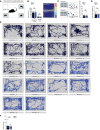
Representative illustration of the pup interaction zones and corner zones. Time spent in pup interaction zone and corner zone was measured during the first 2 min of social investigation in the pup retrieval test (trial 1) in Shank2 +/+ and Shank2 −/− mice.
Left diagram: No significant difference was detected comparing time spent in pup interaction zones (unpaired two‐tailed Student’s t‐test, P = 0.7090), and corner zones (unpaired, two‐tailed Student’s t‐test, P = 0.4597). Right diagram: Representative heat map tracings of Shank2 +/+ and Shank2 −/− mice. Hot colors indicate more time spent in a specific area, while cool colors represent less amount of time.
Left diagram: Sample behavior raster blot showing average duration Shank2 +/+ and Shank2 −/− spent in pup interaction zones. Right diagram: No significant difference was detected comparing the average duration Shank2 +/+ and Shank2 −/− mice spent in the pup interaction zones (Mann–Whitney test, P = 0.3865).
No significant difference was detected comparing the time spent freezing between Shank2 +/+ and Shank2 −/− mice (two‐tailed Student’s t‐test, P = 0.8329).
Tracking paths of Shank2 +/+ and Shank2 −/− mice during the first 2 min of social exposure toward the pups.
No significant difference was detected comparing the average movement velocity when Shank2 +/+ and Shank2 −/− mice did not engage in maternal behavior (trial 5, test day) (Mann–Whitney test, P = 0.8633). All graphs are presented as mean ± SEM, *P < 0.05, **P < 0.01, ***P < 0.001. Shank2 +/+ n = 9, Shank2 −/− n = 9.

- A
Left panel: Maternal behavior can be induced in male mice through repeated social exposure to pups (trial 1–5). After repeated pup exposure (right panel), male mice started to retrieve pups and displayed maternal behavior by crouching over the pups, thereby becoming more attached to the nest location as displayed in the tracking path (right panel).
- B
No significant difference was detected in the latency to approach the provided pups between Shank2 +/+ and Shank2 −/− male mice, Mann–Whitney test, P = 0.662.
- C
Pup‐exposed male Shank2 −/−mice failed to become attached toward the pups after repeated exposure, as demonstrated by the failure to retrieve pups during trial 1–5 compared to Shank2 +/+ males, two‐way mixed ANOVA, effect of genotype: **P < 0.003, effect of trial: *P = 0.032, trial × genotype interaction: *P = 0.032.
- D–H
Upper panel: Representative tracking path of Shank2 +/+ and Shank2 −/− male mice during exposure to pups in the 20 min test session (trial: 5). Shank2 +/+ males spent the majority of their time on the nest location crouching over the pups; Shank2 −/− males displayed no preference for the nest location, or participate in nest building activity (lower panel). Additionally, all major components of maternal care responses (E) pup grooming, Mann–Whitney test, ***P < 0.001, (F) crouching, Mann–Whitney test, **P = 0.007, (G) nest building, Mann–Whitney test, **P = 0.002, (H) paternal interaction, Mann–Whitney test, ***P < 0.001, were significantly reduced in Shank2 −/− male mice. All graphs are presented as mean ± SEM, *P < 0.05, **P < 0.01, ***P < 0.001. Shank2 +/+ n = 8, Shank2 −/− n = 9.

Representative illustration of the neuronal pathway (a–g) involved in the regulation of social attachment behavior. (a) OB, olfactory bulb; (b) AON, anterior olfactory nucleus; (c) AMY, amygdala; (d) MPOA, medial preoptic area; (e) LS, lateral septum, (f) BNST, bed nucleus of the stria terminalis, (g) VTA, ventral tegmental area; (h) NAcc, nucleus accumbens.
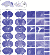
(a–h) Distribution of Shank2 mRNAs in major core regions of the neuronal circuit responsible for the regulation of social attachment behavior. Images shown are coronal brain sections (16 µm) of 8‐week‐old Shank2 +/+ mice, scale bar: 1 mm. In situ hybridization results indicated the expression levels of Shank2 in the (a) olfactory bulb (c) amygdala and (h) nucleus accumbens. GrO, granular cell layer of the olfactory bulb; Mi, mitral cell layer of the olfactory bulb; EPI, external plexiform layer of the olfactory bulb; AOM, anterior olfactory nucleus medial part; AOP, anterior olfactory nucleus posterior part; MePD, medial amygdaloid nucleus; BMA, basomedial amygdaloid nucleus, anterior part; MPOA, medial preoptic area; LSD, lateral septal nucleus, dorsal part; LSI, lateral septal nucleus, intermediate part; LSV, lateral septal nucleus, ventral part; BNST, bed nucleus of the stria terminalis; VTA, ventral tegmental area; AcbC, accumbens nucleus, core; AcbSh, accumbens nucleus, shell.

Representative illustration of the neuronal pathway regulating social attachment behavior in mice. (a) OB, olfactory bulb; (b) AON, anterior olfactory nucleus; (c) AMY, amygdala; (d) MPOA, medial preoptic area; (e) LS, lateral septum, (f) BNST, bed nucleus of the stria terminalis, (g) VTA, ventral tegmental area; (h) NAcc, nucleus accumbens.
Circuit‐specific comparison of the brain morphology between Shank2 +/+ and Shank2 −/− mice in low (left panel) and high (right panel) magnification view (a–f). Scale bars: 1 mm coronal section, 100 µm left magnification, 20 µm right magnification. Nissl‐stained coronal brain sections (16 µm) show similar brain morphology between Shank2 +/+ and Shank2 −/− mice at the age of 8 weeks. No major differences were evident in the neuroanatomical organization of the (a) AON, (b) Amy, (c) LS, (d) MPOA, (e) VTA, and (d) NAcc. AOM, anterior olfactory nucleus medial part; AOP, anterior olfactory nucleus posterior part; MePD, medial amygdaloid nucleus, posterodorsal part; MePV, medial amygdaloid nucleus, posteroventral part; BMA, basomedial amygdaloid nucleus, anterior part; LSD, lateral septal nucleus, dorsal part; LSI, lateral septal nucleus, intermediate part; LSV, lateral septal nucleus; aca, anterior commissure, anterior part; MPOL, medial preoptic nucleus, lateral part; MPOA, medial preoptic area; SNR, substantia nigra, reticular part; VTA, ventral tegmental area; AcbC, accumbens nucleus, core; AcbSh, accumbens nucleus, shell; LAcbSh, lateral accumbens shell.

- A
Shank2 +/+ and Shank2 −/− mice were bilaterally injected with a Cre‐dependent DREADD virus (pAAV‐hSyn‐DIO‐hM3D (Gq)‐mCherry) and (pENN.AAV.hSyn.HI.eGFP‐Cre)) into the MPOA region. After 4 weeks, a pup exposure assay was performed once a day for 5 days after intraperitoneal injection of vehicle or 5 mg/kg CNO (30 min before pup exposure). Maternal behavior was analyzed at the final 20 min of pup exposure on day 5 (test day).
- B
Confocal images showing the location and expression of the stereotaxically injected viral constructs in the MPOA region. (a) Merge of GFP(CRE) and mCherry (DREAAD) (scale bar: 500 µm) (b–d) Magnified views of the MPOA region (scale bar: 20 µm), (b) GFP and mCherry merge, (c) GFP, (d) mCherry. The dotted lines outline the MPOA and the third ventricle (middle line).
- C
A significant genotype × treatment interaction was detected for pup retrieval, two‐way ANOVA, effect of genotype: P = 0.187, effect of treatment: P = 0.759, genotype × treatment interaction: *P = 0.031. Shank2 +/+ vehicle injected n = 6, Shank2 −/− vehicle injected n = 6, Shank2 +/+ CNO injected n = 6, Shank2 −/− CNO injected n = 7.
- D–G
DREADD activation by CNO injection selectively rescues major components of social attachment behavior in Shank2 −/− mice: (D) grooming, two‐way ANOVA, effect of genotype: *P = 0.018, effect of treatment: **P = 0.002, genotype × treatment interaction: ***P = 0.001. (E) Crouching, two‐way ANOVA, effect of genotype: P = 0.091, effect of treatment: P = 0.869, genotype × treatment interaction: P = 0.663, (F) nest building, two‐way ANOVA, effect of genotype: P = 0.415, effect of treatment: P = 0.341 genotype × treatment interaction: P = 0.093, (G) Maternal interaction, two‐way ANOVA, effect of genotype: *P = 0.034, effect of treatment: P = 0.149, genotype × treatment interaction: *P = 0.045. Shank2 +/+ vehicle injected n = 6, Shank2 −/− vehicle injected n = 6, Shank2 +/+ CNO injected n = 6, Shank2 −/− CNO injected n = 7.

Similar articles
-
Cerebellar Shank2 Regulates Excitatory Synapse Density, Motor Coordination, and Specific Repetitive and Anxiety-Like Behaviors.J Neurosci. 2016 Nov 30;36(48):12129-12143. doi: 10.1523/JNEUROSCI.1849-16.2016. J Neurosci. 2016. PMID: 27903723 Free PMC article.
-
Genes showing altered expression in the medial preoptic area in the highly social maternal phenotype are related to autism and other disorders with social deficits.BMC Neurosci. 2014 Jan 14;15:11. doi: 10.1186/1471-2202-15-11. BMC Neurosci. 2014. PMID: 24423034 Free PMC article.
-
NEXMIF/KIDLIA Knock-out Mouse Demonstrates Autism-Like Behaviors, Memory Deficits, and Impairments in Synapse Formation and Function.J Neurosci. 2020 Jan 2;40(1):237-254. doi: 10.1523/JNEUROSCI.0222-19.2019. Epub 2019 Nov 8. J Neurosci. 2020. PMID: 31704787 Free PMC article.
-
Neural basis of maternal behavior in the rat.Psychoneuroendocrinology. 1988;13(1-2):47-62. doi: 10.1016/0306-4530(88)90006-6. Psychoneuroendocrinology. 1988. PMID: 2897700 Review.
-
Molecular neuroanatomy of the mouse medial preoptic area with reference to parental behavior.Anat Sci Int. 2019 Jan;94(1):39-52. doi: 10.1007/s12565-018-0468-4. Epub 2018 Nov 3. Anat Sci Int. 2019. PMID: 30392107 Review.
Cited by
-
Sexual dimorphism in the bed nucleus of the stria terminalis, medial preoptic area and suprachiasmatic nucleus in male and female tree shrews.J Anat. 2022 Mar;240(3):528-540. doi: 10.1111/joa.13568. Epub 2021 Oct 12. J Anat. 2022. PMID: 34642936 Free PMC article.
-
Shankopathies in the Developing Brain in Autism Spectrum Disorders.Front Neurosci. 2021 Dec 22;15:775431. doi: 10.3389/fnins.2021.775431. eCollection 2021. Front Neurosci. 2021. PMID: 35002604 Free PMC article. Review.
-
Automated procedure to assess pup retrieval in laboratory mice.Sci Rep. 2022 Jan 31;12(1):1663. doi: 10.1038/s41598-022-05641-w. Sci Rep. 2022. PMID: 35102217 Free PMC article.
-
Brain region and gene dosage-differential transcriptomic changes in Shank2-mutant mice.Front Mol Neurosci. 2022 Oct 13;15:977305. doi: 10.3389/fnmol.2022.977305. eCollection 2022. Front Mol Neurosci. 2022. PMID: 36311025 Free PMC article.
-
Chemogenetics as a neuromodulatory approach to treating neuropsychiatric diseases and disorders.Mol Ther. 2022 Mar 2;30(3):990-1005. doi: 10.1016/j.ymthe.2021.11.019. Epub 2021 Dec 1. Mol Ther. 2022. PMID: 34861415 Free PMC article. Review.
References
-
- Bauminger N, Shulman C, Agam G (2003) Peer interaction and loneliness in high‐functioning children with autism. J Autism Dev Disord 33: 489–507 - PubMed
-
- Berkel S, Marshall CR, Weiss B, Howe J, Roeth R, Moog U, Endris V, Roberts W, Szatmari P, Pinto D et al (2010) Mutations in the SHANK2 synaptic scaffolding gene in autism spectrum disorder and mental retardation. Nat Genet 42: 489–491 - PubMed
-
- Boeckers TM, Bockmann J, Kreutz MR, Gundelfinger ED (2002) ProSAP/Shank proteins – a family of higher order organizing molecules of the postsynaptic density with an emerging role in human neurological disease. J Neurochem 81: 903–910 - PubMed
Publication types
MeSH terms
Substances
LinkOut - more resources
Full Text Sources
Other Literature Sources
Medical
Molecular Biology Databases

