. 2021 Jan 19;13(1):138.
doi: 10.3390/v13010138.

A Novel In Vitro Culture Model System to Study Merkel Cell Polyomavirus-Associated MCC Using Three-Dimensional Organotypic Raft Equivalents of Human Skin

Affiliations

A Novel In Vitro Culture Model System to Study Merkel Cell Polyomavirus-Associated MCC Using Three-Dimensional Organotypic Raft Equivalents of Human Skin

Amanda S W Loke et al. Viruses. .

Abstract

Merkel cell polyomavirus (MCPyV) is a human polyomavirus causally linked to the development of Merkel cell carcinoma (MCC), an aggressive malignancy that largely arises within the dermis of the skin. In this study, we recapitulate the histopathology of human MCC tumors in vitro using an organotypic (raft) culture system that is traditionally used to recapitulate the dermal and epidermal equivalents of skin in three dimensions (3D). In the optimal culture condition, MCPyV+ MCC cells were embedded in collagen between the epidermal equivalent comprising human keratinocytes and a dermal equivalent containing fibroblasts, resulting in MCC-like lesions arising within the dermal equivalent. The presence and organization of MCC cells within these dermal lesions were characterized through biomarker analyses. Interestingly, co-culture of MCPyV+ MCC together with keratinocytes specifically within the epidermal equivalent of the raft did not reproduce human MCC morphology, nor were any keratinocytes necessary for MCC-like lesions to develop in the dermal equivalent. This 3D tissue culture system provides a novel in vitro platform for studying the role of MCPyV T antigens in MCC oncogenesis, identifying additional factors involved in this process, and for screening potential MCPyV+ MCC therapeutic strategies.

Keywords: DNA tumor virus; Merkel cell carcinoma; Merkel cell polyomavirus; Merkel cells; human polyomavirus; organotypic rafts; skin equivalents.


Conflict of interest statement

The authors declare no conflict of interest.

Figures
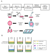
Figure 1
Figure 1
Experimental design of Merkel cell polyomavirus–positive (MCPyV+) Merkel cell carcinoma (MCC) culture in organotypic rafts. (A) General workflow and timeline of 3D organotypic raft co-culture with MCPyV+ MCC. Rafts were harvested at the endpoint using the process depicted; paraffin-embedded and 5 µM-thick sections were cut from a paraffin block and laid onto slides for hematoxylin and eosin (H&E) and immunostaining analyses. (B) Experimental organization/setups of rafts generated. The six conditions tested are depicted. Cell types included in the various raft setups include NIKS keratinocytes (yellow), early passage human foreskin fibroblasts (EF-1-F) fibroblasts (green), MCPyV+ MCC cells without collagen (purple), and MCPyV+ MCC cells in collagen (blue). (C) Experimental workflow for each raft setup depicted in (B). Dermal equivalents were generated with either EF-1-F or EF-1-F/MKL-1 mix and left to culture for 4–7 days. The epithelial layer was then generated by layering NIKS epithelial cells or epithelial cells with MKL-1 cells. For Setup 4, MKL-1 cells were suspended in collagen and added to the dermal equivalent. For Setup 6, an intermediate layer of MKL-1 cells in collagen was added and allowed to settle for 24 h, followed by the addition of NIKS keratinocytes. (D) Immunoblot analysis for the MCPyV large T antigen (LT) and small T antigen (ST) proteins in cell types used in raft studies. Protein lysates from NIKS keratinocytes, EF-1-F fibroblasts, and the MCPyV+ MCC cell line MKL-1 were analyzed by immunoblotting with the Ab5 mouse monoclonal antibody. An immunoblot for β-actin was included as a loading control.
Figure 1
Figure 1
Experimental design of Merkel cell polyomavirus–positive (MCPyV+) Merkel cell carcinoma (MCC) culture in organotypic rafts. (A) General workflow and timeline of 3D organotypic raft co-culture with MCPyV+ MCC. Rafts were harvested at the endpoint using the process depicted; paraffin-embedded and 5 µM-thick sections were cut from a paraffin block and laid onto slides for hematoxylin and eosin (H&E) and immunostaining analyses. (B) Experimental organization/setups of rafts generated. The six conditions tested are depicted. Cell types included in the various raft setups include NIKS keratinocytes (yellow), early passage human foreskin fibroblasts (EF-1-F) fibroblasts (green), MCPyV+ MCC cells without collagen (purple), and MCPyV+ MCC cells in collagen (blue). (C) Experimental workflow for each raft setup depicted in (B). Dermal equivalents were generated with either EF-1-F or EF-1-F/MKL-1 mix and left to culture for 4–7 days. The epithelial layer was then generated by layering NIKS epithelial cells or epithelial cells with MKL-1 cells. For Setup 4, MKL-1 cells were suspended in collagen and added to the dermal equivalent. For Setup 6, an intermediate layer of MKL-1 cells in collagen was added and allowed to settle for 24 h, followed by the addition of NIKS keratinocytes. (D) Immunoblot analysis for the MCPyV large T antigen (LT) and small T antigen (ST) proteins in cell types used in raft studies. Protein lysates from NIKS keratinocytes, EF-1-F fibroblasts, and the MCPyV+ MCC cell line MKL-1 were analyzed by immunoblotting with the Ab5 mouse monoclonal antibody. An immunoblot for β-actin was included as a loading control.
Figure 2
Figure 2
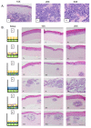
Histological analysis of MCPyV+ MCC-like lesions in organotypic raft cultures. (A) Representative images of H&E-stained tissue sections of human MCC tumors arising in the dermis of human skin (Panel I–III). (B) Representative H&E images of rafts generated using various culture conditions. Each raft setup is shown on the left and cell types are represented using the symbols outlined in Figure 1B. H&E-stained images are shown with 10× magnification in the first two columns (one each of both replicate rafts) and 20× magnification of histology in the rightmost column of one of those replicates (Panels I–XVIII). All scale bars = 100 µM.
Figure 3
Figure 3
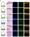
Identification of MCPyV+ MCC cells within raft structures. (A) Tissue sections from all raft setups were subjected to immunofluorescence analysis for LT antigen expression. DAPI counterstain was used to identify cell nuclei (blue, Column II), LT antigen was detected using the CM2B4 antibody (green, Column III), and K14 staining was used as an epithelial marker (yellow, Column IV). Corresponding H&E images are shown in Column I. All images are shown at 20× magnification. Scale bars = 100 µM. (B) High magnification (63×) of representative MCC-lesions that developed in rafts and IF biomarker analysis. Panel I—H&E, Panel II—DAPI (blue), Panel III—LT antigen (green), Panel IV—epithelial marker cytokeratin 14 (yellow), Panel V—cytokeratin 8 (pink). Merged IF images are shown in Panel VI. For rafts that do not develop these MCC lesions (Setup 1 and 2), representative images of the dermal layer are shown. Scale bars = 20 µM. Note in Setup 3 (panel III) LT staining was dimmer than in the MCC-like lesions that arose under other setups (4–6). We believe this is an artifact of the position of the MCC-like lesion under setup 3 toward the edge of the raft, where we often see a dimmer fluorescence signal.
Figure 3
Figure 3
Identification of MCPyV+ MCC cells within raft structures. (A) Tissue sections from all raft setups were subjected to immunofluorescence analysis for LT antigen expression. DAPI counterstain was used to identify cell nuclei (blue, Column II), LT antigen was detected using the CM2B4 antibody (green, Column III), and K14 staining was used as an epithelial marker (yellow, Column IV). Corresponding H&E images are shown in Column I. All images are shown at 20× magnification. Scale bars = 100 µM. (B) High magnification (63×) of representative MCC-lesions that developed in rafts and IF biomarker analysis. Panel I—H&E, Panel II—DAPI (blue), Panel III—LT antigen (green), Panel IV—epithelial marker cytokeratin 14 (yellow), Panel V—cytokeratin 8 (pink). Merged IF images are shown in Panel VI. For rafts that do not develop these MCC lesions (Setup 1 and 2), representative images of the dermal layer are shown. Scale bars = 20 µM. Note in Setup 3 (panel III) LT staining was dimmer than in the MCC-like lesions that arose under other setups (4–6). We believe this is an artifact of the position of the MCC-like lesion under setup 3 toward the edge of the raft, where we often see a dimmer fluorescence signal.
Figure 4
Figure 4
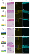
Identification of fibroblasts and differentiated keratinocytes within raft structures. Immunofluorescence analysis of tissue sections from all raft setups was performed using antibodies specific to the fibroblast marker α-smooth muscle actin (SMA) (yellow, Column II) and differentiated epithelial marker cytokeratin 10 (K10) (aqua, Column III). Corresponding H&E images are shown in Column I. Note for Setup 6 Panel II, the fluorescent surrounding the MCC lesion is likely background. Each raft setup is shown on the left and cell types are represented using the symbols outlined in Figure 1B. For SMA, 63× images of a positive fibroblast cell (with DAPI counterstain) are included as inset pictures in the top left corner. All scale bars = 100 µM.
Figure 5
Figure 5
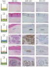
MCPyV+ MCC cells retain LT activity and proliferative capacity in organotypic raft cultures. Representative images showing immunohistochemical analysis for MCM7 (Panel II) and BrdU (Panel III) in raft setups are shown. Positive signal is indicated by brown nuclei staining. Counterstaining was done using hematoxylin (blue). Each raft setup is shown on the left and cell types are represented using the symbols outlined in Figure 1B. Corresponding H&E images are shown in Column I. All scale bars = 100 µM.

Similar articles

Cited by

References

    1. Feng H., Shuda M., Chang Y., Moore P.S. Clonal Integration of a Polyomavirus in Human Merkel Cell Carcinoma. Science. 2008;319:1096. doi: 10.1126/science.1152586. - DOI - PMC - PubMed
    1. Toker C. Trabecular Carcinoma of the Skin. Arch. Dermatol. 1972;105:107–110. doi: 10.1001/archderm.1972.01620040075020. - DOI - PubMed
    1. Becker J.C., Stang A., DeCaprio J.A., Cerroni L., Lebbe C., Veness M., Nghiem P. Merkel cell carcinoma. Nat. Rev. Dis. Prim. 2017;3:17077. doi: 10.1038/nrdp.2017.77. - DOI - PMC - PubMed
    1. Cook L. Polyomaviruses. Microbiol. Spectr. 2016;4 doi: 10.1128/microbiolspec.DMIH2-0010-2015. - DOI - PubMed
    1. DeCaprio J.A., Garcea R.L. A cornucopia of human polyomaviruses. Nat. Rev. Microbiol. 2013;11:264–276. doi: 10.1038/nrmicro2992. - DOI - PMC - PubMed

Publication types

MeSH terms

LinkOut - more resources