. 2021 Aug 17;45(4):fuaa063.
doi: 10.1093/femsre/fuaa063.

The biology of thermoacidophilic archaea from the order Sulfolobales

Affiliations

The biology of thermoacidophilic archaea from the order Sulfolobales

April M Lewis et al. FEMS Microbiol Rev. .

Abstract

Thermoacidophilic archaea belonging to the order Sulfolobales thrive in extreme biotopes, such as sulfuric hot springs and ore deposits. These microorganisms have been model systems for understanding life in extreme environments, as well as for probing the evolution of both molecular genetic processes and central metabolic pathways. Thermoacidophiles, such as the Sulfolobales, use typical microbial responses to persist in hot acid (e.g. motility, stress response, biofilm formation), albeit with some unusual twists. They also exhibit unique physiological features, including iron and sulfur chemolithoautotrophy, that differentiate them from much of the microbial world. Although first discovered >50 years ago, it was not until recently that genome sequence data and facile genetic tools have been developed for species in the Sulfolobales. These advances have not only opened up ways to further probe novel features of these microbes but also paved the way for their potential biotechnological applications. Discussed here are the nuances of the thermoacidophilic lifestyle of the Sulfolobales, including their evolutionary placement, cell biology, survival strategies, genetic tools, metabolic processes and physiological attributes together with how these characteristics make thermoacidophiles ideal platforms for specialized industrial processes.

Keywords: Sulfolobales; Archaea; Thermoacidophiles.


Figures

Figure 1.
Figure 1.
Timeline of thermoacidophile isolations and major events. Timeline contains the organism's name at the time of the associated event. The following are the current classifications: Sulfolobus brierleyi (f. Acidianus brierleyi), Saccharolobus solfataricus (f. Sulfolobus solfataricus), Acidianus ambivalens (f. Desulfurolobus ambivalens), Saccharolobus shibatae (f. Sulfolobus shibatae) Sulfuracidifex metallicus (f. Sulfolobus metallicus), Metallosphaera hakonensis (f. Sulfolobus hakonensis), Sulfurisphaera tokodaii (f. Sulfolobus tokodaii) and Saccharolobus islandicus (f. Sulfolobus islandicus).
Figure 2.
Figure 2.
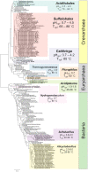
16S phylogeny tree of thermoacidophilic organisms.
Figure 3.
Figure 3.
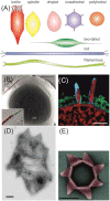
Thermoacidophilic archaeal viruses and their infection mechanisms. (A) Schematic representation of virion morphologies of viruses infecting thermoacidophilic archaea as described in the text. (B) Segmented tomographic volume of an SIRV2 virion (red) attached to a surface filament of Sa. islandicus (green) with help of the three terminal virion fibers (blue). Inset depicts a magnification of the interaction between the tail fibers and the surface structure. Scale bar, 500 nm. (C) Volume segmentations of electron microscopy tomograms showing Sulfolobus spindle-shaped virus 1 maturation and release by budding. Scale bar, 50 nm. (D) Transmission electron micrograph of a thin section of a SIRV2-infected Sa. islandicus cell displaying several pyramidal egress structures. Scale bar, 100 nm. (E)Transmission electron micrographs of an isolated pyramidal egress structure in open conformation isolated after SIRV2 infection of Sa. islandicus. Scale bar, 100 nm. Adapted from Bize et al. (2009); Quax et al. (2011); Quemin et al. (2013, 2016).
Figure 4.
Figure 4.
Main principles in genome organization and genetic information processing in the Sulfolobales. Conceptual schemes representing the major elements and principles of macro-level organization of the genomic DNA (adapted from Takemata, Samson and Bell 2019) (A), micro-level organization of the genomic DNA (partially adapted from Peeters et al. 2015) (B), initiation of replication (C) and initiation of transcription (D).
Figure 5.
Figure 5.
Predicted protein kinase and protein phosphatase homologs in the four different Sulfolobales species: S. acidocaldarius, Sa. islandicus, Sa. solfataricus and Sulfuri. tokodaii. Depicted are the different canonical and non-canonical Hanks-type protein kinases and protein phosphatases with their correspondent domain structure. (Lower Bischoff and Kennelly ; Lower and Kennelly , ; Lower, Potters and Kennelly ; Haile and Kennelly ; Esser and Siebers ; Ray et al. ; Esser et al. ; Hoffmann et al. ; Huang et al. 2017).
Figure 6.
Figure 6.
Representation of known phosphorylation-based regulatory networks in different Sulfolobales strains (Sa. solfataricus in pink, Sa. islandicus in orange, S. acidocaldarius in blue and Sulfuri. tokodaii in gray) with their physiological function. From upper left to right: The FadR transcriptional regulator represses transcription of the fatty acid gene cluster and dissociates from the DNA upon binding to acyl-CoA. Phosphorylation of FadR by the ePK ArnC (Saci_1196) prevents acyl-CoA binding and thus hinders transcription of the gene cluster (Maklad et al. 2020). The archaellum regulatory network consists of the gene cluster arlBXGFHIJ (flaBXGFHIJ), which encodes the motility structure, the archaellum, and is under the control of two promoters, one upstream of arlB (flaB) being induced under starvation and one weak promoter upstream of arlX (flaX). The two negative regulators, ArnA (Saci_1210) and ArnB (Saci_1211), were shown to be phosphorylated by the ePKs ArnC and ArnD (Saci_1694) and dephosphorylated by the PP PP2A (Saci_0884). Deletion of the PP2A led to a hypermotile phenotype suggesting a negative influence on the gene cluster (Reimann et al. ; Hoffmann et al. 2017). The DNA-binding protein AbfR1 (Saci_0446) is a positive regulator of the arlB (flaB) promoter (Orell et al. 2013a). Phosphorylation of AbfR1 inhibits DNA binding and thus regulates biofilm formation and motility (Li et al. 2017). The FHA domain containing protein ST0829 was shown to interact and be phosphorylated by the ePK ST1565 indicating a role in transcription regulation (Duan and He 2011). The Holliday junction resolvase (Hjc) (SiRe_1431) is phosphorylated by the aPK SiRe_0171 facilitating DNA repair (Huang et al. 2019). The phosphohexomutase (SSO0207) exhibited a decreased Vmax value after being phosphorylated (Ray et al. 2005). The Rio kinases (Saci_0796 and Saci_0965) were shown to play a role in the ribosome maturation of the small subunit (Knüppel et al. 2018).
Figure 7.
Figure 7.
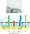
Archaeal cell surface structures involved in planktonic and biofilm growth. Upper image: electronic microscopy image from a S. acidocaldarius cell where archaellum and pilus can be seen. The upper image is reproduced from Albers and Meyer . Lower image: schematic model with all proposed cell surface appendages in the Sulfolobales: the Aap pili (archaeal adhesive pili), the Ups pili (UV-induced pili), the archaellum and the threads. Also depicted is the S-layer and its proteins, SlaA and SlaB.
Figure 8.
Figure 8.
Confocal laser microscopy images from static biofilm from S. acidocaldarius in days 3 to 7 of growth. Cells (DNA) stained with 4′,6-diamidino-2-phenylindole (DAPI; blue); extracellular glucose and mannose residues stained with fluorescently labeled concanavalin A (conA; green); and N-acetyl-d-glucosamine residues stained with fluorescently labeled lectin IB4 (yellow). Scale bars: 20 μm. Reproduced from Koerdt et al..
Figure 9.
Figure 9.
Model of the Sulfolobales S-layer. (A) The Sulfolobales S-layer consists of the two protein subunits. SlaA dimers (red, orange, yellow) form the outer S-layer canopy. Each SlaA protein is predicted to be rich in β-strands. The SlaA dimer has a boomerang-like shape, the angle of which determines the S-layer unit cell size. SlaB trimers (gray) form the membrane anchors of the S-layer. Each SlaB is predicted to consist of an N-terminal transmembrane domain (TMD), a coiled-coil domain (CC) and two to three C-terminal β-sandwich domains (β). SlaA and SlaB proteins are highly glycosylated (green). (B) Electron microscopy image from negatively stained isolated S-layer from S. acidocaldarius. (C)SDS-PAGE (sodium dodecyl sulfate polyacrylamide gel electrophoresis) of the isolation of S-layer from S. acidocaldarius cells using detergent buffers. Lane 1: pellet after incubating the cells with the detergent buffer the first time, the second time (lane 2) and the third time (lane 3). SlaB is being successively washed off and in the last wash a pure SlaA prep is obtained. (D) Subtomogram average of fully assembled S-layer. (E) Subtomogram average of SlaB-depleted S-layer. (F) Difference map (pink) overlaid with the complete S-layer visualizes location of SlaB. (Scale bars, and C–E, 20 nm). (Figure adapted from Gambelli et al. 2019).
Figure 10.
Figure 10.
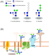
Glycosylation. (A) Comparison of the N-glycan trees of three different Sulfolobus/Saccharolobus species. (B) The current understanding of the N-glycosylation pathway in S. acidocaldarius. The N-glycan biosynthesis is initiated by adding nucleotide-activated monosaccharides sequentially to the lipid carrier dolichol phosphate on the cytoplasmic side of the membrane. The fully assembled dolichol pyrophosphate-linked N-glycan (hexasaccharide) is translocated across the membrane and then transferred by AlgB on the specific N-glycosylation sequons in secreted proteins. Sugar code is shown in (A).
Figure 11.
Figure 11.
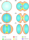
Schematic model of the cell division process in Sulfolobus acidocaldarius. (A)CdvA (red) is the first protein of the S. acidocaldarius cell division machinery that arrives at the future site of cell division, before DNA (light blue) segregation starts. (B) During nucleoid condensation (blue), CdvB (light green) forms a ring-like structure at midcell that is anchored to the membrane by CdvA. (C)CdvB provides a scaffold for CdvB1 and B2 (green) that are positioned at the cell center in ring-like structures. Additionally, CdvC (yellow), a homolog of the hexameric ATPase Vps4, localizes at the septum while nucleoid segregation and initial membrane invagination start. (D) After nucleoid segregation, the CdvB-ring undergoes proteasomal (purple) degradation. (E)Upon CdvB removal, CdvB1 and B2 constrict, leading to the final division of the cell. (F) Directly after fission, the new born cells have an oval shape that rapidly changes to the typical coccoid shape of S. acidocaldarius cells. CdvA and CdvC are organized in a ring-like structure as well. However, for a better overview the organization of both proteins at midcell was only indicated in the figure.
Figure 12.
Figure 12.
Major mechanisms of thermoacidophily. (1) Thermoacidophiles have an inverted membrane potential with a positive charge on the inside of the cellular membrane and a negative charge on the outside to prevent the acidification of the cytoplasm by the passive diffusion of protons. (2) The inverted membrane potential is maintained by transporting cations such as K+ into the cytoplasm. (3) Cyclopentyl ring moieties on tetraether lipids increase packing of the tetraether lipids decreasing the permeability of the membrane by protons and increasing cellular heat stability. (4) Tetraether lipids make a monolayer that is less permeable to protons and more heat stable than diether lipids. (5) Proton pumps export protons from the cytoplasm to prevent the acidification of the cytoplasm. (6) Heat-damaged or protonated proteins can either be degraded via the proteasome or properly refolded by the thermosome.
Figure 13.
Figure 13.
Model of heavy metal resistance in the Sulfolobales. (1) CopA and CopB export Cu outside the cell with ATP consumption. CopM is a metal chaperone that forms part of the Cop system that also includes a transcriptional factor called CopT (not shown). (2)PolyP can sequester cations via its negatively charged surface. (3) PolyP can also be degraded by PPX into inorganic phosphate to be exported outside the cell along with cations via PitA or Pho84 transporters. (4) Some proteins also act by sequestering metal ions, for example Dps. The mechanism for which metals enter the cell is still unknown.
Figure 14.
Figure 14.
An overview of the central metabolism in Sulfolobales. Dashed arrows indicate pathways, which have not yet been experimentally demonstrated. Abbreviations: F6P, fructose 6-phosphate; DHAP, dihydroxyacetone phosphate; GAP, glyceraldehyde 3-phosphate; D-KDG, 2-keto-3-deoxy-d-gluconate; D-KDGal, 2-keto-3-deoxy-d-galactonate; D-KDA, 2-keto-3-deoxy-d-arabinoate; L-KDA, 2-keto-3-deoxy-l-arabinoate; D-KDX, 2-keto-3-deoxy-d-xylonate; AA, amino acid; ED, Entner–Doudoroff pathway; EMP, Embden–Meyerhof–Parnas pathway; RuMP, reversed ribulose monophosphate pathway; TCA, tricarboxylic acid cycle; 3HP/4HB, 3-hydroxyproprionate/4-hydroxybutyrate cycle; ABC, ATP-binding cassette transporters; RC, respiratory chain.
Figure 15.
Figure 15.
The enzymatic pathway of the 3-hydroxypropionate/4-hydroxybutyrate (3-HP/4-HB) cycle as characterized in Metallosphaera sedula. Enzyme names are contained within the yellow oval with arrows indicating reactions for which they have known catalytic activity. Enzymes in pink have shown activity for only a single reaction in the 3-HP/4-HB cycle; enzymes in green exhibit activity on multiple steps in the cycle.
Figure 16.
Figure 16.
Current knowledge of the mechanism of sulfur oxidation and reduction in the Sulfolobales. (A) Sulfur reduction and (B) sulfur oxidation. Solid arrows indicate involvement in a reaction; dotted arrows represent transport of species; and dashed lines indicate that the function is suspected but has not been demonstrated experimentally in the Sulfolobales. Enzyme colors indicate general grouping of function: coupled to electron transport chain (blue), involved in transporting or trafficking sulfur species (yellow), transformation of sulfur species with no energy conservation (orange) and transformation of sulfur species directly coupled to energy-conserving biomolecules (green). Abbreviations: sulfur reductase (Sre), hydrogenase (Hyn), heterodisulfide reductase (Hdr), tetrathionate hydrolase (TetH), sulfide:quinone oxidoreductase (SQO), sulfite:acceptor oxidoreductase (SAOR), thiosulfate:quinone oxidoreductase (TQO), sulfur oxygenase reductase (SOR), adenosine-5′-phosphosulfate reductase (APSR), adenosine-5′-phosphosulfate (APS), adenylylsulfate:phosphate adenylyltransferase (APAT), ATP sulfurylase (ATPS), adenylate kinase (AK).
Figure 17.
Figure 17.
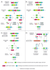
Mechanisms for the generation of markerless deletion mutants.(A)Plasmid integration occurs via single crossover, resulting in a merodiploidal form. After counterselection with 5-FOA, the pyrEF marker cassette is looped out, either with or without the GOI, resulting in a theoretical ration of one to one in mutated and wild-type cells. Double crossover is feasible by introducing a linearized vector. Depending on the experimental design, either parts of the GOI (B) or an upstream (US) region (C) are introduced for recombination. Counterselection with 5-FOA produces marker-free deletion mutants. (D) A plasmid containing a CRISPR array and a repair fragment with homologous sequences to the GOI are introduced into a recipient strain. Upon induction, crRNA is transcribed and forms a ribonucleoprotein complex with the endogenous Cas protein, scanning the genomic DNA for the spacer sequence and cutting it. Only colonies that conducted recombination with the repair fragment survive. GOI, gene of interest; US, upstream; DS, downstream; pyrEF, pyrEF marker cassette; crRNA, CRISPR RNA; crRNP, ribonucleoprotein complex consisting of crRNA and Cas protein.
Figure 18.
Figure 18.
Mechanisms of sulfidic ore dissolution. (A)Thiosulfate mechanism(B) and polysulfide mechanism. Green dashed arrows indicate biological steps; solid arrows indicate spontaneous abiotic reactions; blue dashed-dotted arrows indicate an overall transformation involving multiple reaction steps; yellow dashed-dotted arrows represent phase transition. Bold box around a species indicates that this is the dominant sulfur product of the dissolution process.

Similar articles

Cited by

References

    1. Aagaard C, Leviev I, Aravalli RN et al. General vectors for archaeal hyperthermophiles: strategies based on a mobile intron and a plasmid. FEMS Microbiol Rev. 1996;18:93–104. - PubMed
    1. Aagaard C, Phan H, Trevisanato S et al. A spontaneous point mutation in the single 23S rRNA gene of the thermophilic arachaeon Sulfolobus acidocaldarius confers multiple drug resistance. J Bacteriol. 1994;176:7744–7. - PMC - PubMed
    1. Ahmed H, Ettema TJG, Tjaden B et al. The semi-phosphorylative Entner–Doudoroff pathway in hyperthermophilic archaea: a re-evaluation. Biochem J. 2005;390:529–40. - PMC - PubMed
    1. Albers S-V, Driessen AJM. Conditions for gene disruption by homologous recombination of exogenous DNA into the Sulfolobus solfataricus genome. Archaea. 2007;2:145–9. - PMC - PubMed
    1. Albers S-V, Jarrell KF. The archaellum: how Archaea swim. Front Microbiol. 2015;6:1–12. - PMC - PubMed

Publication types