. 2021 Mar 10;41(10):2245-2263.
doi: 10.1523/JNEUROSCI.0580-20.2021. Epub 2021 Jan 20.

Heparanome-Mediated Rescue of Oligodendrocyte Progenitor Quiescence following Inflammatory Demyelination

Affiliations

Heparanome-Mediated Rescue of Oligodendrocyte Progenitor Quiescence following Inflammatory Demyelination

Darpan Saraswat et al. J Neurosci. .

Abstract

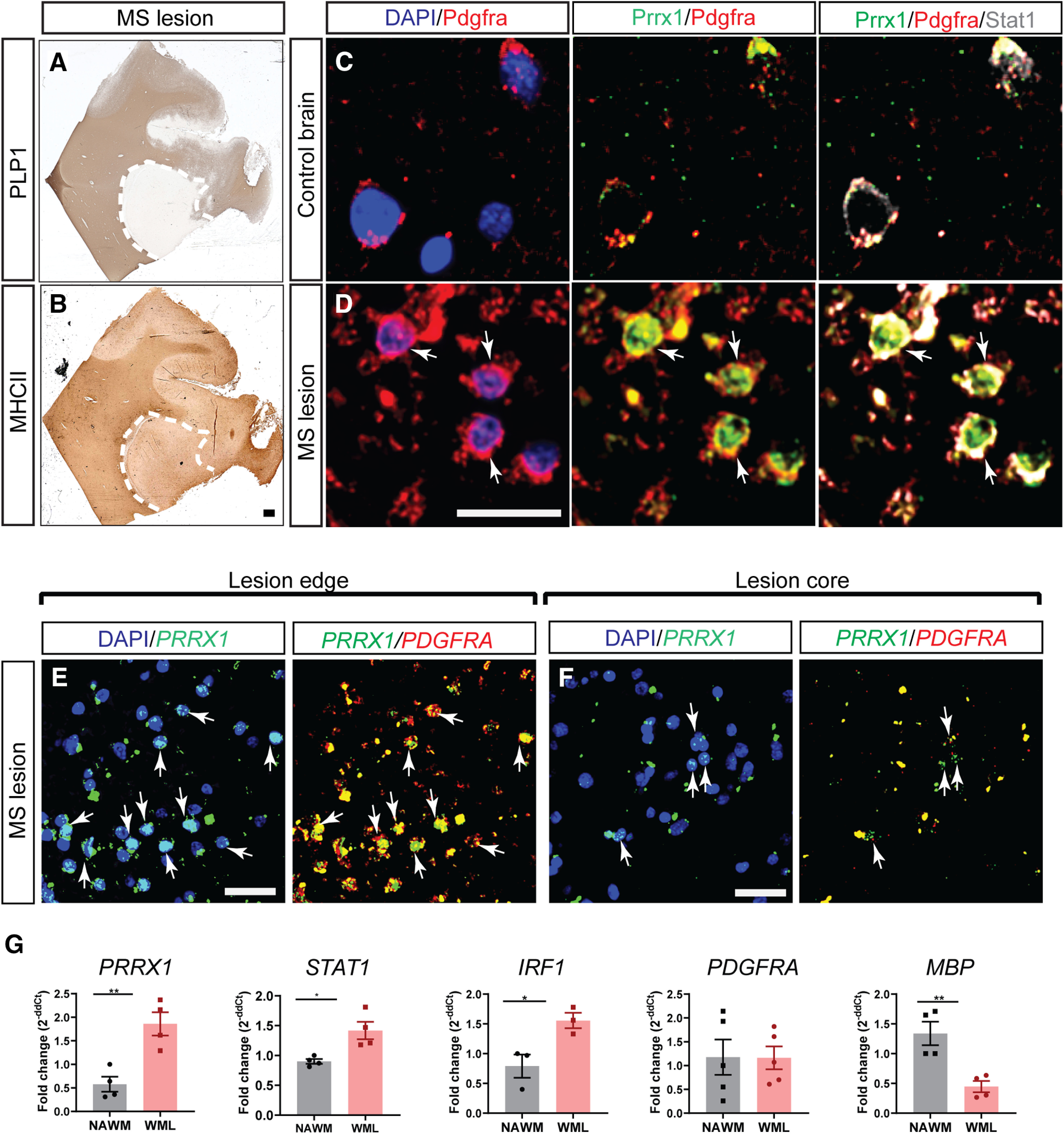
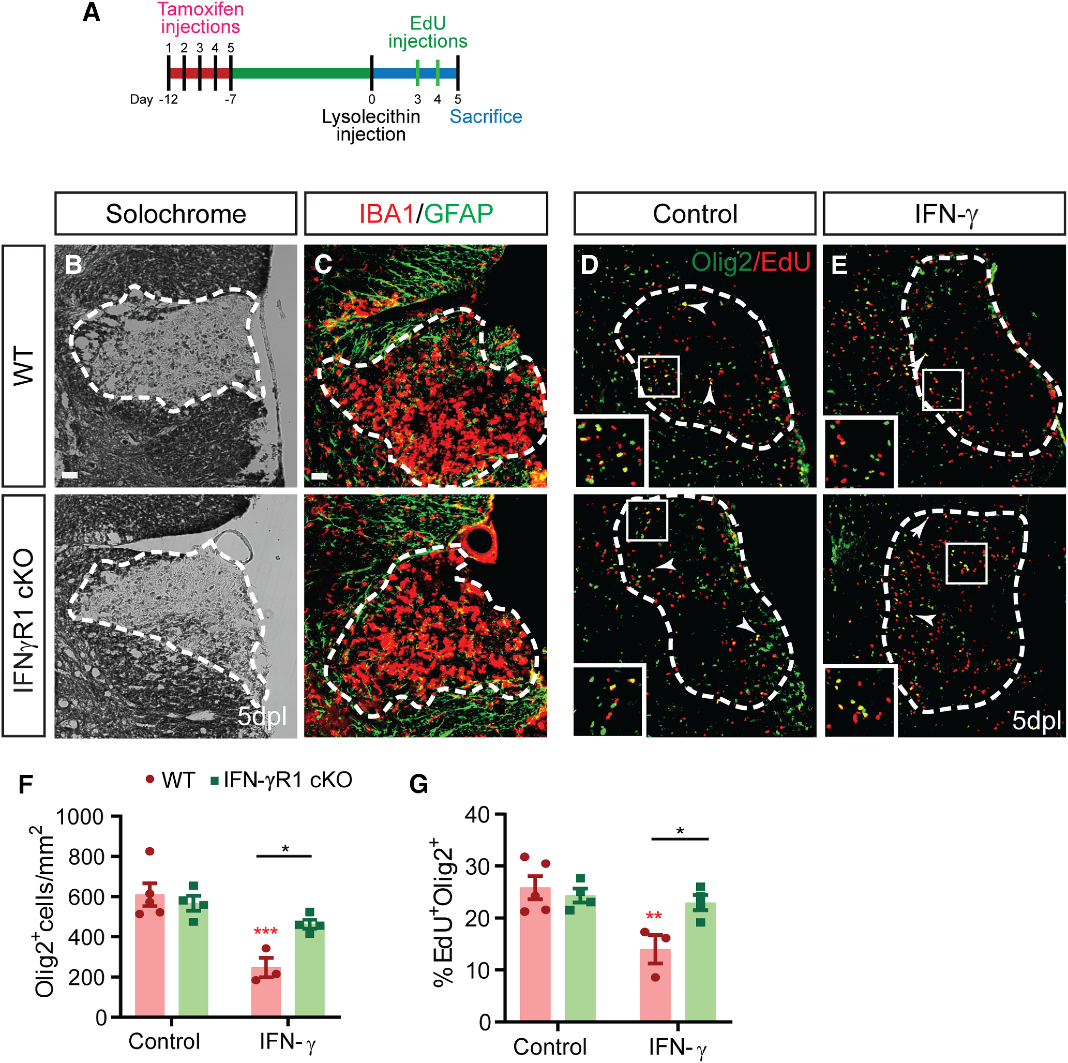
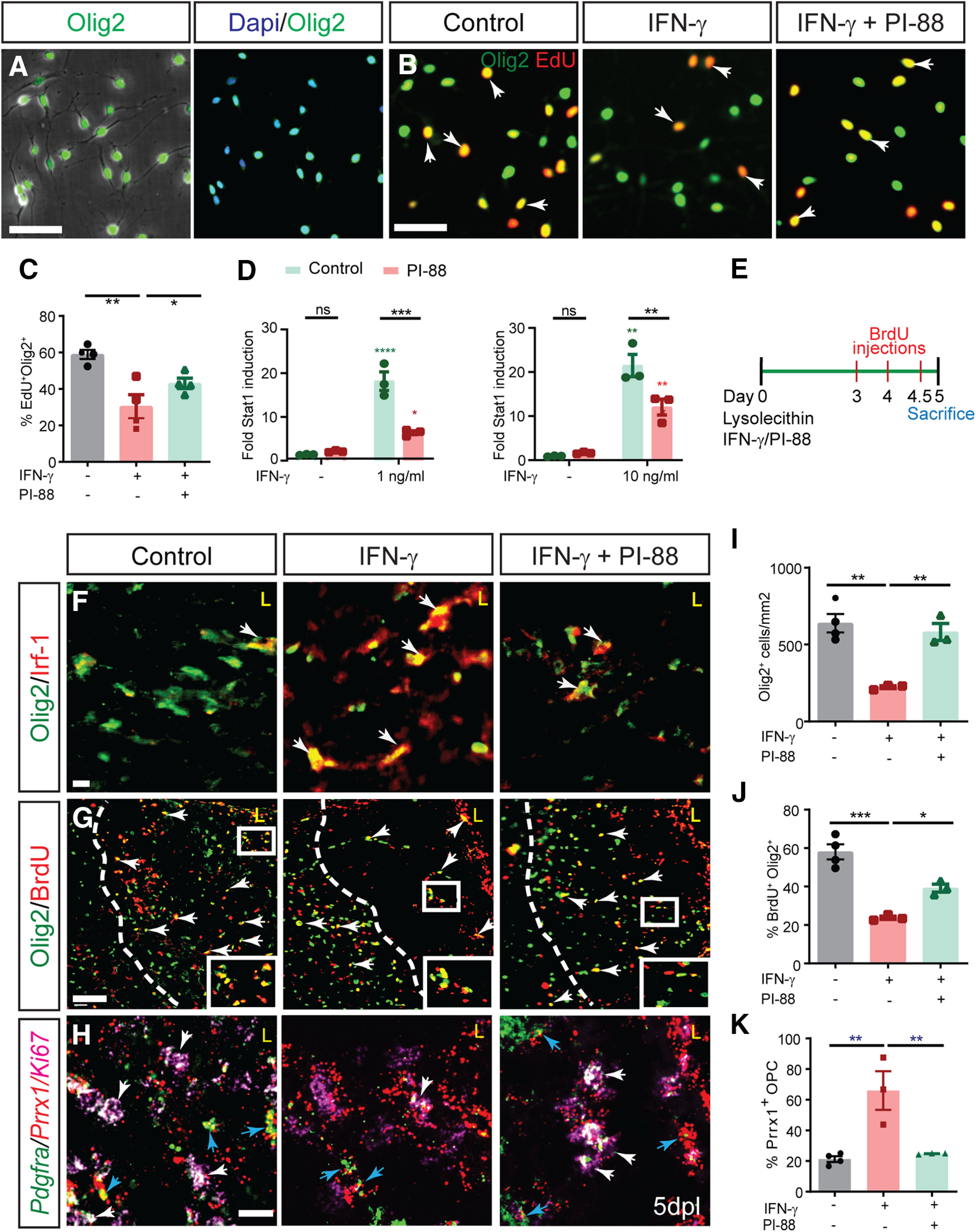
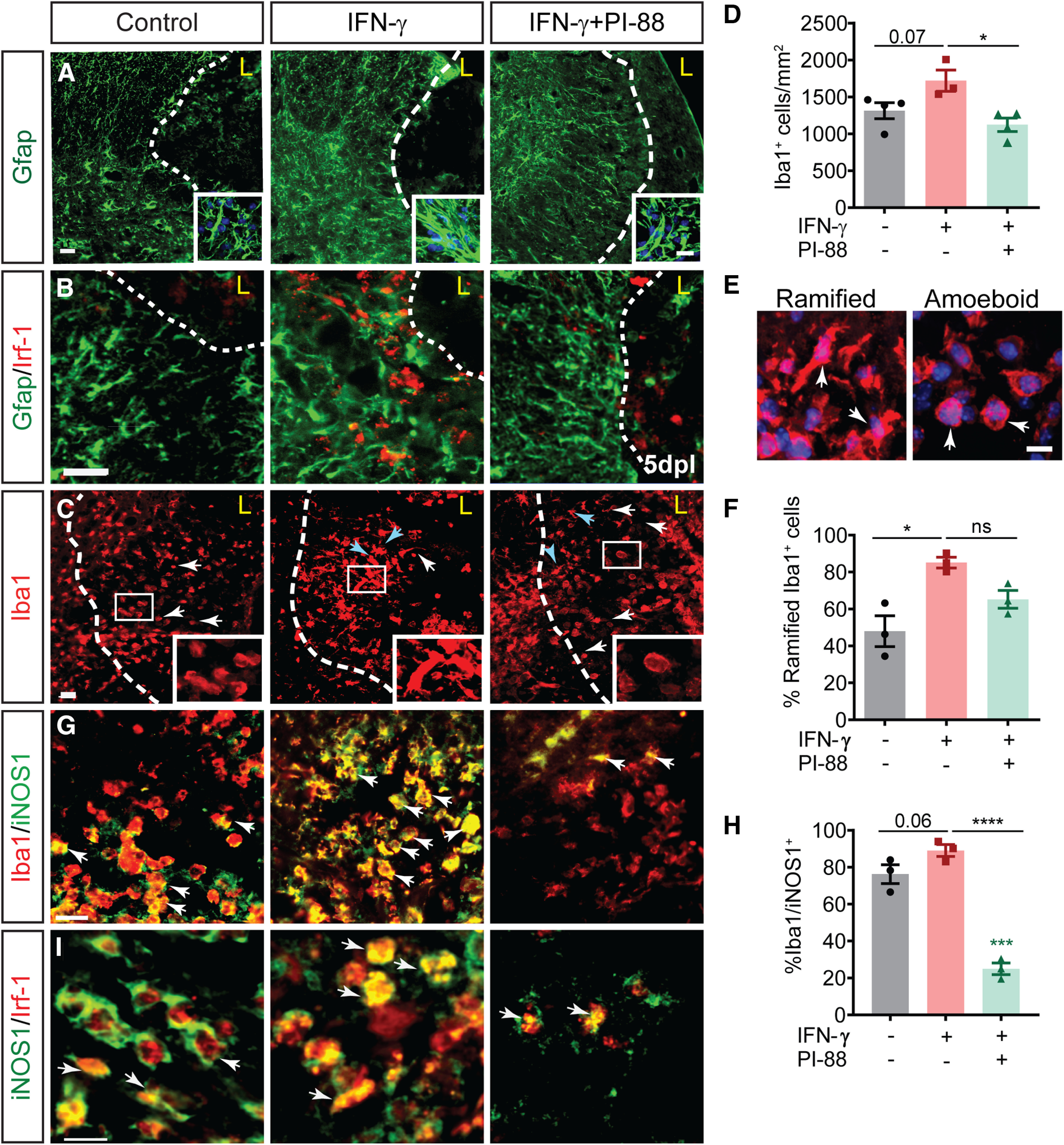
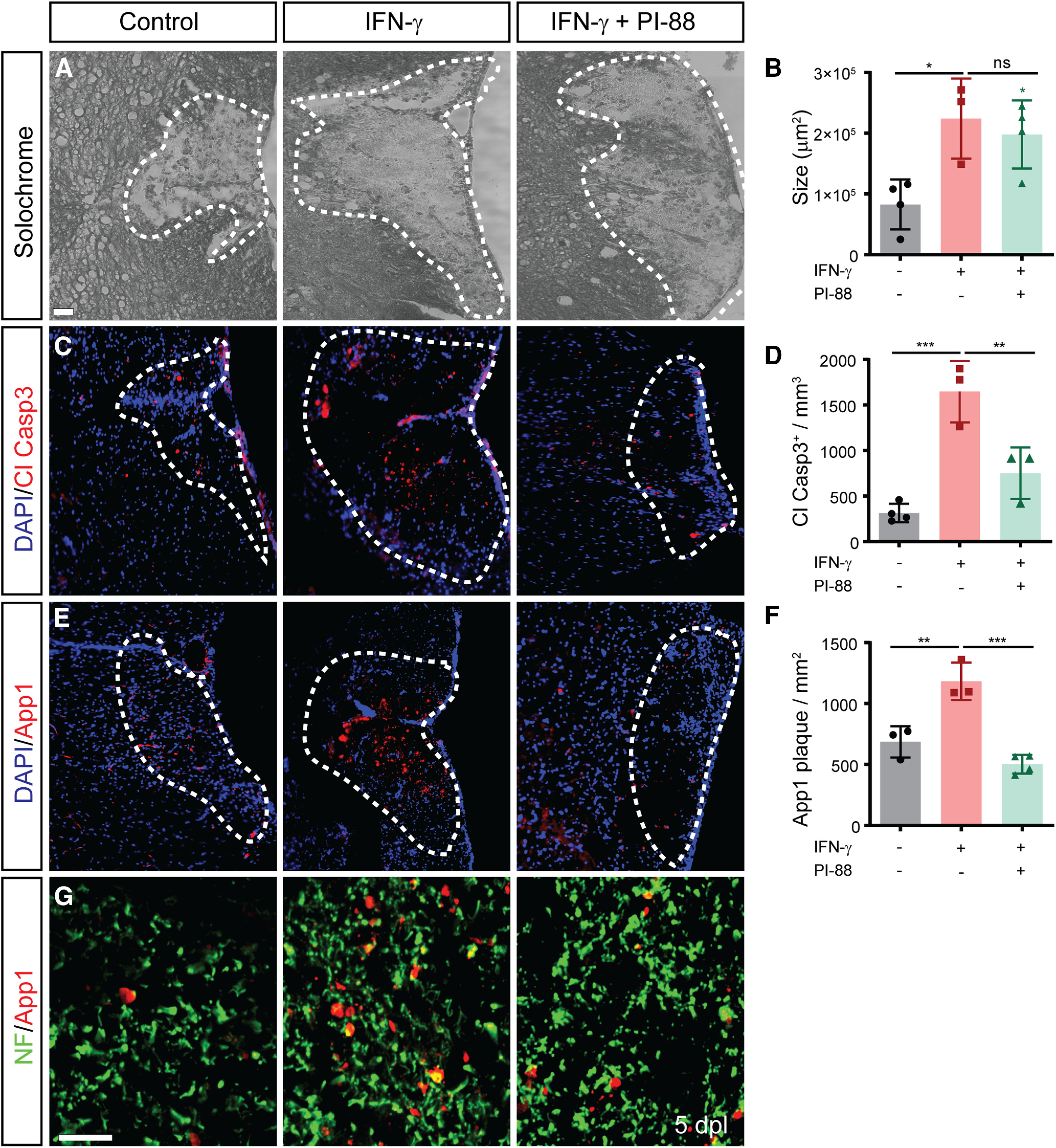
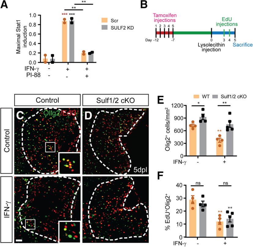
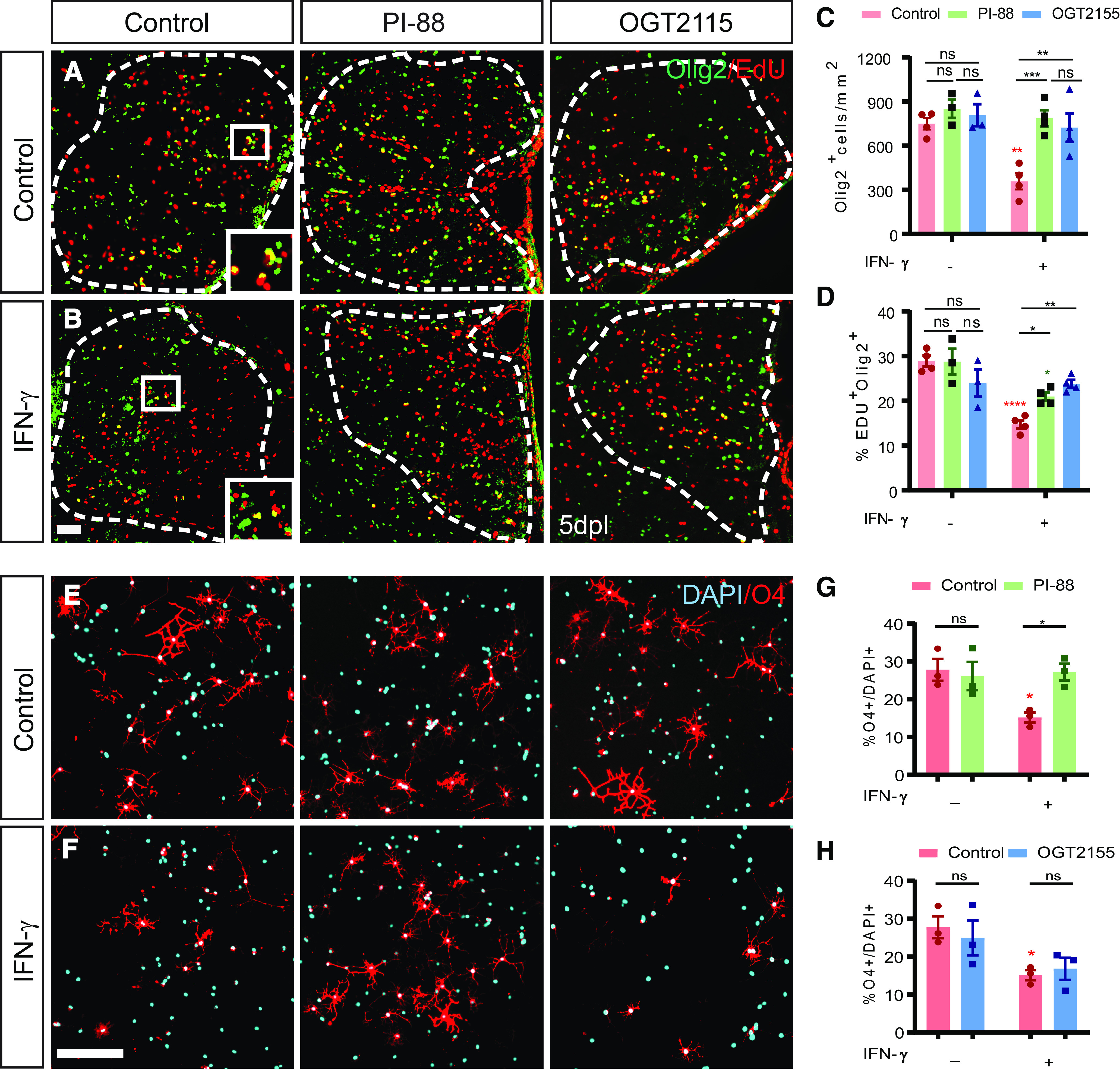
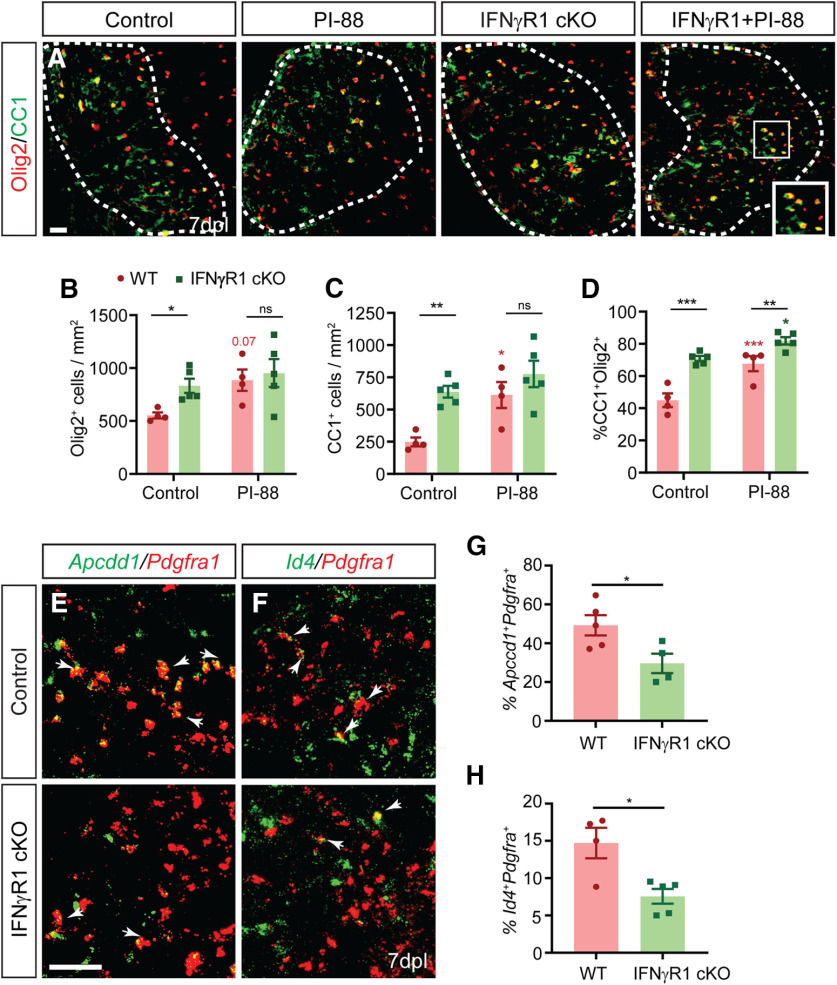
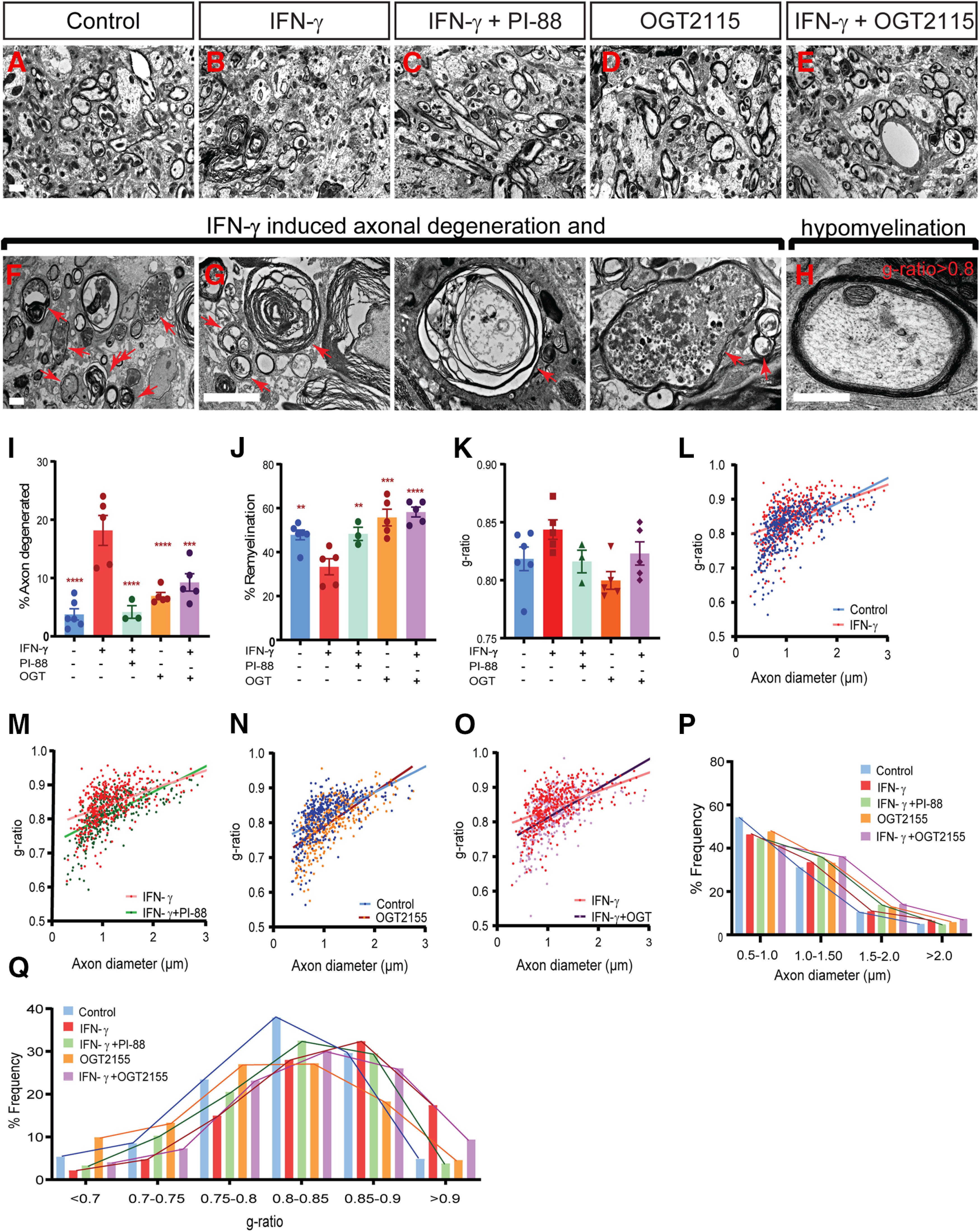
The proinflammatory cytokine IFN-γ, which is chronically elevated in multiple sclerosis, induces pathologic quiescence in human oligodendrocyte progenitor cells (OPCs) via upregulation of the transcription factor PRRX1. In this study using animals of both sexes, we investigated the role of heparan sulfate proteoglycans in the modulation of IFN-γ signaling following demyelination. We found that IFN-γ profoundly impaired OPC proliferation and recruitment following adult spinal cord demyelination. IFN-γ-induced quiescence was mediated by direct signaling in OPCs as conditional genetic ablation of IFNγR1 (Ifngr1) in adult NG2+ OPCs completely abrogated these inhibitory effects. Intriguingly, OPC-specific IFN-γ signaling contributed to failed oligodendrocyte differentiation, which was associated with hyperactive Wnt/Bmp target gene expression in OPCs. We found that PI-88, a heparan sulfate mimetic, directly antagonized IFN-γ to rescue human OPC proliferation and differentiation in vitro and blocked the IFN-γ-mediated inhibitory effects on OPC recruitment in vivo Importantly, heparanase modulation by PI-88 or OGT2155 in demyelinated lesions rescued IFN-γ-mediated axonal damage and demyelination. In addition to OPC-specific effects, IFN-γ-augmented lesions were characterized by increased size, reactive astrogliosis, and proinflammatory microglial/macrophage activation along with exacerbated axonal injury and cell death. Heparanase inhibitor treatment rescued many of the negative IFN-γ-induced sequelae suggesting a profound modulation of the lesion environment. Together, these results suggest that the modulation of the heparanome represents a rational approach to mitigate the negative effects of proinflammatory signaling and rescuing pathologic quiescence in the inflamed and demyelinated human brain.SIGNIFICANCE STATEMENT The failure of remyelination in multiple sclerosis contributes to neurologic dysfunction and neurodegeneration. The activation and proliferation of oligodendrocyte progenitor cells (OPCs) is a necessary step in the recruitment phase of remyelination. Here, we show that the proinflammatory cytokine interferon-γ directly acts on OPCs to induce pathologic quiescence and thereby limit recruitment following demyelination. Heparan sulfate is a highly structured sulfated carbohydrate polymer that is present on the cell surface and regulates several aspects of the signaling microenvironment. We find that pathologic interferon-γ can be blocked by modulation of the heparanome following demyelination using either a heparan mimetic or by treatment with heparanase inhibitor. These studies establish the potential for modulation of heparanome as a regenerative approach in demyelinating disease.

Keywords: demyelination; human; interferon; oligodendrocyte progenitor; quiescence; remyelination.


Figures

Figure 1.
Figure 1.
PRRX1 mRNA is enriched in OPCs located within multiple sclerosis lesions. A, B, IHC for PLP1 and MHCII for characterization of chronic lesion in the multiple sclerosis patient brain. C, D, PDGFRA, PRRX1, and STAT1 immunofluorescence in control brain and MS lesion brain (PDGFRA, red; PRRX1, green; STAT1, gray). NAWM from control brain expressed low levels of PRRX1 and STAT1. Both PRRX1 and STAT1 proteins were upregulated in OPCs within a chronic active lesion from a secondary progressive MS patient (arrows indicate colocalization of PRRX1/STAT1 with PDGFRA. E, Fluorescence RNA-scope in situ hybridization illustrates colocalization of PRRX1 mRNA with PDGFRA mRNA-expressing OPCs found at lesion edges. F, Despite being less frequent, PDGFRA+ OPCs in the lesion core also expressed PRXX1+. Arrows represents colocalization of PRRX1 with PDGFRA. G, Quantitative PCR analyses of PRRX1, IRF1, STAT1, PDGFRA, and MBP on RNA extracted from chronic active MS lesions and NAWM (mean ± SEM, n = 3–4 SPMS patients). GAPDH was used as internal control. Student's t test: *p < 0.05 and **p < 0.01, respectively. Scale bars: A, B, 100 µm; C, D, 10 µm; E, F, 20 µm.
Figure 2.
Figure 2.
Conditional IFNγR1 knockout in OPCs enhances progenitor recruitment and proliferation following focal demyelination. A, OPC-specific IFNγR1 conditional knockout was induced in adult NG2CreERT2: Rosa-YFP: IFNγR1fl/fl mice by daily intraperitoneal administration of 200 mg/kg tamoxifen for 5 d. Animals underwent spinal cord demyelination 1 week later with (E) or without (B–D) simultaneous administration of 3 ng of IFN-γ directly into the lesion. Animals were killed at 5 dpl to assess the effects on OPC recruitment. B, Solochrome cyanine was used to identify demyelinated lesions. C, The innate inflammatory response was assessed by Gfap and Iba1 immunofluorescence. DF, In control (D) and IFN-γ-treated (E) lesions, OPC recruitment was assessed using the oligodendrocyte lineage marker Olig2, and Olig2+ cell density quantified in F (cells/mm2, mean ± SEM). G, The percentage of EdU+ cells among Olig2+ cells was determined as a measure of OPC proliferation. Arrowhead and inserts indicate proliferating EdU+ OPCs. IFN-γ treatment significantly reduced OPC recruitment and proliferation. These effects were dependent on IFNγR1 expression in OPC, as IFNγR1 mice were unresponsive to IFN-γ treatment (n = 3–4 mice/group; two-way ANOVA interaction, p < 0.05 for Olig2+ cell density and p < 0.05 for the percentage of proliferating OPCs). *p < 0.05, Holm–Sidak multiple-comparisons post-test). Red ** and *** indicate Holm-Sidak p < 0.01 and p < 0.0001 respectively compared to matched wildtype control. Two-way ANOVA (Extended Data Fig. 2-1). Scale bar, 20 µm.
Figure 3.
Figure 3.
IFN-γ-mediated quiescence of human and mouse OPCs is rescued by heparan sulfate mimetic PI-88. A, Phase contrast image of CD140a/PDGFRA+ OPCs isolated from human fetal brain illustrates the typical bipolar morphology, and >95% isolated cells were positive for Olig2, an oligodendrocyte lineage marker. IFN-γ treatment of hOPCs was found to regulate a wide variety of genes associated with heparan sulfate proteoglycan synthesis and modulation (Extended Data Fig. 3-1). B, hOPCs were treated with or without heparan sulfate mimetic PI-88 (2 µg/ml) for 0.5 h and then exposed to IFN-γ (10 ng/ml). C, At 24 h, cells were pulsed with EdU for an additional 24 h and the proportion of EdU+ cells was quantified. IFN-γ treatment significantly reduced OPC proliferation (n = 4 fetal human samples, one-way ANOVA with Holm–Sidak post-test, p < 0.01). PI-88 rescued the negative effects of IFN-γ on proliferation (p < 0.05). D, The activity of a luciferase-expressing STAT1 reporter in transduced hOPCs was assessed following 24 h of IFN-γ treatment (1 and 10 ng/ml) with or without PI-88 (2 μg/ml). Luciferase activity was measured in triplicate for each human sample (n = 3 fetal human samples). Holm–Sidak test: *p < 0.05, **p < 0.01, ***p < 0.001, ****p < 0.0001 for each pairwise comparison following two-way ANOVA. Green indicates comparison with untreated cells, red indicates comparison with PI-88 control. Two-way ANOVA (Extended Data Fig. 3-2). E, Injection of 10 ng of IFN-γ with or without 10 μg/ml PI-88 directly into demyelinated lesion in 8-week-old mice. F, The effect of PI-88 on oligodendrocyte lineage cell density and IFN-γ/STAT1 pathway activity at 5 dpl was assessed by immunofluorescence for Olig2 (green) and Irf-1 (red), respectively. PI-88 treatment decreased colocalization of Irf1 in Olig2+ cells. G, I, J, OPC density and proliferation was assessed by Olig2 and by determining the proportion of BrdU+Olig2+ OPCs (G; quantified in I and J; mean ± SEM, n = 3–4). I, J, IFN-γ treatment significantly decreased OPC recruitment (I) and proliferation (J), while PI-88 injections rescued the effects of IFN-γ. Holm–Sidak test: *p < 0.05, **p < 0.01, ***p < 0.001 following one-way ANOVA. H, Fluorescence in situ hybridization at 5 dpl using Prrx1, Pdgfra, and Ki67-specific mRNA probes (red, green, and pink, respectively). Prrx1 expression was upregulated following demyelination in the presence of IFN-γ and PI-88 treatment decreased Prrx1 expression within the lesion. White arrows denote proliferating OPCs (Ki67+Pdgfra+), which were typically Prrx1 low/negative cells, while blue arrows denote nonproliferating (Ki67) Pdgfra+ OPCs that expressed high levels of Prrx1 mRNA. A subset of astrocytes expressed Prrx1 following IFN-γ-augmented demyelination, but colocalization with Iba+ microglia was not observed (Extended Data Fig. 3-3). K, IFN-γ treatment significantly increased Prrx1 expression in Pdgfra+ OPCs, while PI-88 treatment decreased expression of Prrx1 in OPCs. Holm–Sidak test: *p < 0.05, **p < 0.01, ***p < 0.001 following one-way ANOVA. Scale bars: A, B, 50 µm; and F–H, 20 µm. ns denotes not significant.
Figure 4.
Figure 4.
PI-88 rescues IFN-γ-mediated proinflammatory effects on glial cells following demyelination. A, Following demyelination, astrogliosis was assessed by Gfap immunofluorescence at 5 dpl (inserts shows higher-magnification confocal image). Gfap was substantially increased by IFN-γ, while PI-88 treatment decreased reactive astrogliosis (n = 3–4/group). B, IFN-γ-dependent signaling in Gfap+ astrocytes was not apparent as Irf1 immunoreactivity was not colocalized with Gfap. C, D, Microglial infiltration was assessed by Iba1 immunofluorescence in lesion (C), quantified in D. Density of Iba1+ microglial cells increased following IFN-γ treatment, whereas PI-88 treatment decreased microglial infiltration (mean ± SEM, n = 3–4). Holm–Sidak test: *p < 0.05, ***p < 0.001, ****p < 0.0001 following one-way ANOVA, respectively. White arrows denote amoeboid shaped microglial cells, while blue arrows indicate reactive ramified. E, F, Confocal images showing examples of ramified and amoeboid shape microglia, quantified in F. G, The presence of proinflammatory M1 microglia was assessed by iNOS1 immunofluorescence. White arrows indicate double-positive cells. H, Percentage of iNOS+Iba1+ microglial cells among total Iba1+ cells (Holm–Sidak post-test shown). I, IFN-γ signaling was assessed by colocalization of Irf-1 and iNOS+ proinflammatory microglia. Scale bar, 20 µm. ns denotes not significant.
Figure 5.
Figure 5.
IFN-γ exacerbates cell and axonal injury following demyelination which was rescued by PI-88 treatment. A, Lesion size was assessed by Solochrome cyanine lipid stain at 5 dpl. B, IFN-γ treatment increased lesion size, which was not significantly affected by PI-88 treatment (area: mean ± SEM, n = 3–4/group). C, D, Cell death was assessed by analysis of cleaved caspase 3-stained particles (C) and quantified in D. E, F, Axon damage was assessed by the accumulation of App1 following demyelination (E) and quantified in F. Holm–Sidak test: *p < 0.05, **p < 0.01, ***p < 0.001 following one-way ANOVA. Cell death and axon injury was exacerbated by IFN-γ, and both of these measures of tissue injury were rescued by concurrent PI-88 treatment. G, App1 was restricted to neurofilament-positive axons (SMI31/32). Scale bars: A–C, 50 µm; D, 20 µm. ns denotes not significant.
Figure 6.
Figure 6.
IFN-γ signaling in OPCs following demyelination was not influenced by sulfatases (Sulf1/2). A, The activity of a STAT1-dependent luciferase reporter in human OPCs following 24 h of IFN-γ treatment (10 ng/ml) with or without PI-88 (2 μg/ml) and following infection with lentiviral SULF2 or control scrambled shRNAi was assessed. Luciferase activity was measured in triplicate for each human sample (n = 2 human fetal samples). Holm–Sidak test: *p < 0.05, **p < 0.01, ***p < 0.001 for each pairwise comparison following two-way ANOVA (mean ± SEM normalized to maximal response). SULF2 KD did not influence IFN-γ-dependent STAT activity, two-way ANOVA (Extended Data Fig. 6-1). B, OPC-specific Sulf1/2 knockout was induced in adult NG2: CreER; Rosa26IsIYFP; Sulf1fl/fl-Sulf2fl/fl (Sulf1/2 cKO) mice by daily intraperitoneal administration of 200 mg/kg tamoxifen for 5 d. Littermate cre-negative mice were similarly injected with tamoxifen as controls. Animals were lesioned 1 week after the last day of injection with or without simultaneous administration of 3 ng of IFN-γ directly into the lesion site. Animals were killed at 5 dpl, and spinal cord tissue was processed. C, D, Olig2/EdU immunofluorescence of wild-type control and Sulf1/2 cKO mice with or without IFN-γ treatment (n = 4–5 mice/group). Insert shows colocalization of EdU (red) in a subset of Olig2+ cells (green). E, F, Sulf1/2 cKO did not influence the effects of IFN-γ on OPC recruitment (Olig2+ cells/mm2; E) and proliferation (percentage EdU among Olig2+ cells; F). Examination of the interaction of these variables by two-way ANOVA did not reveal a significant effect, suggesting independent modes of action for Sulf1/2 and IFN-γ (p = 0.17, p = 0.31). Two-way ANOVA (Extended Data Fig. 6-2). Scale bar, 20 µm. ns denotes not significant.
Figure 7.
Figure 7.
PI-88 acts via heparanase inhibition to rescue IFN-γ-mediated quiescence of OPCs. A, B, Control (A) and IFN-γ (B) mouse groups underwent focal spinal cord demyelination with or without PI-88 or OGT2115 injection and were killed at 5 dpl. Immunofluorescence for Olig2 (green) and EdU (red) was performed. C, D, OPC recruitment (Olig2+ cells/mm2; C) and proliferation (EdU percentage among Olig2+ cells; D) were quantified. Both PI-88 and OGT2115 treatment rescued the negative effects of IFN-γ on OPC recruitment (G) and proliferation (H) following demyelination (mean ± SEM; n = 3–4 mice/group). Two-way ANOVA interaction for Olig2+ cell density and percentage EdU OPCs were Holm–Sidak test: *p < 0.05, **p < 0.01 (Extended Data Fig. 7-1). Red and green denotes p value compared to matched untreated controls. E, F, at 0.5 h following treatment with or without PI-88 (2 μg/ml) or OGT2155 (0.4 μM) hOPCs were treated with vehicle (E) or IFN-γ (F) (10 ng/ml). O4+ oligodendrocyte (red) differentiation was assessed at 48 h and the proportion of O4+ cells quantified (mean ± SEM, n = 3 fetal samples) following PI-88 (G) or OGT2155 (H). IFN-γ has a significant inhibitory effect of differentiation (red, *), which is rescued by PI-88 (two-way ANOVA; Extended Data Fig. 7-2) but not OGT2115 treatment (two-way ANOVA; Extended Data Fig. 7-3). Holm–Sidak test: *p < 0.05 following two-way ANOVA. Scale bars: A, B, 20 µm; E, F, 50 µm. ns denotes not significant. ***p < 0.001, ****p < 0.0001.
Figure 8.
Figure 8.
Excessive IFN-γ signaling blocks OPC differentiation following demyelination in a cell-autonomous manner and is associated with the activation of Wnt/Bmp target genes in OPCs. A, Wild-type and IFNγR1 cKO were subjected to lysolecithin-mediated focal demyelination of the spinal cord with or without coadministration of 10 μg/ml PI-88. To assess the formation of newly generated oligodendrocytes, animals were killed at 7 d post-lesion (7 dpl). Olig2 (Red) and CC1 (green) colocalization was used to assess OL differentiation. B–D, The densities of Olig2+ oligodendrocytes lineage cells (cells/mm2; B) and Olig2+CC1+ postmitotic oligodendrocytes (C) was quantified along with the percentage of CC1+ oligodendrocytes (D; n = 3–5 mice/group; mean ± SEM). Holm–Sidak test: *p < 0.05, **p < 0.01, ***p < 0.001 for each pairwise comparison following two-way ANOVA. Red denotes p values compared with control wild-type, while green denotes p values compared with IFNγR1 cKO group. E, F, Two-way ANOVA did not reveal a significant interaction between genotype and PI-88 treatment on all parameters (p = 0.28, p = 0.17, p = 0.09; Extended Data Fig. 8-1). G, H, Quantification of Wnt target gene expression Apcdd1 (G) and BMP target gene Id4 (H) was determined in Pdgfra+ OPCs at 7 dpl (n = 4–5 mice/group, mean ± SEM) and at 5 dpl (Extended Data Fig. 8-2). t test: *p < 0.05. Both Wnt and Bmp target genes were regulated in an IFNγR1-dependent manner in OPCs. Scale bar, 20 μm. ns denotes not significant.
Figure 9.
Figure 9.
Heparanase inhibitors PI-88 and OGT2115 rescue remyelination and reduce axonal injury induced by IFN-γ treatment. A–E, Injection of 3 ng of IFN-γ with or without PI-88 (10 μg/ml) or OGT2155 (0.4 μm) directly into demyelinated lesions in mice (8–10 weeks of age). Animals were killed at 14 dpl, and spinal cord tissue was processed for electron microscopy. Representative fields shown within the lesion periphery. F, G, IFN-γ treatment induces various forms of axonal injury and degeneration. H, Several remyelinated axons also exhibit very thin remyelination following IFN-γ treatment. Arrows represent degenerating axons. I–K, The proportions of degenerating axons among total axons (I), the proportion of remyelinated axons among healthy-appearing axons (J), and the g-ratio of normal-appearing remyelinating axons (K) were calculated in each animal (mean ± SEM; n = 3–6 animals/group). I, J, Both PI-88 and OGT2115 treatment rescued the negative effects of IFN-γ on axonal damage (I) and the percentage of remyelination (J) following demyelination. L–O, Relationship between axon diameter and g-ratio (linear regression shown) between groups (L; control vs IFN-γ; M, IFN-γ vs IFN-γ + PI-88; N, control vs OGT2155; and O, IFN-γ vs IFN-γ + OGT2155). P, Q, Frequency distribution of axonal diameter (P) and g-ratio (Q) in lesion (n = 3–6 animals/group, ≥400 axons). Holm–Sidak test: **p < 0.01, ***p < 0.001, ****p < 0.0001 for each pairwise comparison following one-way ANOVA. Scale bar, 2 μm.

Similar articles

Cited by

References

    1. Abiraman K, Pol SU, O'Bara MA, Chen GD, Khaku ZM, Wang J, Thorn D, Vedia BH, Ekwegbalu EC, Li JX, Salvi RJ, Sim FJ (2015) Anti-muscarinic adjunct therapy accelerates functional human oligodendrocyte repair. J Neurosci 35:3676–3688. 10.1523/JNEUROSCI.3510-14.2015 - DOI - PMC - PubMed
    1. Agresti C, D'Urso D, Levi G (1996) Reversible inhibitory effects of interferon-gamma and tumour necrosis factor-alpha on oligodendroglial lineage cell proliferation and differentiation in vitro. Eur J Neurosci 8:1106–1116. 10.1111/j.1460-9568.1996.tb01278.x - DOI - PubMed
    1. Arellano G, Ottum PA, Reyes LI, Burgos PI, Naves R (2015) Stage-specific role of interferon-gamma in experimental autoimmune encephalomyelitis and multiple sclerosis. Front Immunol 6:492. 10.3389/fimmu.2015.00492 - DOI - PMC - PubMed
    1. Baerwald KD, Popko B (1998) Developing and mature oligodendrocytes respond differently to the immune cytokine interferon-gamma. J Neurosci Res 52:230–239. 10.1002/(SICI)1097-4547(19980415)52:2<230::AID-JNR11>3.0.CO;2-B - DOI - PubMed
    1. Balabanov R, Strand K, Kemper A, Lee JY, Popko B (2006) Suppressor of cytokine signaling 1 expression protects oligodendrocytes from the deleterious effects of interferon-gamma. J Neurosci 26:5143–5152. 10.1523/JNEUROSCI.0737-06.2006 - DOI - PMC - PubMed

Publication types

MeSH terms

Substances

LinkOut - more resources