Functional characterization of DLK1/MEG3 locus on chromosome 14q32.2 reveals the differentiation of pituitary neuroendocrine tumors
- PMID: 33472171
- PMCID: PMC7835058
- DOI: 10.18632/aging.202376
Functional characterization of DLK1/MEG3 locus on chromosome 14q32.2 reveals the differentiation of pituitary neuroendocrine tumors
Abstract
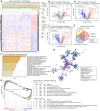
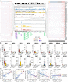
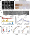
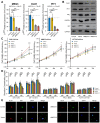
Pituitary neuroendocrine tumors (PitNETs) represent the neoplastic proliferation of the anterior pituitary gland. Transcription factors play a key role in the differentiation of PitNETs. However, for a substantial proportion of PitNETs, the etiology is poorly understood. According to the transcription data of 172 patients, we found the imprinting disorders of the 14q32.2 region and DLK1/MEG3 locus associated with the differentiation of PitNETs. DLK1/MEG3 locus promoted somatotroph differentiation and inhibited tumor proliferation in PIT1(+) patients, furthermore, the level of DLK1 played a critical role in the trend of somatotroph or lactotroph differentiation. Anti-DLK1 monoclonal antibody blockade or siMEG3 both indicated that the DLK1/MEG3 significantly promoted the synthesis and secretion of GH/IGF-1 and inhibited cell proliferation. In addition, loss of DLK1 activated the mTOR signaling pathway in high DLK1-expressing and PIT1(+) GH3 cell lines, a mild effect in the low DLK1-expressing and PIT1(+) MMQ cell lines and no effect in the PIT1(-) ATT20 cell line. These findings emphasize that expression at the DLK1/MEG3 locus plays a key role in the differentiation of PitNETs, especially somatotroph adenomas, and provide potential molecular target data for patient stratification and treatment in the future.
Keywords: DLK1/MEG3 locus; differentiation; growth hormone secreting; pituitary neuroendocrine tumors; somatotroph adenomas.
Conflict of interest statement
Figures


Similar articles
-
Silencing of the imprinted DLK1-MEG3 locus in human clinically nonfunctioning pituitary adenomas.Am J Pathol. 2011 Oct;179(4):2120-30. doi: 10.1016/j.ajpath.2011.07.002. Epub 2011 Aug 24. Am J Pathol. 2011. PMID: 21871428 Free PMC article.
-
Selective loss of MEG3 expression and intergenic differentially methylated region hypermethylation in the MEG3/DLK1 locus in human clinically nonfunctioning pituitary adenomas.J Clin Endocrinol Metab. 2008 Oct;93(10):4119-25. doi: 10.1210/jc.2007-2633. Epub 2008 Jul 15. J Clin Endocrinol Metab. 2008. PMID: 18628527 Free PMC article.
-
Molecular Biology of Pituitary Adenomas.Neurosurg Clin N Am. 2019 Oct;30(4):391-400. doi: 10.1016/j.nec.2019.05.001. Epub 2019 Jul 5. Neurosurg Clin N Am. 2019. PMID: 31471046 Review.
-
Chromosomal instability in the prediction of pituitary neuroendocrine tumors prognosis.Acta Neuropathol Commun. 2020 Nov 10;8(1):190. doi: 10.1186/s40478-020-01067-5. Acta Neuropathol Commun. 2020. PMID: 33168091 Free PMC article.
-
Histopathology of growth hormone-secreting pituitary tumors: State of the art and new perspectives.Best Pract Res Clin Endocrinol Metab. 2024 May;38(3):101894. doi: 10.1016/j.beem.2024.101894. Epub 2024 Apr 2. Best Pract Res Clin Endocrinol Metab. 2024. PMID: 38614953 Review.
Cited by
-
The Role of Long Non-coding RNAs in Human Imprinting Disorders: Prospective Therapeutic Targets.Front Cell Dev Biol. 2021 Oct 25;9:730014. doi: 10.3389/fcell.2021.730014. eCollection 2021. Front Cell Dev Biol. 2021. PMID: 34760887 Free PMC article. Review.
-
Clinical Biology of the Pituitary Adenoma.Endocr Rev. 2022 Nov 25;43(6):1003-1037. doi: 10.1210/endrev/bnac010. Endocr Rev. 2022. PMID: 35395078 Free PMC article. Review.
References
-
- Lloyd R, Osamura R, Klöppel G, Rosai J. (2017) WHO classification of tumours of the endocrine organs, 4th edn. Lyon: International Agency for Research on Cancer;
Publication types
MeSH terms
Substances
LinkOut - more resources
Full Text Sources
Medical
Molecular Biology Databases
Miscellaneous

