. 2021 Mar 4;81(5):1100-1115.e5.
doi: 10.1016/j.molcel.2020.12.033. Epub 2021 Jan 19.

Structural basis for self-cleavage prevention by tag:anti-tag pairing complementarity in type VI Cas13 CRISPR systems

Affiliations

Structural basis for self-cleavage prevention by tag:anti-tag pairing complementarity in type VI Cas13 CRISPR systems

Beibei Wang et al. Mol Cell. .

Abstract

Bacteria and archaea apply CRISPR-Cas surveillance complexes to defend against foreign invaders. These invading genetic elements are captured and integrated into the CRISPR array as spacer elements, guiding sequence-specific DNA/RNA targeting and cleavage. Recently, in vivo studies have shown that target RNAs with extended complementarity with repeat sequences flanking the target element (tag:anti-tag pairing) can dramatically reduce RNA cleavage by the type VI-A Cas13a system. Here, we report the cryo-EM structure of Leptotrichia shahii LshCas13acrRNA in complex with target RNA harboring tag:anti-tag pairing complementarity, with the observed conformational changes providing a molecular explanation for inactivation of the composite HEPN domain cleavage activity. These structural insights, together with in vitro biochemical and in vivo cell-based assays on key mutants, define the molecular principles underlying Cas13a's capacity to target and discriminate between self and non-self RNA targets. Our studies illuminate approaches to regulate Cas13a's cleavage activity, thereby influencing Cas13a-mediated biotechnological applications.

Keywords: CRISPR-Cas; Cas13; RNA cleavage; cryo-EM structure; inhibition mechanism; target discrimination.


Conflict of interest statement

Declaration of interests The authors declare no competing interests.

Figures

Figure 1.
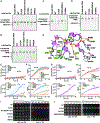
Figure 1.. Extended tag:anti-tag pairing prevents RNA cleavage by Cas13a
(A and C) Schematic of target RNAs designed for in vitro cis-RNase and trans-RNase cleavage assays. The spacer and repeat of crRNA are shown in magenta and pink, respectively. The target segment is shown in gray, with anti-tag indicated in red. (B and D) In vitro cleavage assays by LshCas13a and LbuCas13a monitoring substrate RNA degradation on formation of target RNA (B) and non-target substrate RNA (D) ternary complexes in the presence or absence of anti-tag. Two reported dead mutations, LshCas13a-R1278A and LbuCas13a-R472A/H477A, are used as negative controls and labeled as dCas13a. The sequence and schematic of crRNA and target RNAs are shown in (A), (C), and Figures S1B and S1C. (E and G) Quantification of EYFP mRNA knockdown by LshCas13a (E) and LbuCas13a (G) in E. coli cells. EYFP mRNA contains a 28 nt segment complementary to spacer followed by an 8 nt segment with or without anti-tag. Transcription of EYFP mRNA was induced by tetracycline. Error bars represent standard error of mean (SEM) of three biological replicates. (F and H) Effect on growth rate of E. coli cells upon EYFP mRNA interference by LshCas13a (F) and LbuCas13a (H). Error bars represent SEM of three biological replicates. (I and J) Elution profiles run from a Superdex 200 10/300 GL size exclusion column in the presence or absence of anti-tag, showing that anti-tag has no effect on target RNA loading and formation of LshCas13a-crRNA-target RNA (I) and LbuCas13a-crRNA-target RNA (J) ternary complexes. See also Figure S1.
Figure 2.
Figure 2.. Overall structure of LshCas13a-crRNA-anti-tag RNA complex
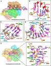
(A) Domain organization of LshCas13a. The NTD domain of LshCas13a has no clear density and is indicated by the dashed box. (B) Ribbon representation of LshCas13a-crRNA-anti-tag RNA6 ternary complex. Color codes of RNA and Cas13a are defined as in Figure 1C and (A), respectively. (C and D) Ribbon representation and schematic of crRNA:target RNA duplex. The anti-tag is complementary with 3′-flank of crRNA repeat and forms an extended A-form RNA duplex beyond the guide:target duplex. Nucleotides not observed in the structures are colored gray in (D). (E) Schematic representation of the conformational changes occurring in crRNA upon anti-tag RNA or target RNA loading. Nucleotides not observed in the structures are indicated by the dashed box. The RNA duplexes inside the binding channel are indicated by yellow boxes. See also Figures S1–S3 and Table S1.
Figure 3.
Figure 3.. Detailed interactions of LshCas13a with crRNA:anti-tag RNA duplex
(A) Surface views of the interfaces between Cas13a and duplex formed by crRNA and anti-tag RNA. (B) Recognition of tag:anti-tag duplex by the Linker and Helical-II domains. (C and D) Interactions between Cas13a and base pairs 2–6 (C) and 7–15 (D) in the guide:target duplex. (E) Surface views of pre-target-bound LshCas13a-crRNA complex, with interactions between crRNA tag region and LshCas13a shown in the zoomed-in panel. See also Figure S3.
Figure 4.
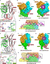
Figure 4.. Comparison of HEPN pocket alignments and global folds between ternary complexes involving bound anti-tag (LshCas13) and target (LbuCas13) RNAs
(A) Structural comparisons of two HEPN domains of LshCas13a in anti-tag-bound (HEPN-I in green, HEPN-II in salmon, target segment in gray, and anti-tag in red) and pre-target-bound (in silver) states by superposing the HEPN-II domains. For simplicity, only target RNA is shown. Comparison of the positioning of the four catalytic residues from the pair of HEPN domains is shown in the zoomed-in segment (inset). (B and C) Surface of LshCas13a showing domain rearrangements to generate crRNA:target RNA duplex binding channel from pre-target-bound (B) to anti-tag-bound (C) states. Black arrows in (B) show the directions of domain movements on ternary complex formation with anti-tag RNA. (D) Surface views of the interfaces between crRNA-bound LshCas13a and duplex formed by bound anti-tag RNA. The guide:target duplex segment is boxed. (E) Structural comparisons of two HEPN domains of LbuCas13a in target-bound (HEPN-I in green, HEPN-II in salmon, target segment in gray) and pre-target-bound (in silver) states by superposing the HEPN-II domains (Liu et al., 2017a). For simplicity, only target RNA is shown. Double mutation R1048A/H1053A was used for structural studies of target RNA-bound LbuCas13a. Comparison of the positioning of the four catalytic residues from the pair of HEPN domains is shown in the zoomed-in segment (inset). (F and G) Surface of LbuCas13a showing domain rearrangements to generate crRNA:target RNA duplex binding channel from pre-target-bound (F) to target-bound (G) states. Black arrows in (F) show the directions of domain movements on ternary complex formation with target RNA. (H) Surface views of the interfaces between crRNA-bound LbuCas13a and duplex formed by bound target RNA. The guide-target duplex segment is boxed. See also Figure S4.
Figure 5.
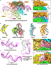
Figure 5.. Conformational changes in LshCas13 upon anti-tag RNA loading
(A) Structural comparison between LshCas13a-crRNA binary complex and LshCas13a-crRNA-anti-tag RNA ternary complex. Vector lengths correlate with the domain motion scales. Arrows show the directions of domain movement from pre-target-bound to anti-tag-bound states. (B) Structural comparison of the Helical-I, HEPN-I, HEPN-II, Linker, and Helical-II domains between LshCas13a-crRNA binary (in silver) and LshCas13a-crRNA-anti-tag RNA ternary (in color) complexes. Arrows indicate the domain movements. The key catalytic residues from the pair of HEPN domains are indicated by black (anti-tag-bound) and red (pre-target-bound) asterisks, respectively. (C and D) Binding with target RNA harboring anti-tag widens the guide:target duplex-binding channel on proceeding from LshCas13a-crRNA binary complex (C) to the LshCas13a-crRNA-anti-tag RNA ternary complex (D). (E–G) Architectures of crRNA in LshCas13a-crRNA binary (E) and LshCas13a-crRNA-anti-tag RNA ternary (F) complexes. The details of tag region are shown in (G), with anti-tag-bound crRNA in color and pre-target-bound crRNA in silver. (H and I) Comparisons of the tag region of crRNA in LshCas13a-crRNA binary (H) and LshCas13a-crRNA-anti-tag RNA ternary (I) complexes. The Helical-II domain, which interacts with and covers the tag region in LshCas13a-crRNA complex, is hidden in (H) to show the position of tag region. See also Figure S4.
Figure 6.
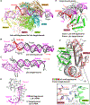
Figure 6.. Structural comparison between target-bound (LbuCas13a) and anti-tag-bound (LshCas13a) ternary complexes
(A) Structural comparison between LshCas13a-crRNA-anti-tag RNA (this study) and LbuCas13a-crRNA-target RNA (Liu et al., 2017a) ternary complexes. Vector lengths correlate with the domain motion scales. Arrows show the directions of domain movement from anti-tag-bound to target-bound states. (B–D) Architectures of crRNA in LshCas13a-crRNA-anti-tag RNA (B) and LbuCas13a-crRNA-target RNA (C) ternary complexes. The positions of 3′ end of target RNAs are indicated by black arrows. The extension directions of RNA duplexes are indicated by red arrows. The details conformational changes of the tag region are shown in (D), with anti-tag bound crRNA in color and target-bound crRNA in silver. (E) Superposition of LshCas13a-crRNA-anti-tag RNA (in color) and LbuCas13a-crRNA-target RNA (in silver) ternary complexes with the focus on HEPN-I and HEPN-II domains. The key catalytic residues in HEPN domains are highlighted in red. The black arrow indicates the movements of HEPN-I domain toward the HEPN-II domain from anti-tag-bound to target-bound states. The red arrows indicates the steric clashes between crRNA:anti-tag RNA duplex and the HEPN domains in target-bound state, indicating that the formation of tag:anti-tag RNA duplex prevents the movements of HEPN domains to generate a competent composite catalytic pocket. (F) Structural comparisons of two HEPN domains LshCas13a in anti-tag-bound (HEPN-I in green, HEPN-II in salmon, target segment in gray, and anti-tag in red) and LbuCas13a in target-bound bound (in silver) states by superposing the HEPN-II domains. For simplicity, only target RNA is shown. Comparison of the positioning of the four catalytic residues from the pair of HEPN domains is shown in the zoomed-in segment (inset). See also Figure S4.
Figure 7.
Figure 7.. Inhibition of RNA cleavage by blockage of conformational changes in tag segment
(A and B) Cleavage assays monitoring degradation on target RNA (A) and substrate RNA (B) with Lsh-crRNA or LshCas13a mutants. (C and D) Cleavage assays by LbuCas13a monitoring degradation on target RNA (C) and substrate RNA (D). (E and G) In vivo RNA knockdown assays of indicated E. coli strains expressing various LshCas13a (E) and LbuCas13a (G). Error bars represent SEM of three biological replicates. (F and H) Liquid growth assays for LshCas13a (F) and LbuCas13a (H) illustrating growth defects upon Cas13a-dependent RNA interference. Error bars represent SEM of three biological replicates. (I and J) Plasmids interference assays for E. coli strains co-transformed with various LshCas13a (I) or LbuCas13a (J) plasmids as well as a tet-induced EYFP plasmid. Ten-fold serial dilution of cells as indicated were spotted onto plates. (K) Recognition of crRNA 3′-flank (positions −3 to −1) by LbuCas13a (HEPN-I in green, HEPN-II in salmon, and crRNA tag segment in violet) and modeled LshCas13a (in silver). See also Figure S4.

Similar articles

Cited by

References

    1. Abudayyeh OO, Gootenberg JS, Konermann S, Joung J, Slaymaker IM, Cox DB, Shmakov S, Makarova KS, Semenova E, Minakhin L, et al. (2016). C2c2 is a single-component programmable RNA-guided RNA-targeting CRISPR effector. Science 353, aaf5573. - PMC - PubMed
    1. Abudayyeh OO, Gootenberg JS, Essletzbichler P, Han S, Joung J, Belanto JJ, Verdine V, Cox DBT, Kellner MJ, Regev A, et al. (2017). RNA targeting with CRISPR-Cas13. Nature 550, 280–284. - PMC - PubMed
    1. Adams PD, Afonine PV, Bunkóczi G, Chen VB, Davis IW, Echols N, Headd JJ, Hung LW, Kapral GJ, Grosse-Kunstleve RW, et al. (2010). PHENIX: a comprehensive Python-based system for macromolecular structure solution. Acta Crystallogr. D Biol. Crystallogr 66, 213–221. - PMC - PubMed
    1. Barrangou R, Fremaux C, Deveau H, Richards M, Boyaval P, Moineau S, Romero DA, and Horvath P (2007). CRISPR provides acquired resistance against viruses in prokaryotes. Science 315, 1709–1712. - PubMed
    1. Chen S, McMullan G, Faruqi AR, Murshudov GN, Short JM, Scheres SH, and Henderson R (2013). High-resolution noise substitution to measure overfitting and validate resolution in 3D structure determination by single particle electron cryomicroscopy. Ultramicroscopy 135, 24–35. - PMC - PubMed

Publication types

MeSH terms

Supplementary concepts