. 2021 Jan 15;11(1):1492.
doi: 10.1038/s41598-021-81077-y.

Histamine H1 receptor antagonists selectively kill cisplatin-resistant human cancer cells

Affiliations

Histamine H1 receptor antagonists selectively kill cisplatin-resistant human cancer cells

Nobuki Matsumoto et al. Sci Rep. .

Abstract

Cancer therapy is often hampered by the disease's development of resistance to anticancer drugs. We previously showed that the autonomously upregulated product of fibroblast growth factor 13 gene (FGF13; also known as FGF homologous factor 2 (FHF2)) is responsible for the cisplatin resistance of HeLa cisR cells and that it is likely responsible for the poor prognosis of cervical cancer patients treated with cisplatin. Here we show that cloperastine and two other histamine H1 receptor antagonists selectively kill HeLa cisR cells at concentrations that little affect parental HeLa S cells. The sensitivity of HeLa cisR cells to cloperastine was abolished by knocking down FGF13 expression. Cisplatin-resistant A549 cisR cells were similarly susceptible to cloperastine. H2, H3, and H4 receptor antagonists showed less or no cytotoxicity toward HeLa cisR or A549 cisR cells. These results indicate that histamine H1 receptor antagonists selectively kill cisplatin-resistant human cancer cells and suggest that this effect is exerted through a molecular mechanism involving autocrine histamine activity and high-level expression of FGF13. We think this represents a potential opportunity to utilize H1 receptor antagonists in combination with anticancer agents to treat cancers in which emergent drug-resistance is preventing effective treatment.


Conflict of interest statement

The authors declare no competing interests.

Figures
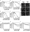
Figure 1
Figure 1
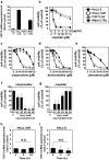
Cloperastine and other H1 receptor antagonists selectively kill HeLa cisR cells. (a,b,dh) HeLa cisR cells (open squares) and HeLa S cells (filled circles) were cultured for 3 days with or without the indicated concentrations of cisplatin (a), the histamine H1 receptor antagonists cloperastine (b), desloratadine (d), or clemastine (e), or the H2 receptor antagonist nizatidine (f), H3 receptor antagonist pitolisant (g), or H4 receptor antagonist JNJ-7777120 (h). Cell viability during the final 5 h of the culture period was evaluated using WST-8 colorimetric assays. Open squares, HeLa cisR cells; filled circles, HeLa S cells. The results are presented as means ± S.D. of sextuplicate (n = 6) (a,b,d,e) or quadruplicate (n = 4) (fh) samples. Four separate experiments yielded essentially the same results. Note that only H1 receptor antagonists resulted in 0% cell viability. Also note that error bars are not visible in many places due to their small size. The significance of the difference between the results from HeLa S and HeLa cisR cells at the same concentration of the indicated compound was determined with ANOVA (***p < 0.001; **p < 0.01; *p < 0.05). (c) HeLa cisR and HeLa S cells cultured for 3 days with 10 µg/ml cisplatin or 100 µM cloperastine were photographed under a phase contrast microscope. Bar, 50 µm.
Figure 2
Figure 2
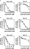
Cisplatin cytotoxicity toward HeLa cisR cells is not synergistically enhanced by H1 receptor antagonists. HeLa cisR cells were cultured for 3 days in the absence (closed circles) or presence (open square) of 40 µM cloperastine (a), 30 µM desloratadine (b), or 10 µM clemastine (c) with the indicated concentrations of cisplatin. Cell numbers during the final 5 h of the culture were evaluated using WST-8 colorimetric assays. The results are presented as means ± S.D. of quintuplicate (n = 5) samples. Three separate experiments yielded essentially the same results.
Figure 3
Figure 3
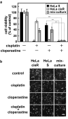
Combined use of cloperastine and cisplatin effectively kill both cisplatin-resistant and cisplatin-sensitive cancer cells in a mixed population. (a) Cytotoxicities of cloperastine and/or cisplatin toward HeLa S cells and HeLa cisR cells were analyzed separately or together as a mixed population. For the separate cultures, 4 × 103 HeLa S cells/well, or 1.5 × 104 HeLa cisR cells/well were analyzed. For the mixed population (combined culture), HeLa S cells (2 × 103 cells/well) and HeLa cisR cells (7.5 × 103 cells/well) were mixed and seeded into each well of a 96-well plate. After incubation for 1 day to allow the cells to attach, 100 µM cloperastine and/or 10 µg/ml (33 µM) cisplatin were added, and the cells were incubated for an additional 3 days, after which cell viability was measured. The results are presented as means ± S.D. of sextuplicate samples. Four separate experiments yielded essentially the same results. The significance of the difference between the selected pairs of results was determined with ANOVA (***p < 0.001). (b) Cells treated as in (a) were photographed under a phase contrast microscope. Bar, 100 µm.
Figure 4
Figure 4
Cytotoxicity of H1 receptor antagonists is dependent on the levels of FGF13 expression and cisplatin resistance. (a) Relative expression level of FGF13 in each cell line. (be) Cytotoxicities of cisplatin (b) and H1 receptor antagonists cloperastine (c), desloratadine (d), and clemastine (e) toward HeLa S cells, HeLa cisR cells, HeLa cisR cells in which FGF13 expression was suppressed by targeted siRNA expression (FGF13kd), and HeLa cisR cells in which degenerate siRNA was expressed (RNAi CTRL). Open squares, HeLa cisR cells; filled circles, HeLa S cells; open triangles, FGF13kd cells; filled diamonds, RNAi CTRL cells. (f) Cytotoxicity of 100 µM cloperastine toward HeLa S cells and five intermediate HeLa cisR cell lines, which were collected during the process of establishing the HeLa cisR cells (#1 to #5, adapted to 0.42, 0.84, 2.0, 6.0 and 8.0 µg/ml cisplatin in this order) and which differ in their expression of FGF13 mRNA. (g) Cytotoxicity of 10 µg/ml (33 µM) cisplatin toward HeLa S cells and the five intermediate HeLa cisR cell lines. (h,i) Separate cultures of HeLa cisR cells (h) and HeLa S cells (i) were treated with cloperastine for the indicated periods, after which relative expression of FGF13 mRNA was quantitated using RT-qPCRs. The results are presented as mean ± S.D. of sextuplicate (be) or triplicate (a,fi) samples. Three separate experiments yielded essentially the same results. In panels (bg) the significance of the difference between the results from HeLa S and HeLa cisR cells was determined with ANOVA (***p < 0.001; **p < 0.01; *p < 0.05).
Figure 5
Figure 5
Expression of histamine H1 receptors is upregulated in HeLa cisR cells, and histamine enhances HeLa cisR cell proliferation. (a) Relative expression of histamine H1 receptor mRNA in HeLa S cells, HeLa cisR cells, FGF13kd cells, and HeLa cisR RNAi CTRL cells. (b,d) Effects of histamine on cell proliferation were examined in the culture media containing the indicated concentrations of FBS. Filled circles, 0 µg/ml; open squares, 50 µg/ml; open triangles, 100 µg/ml; filled diamonds, 200 µg/ml. (c,e) The data collected in the 1% FBS-containing medium in (b,d) are plotted to help comparison. (f) Effects of 500 µg/ml (2.7 mM) histamine on the viability of HeLa cisR and HeLa S cells in the absence or presence of 120 µM cloperastine. In this experiment, attached cells in the 10% FBS-containing medium were first treated with histamine for 1 day and then incubated in the absence or presence of cloperastine for 2 days, after which cell viability was measured as described in the methods. The results are presented as means ± S.D. of triplicate (a), quadruplicate (bd), or sextuplicate (f) samples. Three separate experiments yielded essentially the same results. The significance of the difference between the selected pairs of results was determined with ANOVA (***p < 0.001; **p < 0.01; *p < 0.05; N.S., not significant).
Figure 6
Figure 6
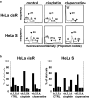
Cloperastine increases the HeLa cisR cell fraction in sub G1 phase. (a) Flow cytometric analysis of the cell cycle. Growing HeLa cisR and HeLa S cells were cultured for 3 days in their respective growth media (control) or in medium containing 1 µg/ml cisplatin or 100 µM cloperastine. The cells were then fixed, the cellular DNA stained with propidium iodide (PI), and the fluorescence intensity and frequency analyzed using a flow cytometer and plotted. Arrowheads and brackets indicate sub G1 phase (sb), G1 phase (2n), S phase (S), and G2/M phase (4n) for each cell line and treatment. Two separate experiments yielded essentially the same results. (b) Relative cell numbers in each cell cycle phase in panel a were quantified using ImageJ software and plotted as percentages of the total cells. The labels indicate sub G1 phase (sb), G1 phase (2), S phase (S), and G2/M phase (4) for each cell line and treatment.
Figure 7
Figure 7
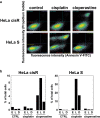
Cloperastine increases the HeLa cisR cell fraction undergoing apoptosis/cell death. (a) Flow cytometric analysis of cell death. Growing HeLa cisR and HeLa S cells were cultured for 24 h in their respective growth media (control) or in medium containing 1 µg/ml cisplatin or 100 µM cloperastine. The cells were stained with Annexin V-FITC and PI, and the intensities of the FITC and PI fluorescences were analyzed using a flow cytometer and plotted. In each panel, the bottom left section represents Annexin VLOWPILOW (live) cells, the bottom right section Annexin VHIGHPILOW cells (early phase apoptosis), the top right section Annexin VHIGHPIHIGH cells (late phase apoptosis), and the top left section Annexin VLOWPIHIGH cells (plasma membrane disintegration). (b) Relative cell numbers in each phase of apoptosis/cell death in panel a were quantified using ImageJ software and plotted as percentages of the total cells. The labels indicate early phase apoptosis (E), late phase apoptosis (L), and plasma membrane disintegration (D) for each cell line and treatment.
Figure 8
Figure 8
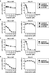
MEK and p38 MAPK signal transduction pathways are not involved in the cloperastine cytotoxicity toward HeLa cisR cells. (a,b) Effects of inhibitors of MEK on the cytotoxicity of cisplatin (a) and cloperastine (b) toward HeLa cisR and HeLa S cells. Filled circles, control; open squares, PD98059; filled triangles, U-0126. c and d, Effects of an inhibitor of p38 MAPK on the cytotoxicity of cisplatin (c) and cloperastine (d). Filled circles, control; open squares, SB203580. The results are presented as means ± S.D. of triplicate (a and b) or quadruplicate (c and d) samples. Three separate experiments yielded essentially the same results.
Figure 9
Figure 9
Weak enhancement of the cytotoxicity of H1 receptor antagonists by verapamil is not selective for HeLa cisR or HeLa S cells. HeLa cisR and HeLa S cells were cultured for 3 days with cloperastine (a), desloratadine (b), or clemastine (c) in the absence (filled circles) or presence (open squares) of 50 µM verapamil. Cell viability during the final 5 h of the culture was evaluated in WST-8 colorimetric assays. The results are presented as means ± S.D. of quadruplicate samples. Three separate experiments yielded essentially the same results. The significance of the difference between the results from control cells and verapamil-treated cells at the same concentration of the indicated compound was determined with ANOVA (***p < 0.001; **p < 0.01; *p < 0.05).
Figure 10
Figure 10
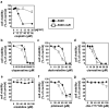
Cisplatin-resistant A549 cisR cells are selectively killed by histamine H1 receptor antagonists. A549 cisR cells were established from parental A549 lung cancer cells and subcultured in the presence of 3 µg/ml cisplatin. A549 cisR cells (open squares) and A549 cells (filled circles) were cultured for 3 days with the indicated concentrations of cisplatin (a); the histamine H1 receptor antagonist cloperastine (b), desloratadine (c), or clemastine (d); the H2 receptor antagonist nizatidine (e); H3 receptor antagonist pitolisant (f); or H4 receptor antagonist JNJ-7777120 (g). Cell viability was then assessed during the final 5 h of the culture using WST-8 colorimetric assays. The results are presented as means ± S.D. of sextuplicate (ad) or quadruplicate (eg) samples. Four separate experiments yielded essentially the same results. Note that only H1 receptor antagonists resulted in 0% cell viability. The significance of the difference between the results from A549 and A549 cisR cells at the same concentration of the indicated compound was determined with ANOVA (***p < 0.001; **p < 0.01; *p < 0.05).

Similar articles

Cited by

References

    1. Okada T, Murata K, Hirose R, Matsuda C, Komatsu T, Ikekita M, Nakawatari M, Nakayama F, Wakatsuki M, Ohno T, Kato S, Imai T, Imamura T. Upregulated expression of FGF13/FHF2 mediates resistance to platinum drugs in cervical cancer cells. Sci. Rep. 2013;3:2899. doi: 10.1038/srep02899. - DOI - PMC - PubMed
    1. Bublik DR, Bursać S, Sheffer M, Oršolić I, Shalit T, Tarcic O, Kotler E, Mouhadeb O, Hoffman Y, Fuchs G, Levin Y, Volarević S, Oren M. Regulatory module involving FGF13, miR-504, and p53 regulates ribosomal biogenesis and supports cancer cell survival. Proc. Natl. Acad. Sci. U.S.A. 2017;114:E496–E505. doi: 10.1073/pnas.1614876114. - DOI - PMC - PubMed
    1. Redmer T, Walz I, Klinger B, Khouja S, Welte Y, Schäfer R, Regenbrecht C. The role of the cancer stem cell marker CD271 in DNA damage response and drug resistance of melanoma cells. Oncogenesis. 2017;6:e291. doi: 10.1038/oncsis.2016.88. - DOI - PMC - PubMed
    1. Johnstone CN, Pattison AD, Harrison PF, Powell DR, Lock P, Ernst M, Anderson RL, Beilharz TH. FGF13 promotes metastasis of triple-negative breast cancer. Int. J. Cancer. 2020;147:230–243. doi: 10.1002/ijc.32874. - DOI - PubMed
    1. Jayasimha RD, Basha S, Kotha P, Katike U. Designing, docking and molecular dynamics simulation studies of novel cloperastine analogues as anti-allergic agents: Homology modeling and active site prediction for the human histamine H1 receptor. RSC Adv. 2020;10:4745–4754. doi: 10.1039/C9RA09245E. - DOI - PMC - PubMed

Publication types

MeSH terms

LinkOut - more resources