. 2021 Apr;19(4):688-701.
doi: 10.1158/1541-7786.MCR-20-0863. Epub 2020 Dec 22.

Identification and Characterization of Cancer Cells That Initiate Metastases to the Brain and Other Organs

Affiliations

Identification and Characterization of Cancer Cells That Initiate Metastases to the Brain and Other Organs

Anna S Berghoff et al. Mol Cancer Res. 2021 Apr.

Abstract

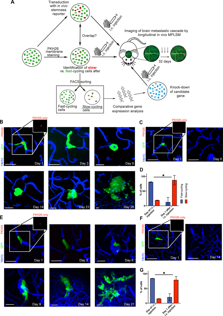
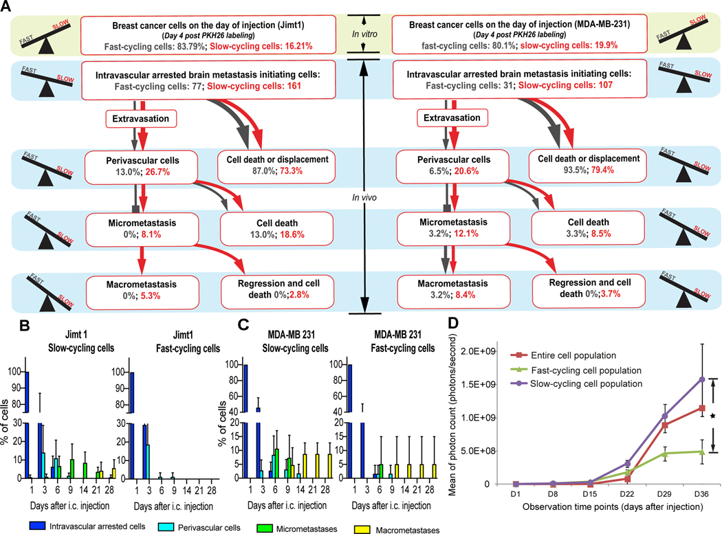
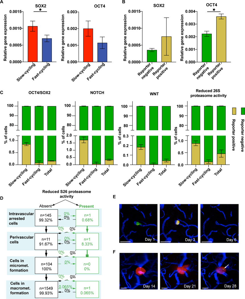
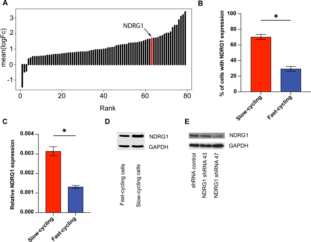
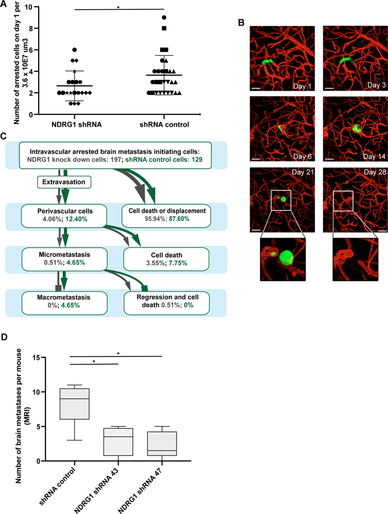
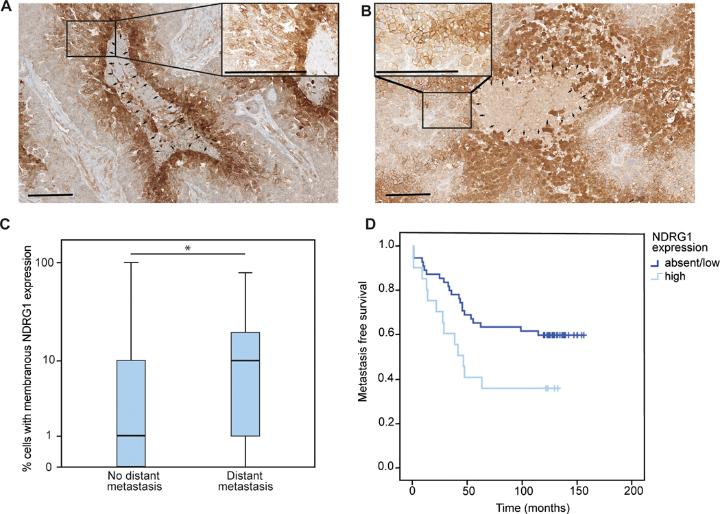
Specific biological properties of those circulating cancer cells that are the origin of brain metastases (BM) are not well understood. Here, single circulating breast cancer cells were fate-tracked during all steps of the brain metastatic cascade in mice after intracardial injection over weeks. A novel in vivo two-photon microscopy methodology was developed that allowed to determine the specific cellular and molecular features of breast cancer cells that homed in the brain, extravasated, and successfully established a brain macrometastasis. Those BM-initiating breast cancer cells (BMIC) were mainly originating from a slow-cycling subpopulation that included only 16% to 20% of all circulating cancer cells. BMICs showed enrichment of various markers of cellular stemness. As a proof of principle for the principal usefulness of this approach, expression profiling of BMICs versus non-BMICs was performed, which revealed upregulation of NDRG1 in the slow-cycling BMIC subpopulation in one BM model. Here, BM development was completely suppressed when NDRG1 expression was downregulated. In accordance, in primary human breast cancer, NDRG1 expression was heterogeneous, and high NDRG1 expression was associated with shorter metastasis-free survival. In conclusion, our data identify temporary slow-cycling breast cancer cells as the dominant source of brain and other metastases and demonstrates that this can lead to better understanding of BMIC-relevant pathways, including potential new approaches to prevent BM in patients. IMPLICATIONS: Cancer cells responsible for successful brain metastasis outgrowth are slow cycling and harbor stemness features. The molecular characteristics of these metastasis-initiating cells can be studied using intravital microscopy technology.


Figures

Figure 1:
Figure 1:. Combined in vitro / in vivo model to study the role of slow- and fast-cycling cancer cells in brain metastasis formation
(A) Experimental strategy and procedures. Different cancer cell subpopulations are labeled in vitro (fast- vs. slow-cycling cells by PKH26, and live stemness reporters), and their characteristics is further analyzed in vitro (overlap with other labels indicating a certain population or a certain relative gene expression, cell growth), and in vivo (likelihood to master all steps of the brain metastatic cascade; overall brain metastatic efficacy). Results from different brain metastatic capacities of slow- vs. fast-cycling cancer cells are then used for identification of key molecular differences of these cell populations, finally used for knock-down studies of candidate gene(s) that can be tested regarding their influence on the brain metastatic cascade. MPLSM: multiphoton laser-scanning microscopy.(B) Representative images of a slow-cycling MDA-MB-231 breast cancer cell (remaining PKH26 staining; arrow), followed by repetitive in vivo MPLSM through all steps of the brain metastatic cascade: intravascular arrest (Days 1,3 – single cells), extravasation and colonization of the perivascular niche (Day 6 – up to 5 cells), micro- (Days 14, 21 – up to 50 cells) and macrometastasis (Day 28 - > 50 cells) formation. Green, cytoplasm of GFP-positive cancer cell(s); blue, brain microvessels labeled by TRITC angiogram; red, PKH26 staining labeling slow-cycling cancer cells. scale bars: 30μm. (C) Representative images of a fast-cycling MDA-MB-231 cell (absence of PKH26 staining) through the early steps of the brain metastatic cascade (Day 1 – single cells), until its death on day 6. scale bars: 30μm. (D) Percentage of all slow-cycling and fast-cycling MDA-MB-231 breast cancer cells, in vitro at the day of intracardial injection, and in vivo one day after. At day 1, all cancer cells were still in the state of intravascular arrest. Included cells day 0 MDA-MB-231 n=784; included cells on day 1 MDA-MB-231 n=138 (p<0.001; Chi Square test). (E) Slow-cycling JIMT1 breast cancer cell mastering all steps of the brain metastatic cascade; intravascular arrest (day 1 & day 3), extravasation and colonization of the perivascular niche (day 6), micrometastasis (day 9), macrometastasis (day 14 & day 21 & day 28). Green, cancer cell(s); blue, brain microvessels. scale bars: 30μm. (F) Fast-cycling JIMT1 breast cancer cell mastering intravascular arrest (day 1) and extravasation (day 6), but disappears afterwards until day 14. (G) Percentage of all slow-cycling and fast-cycling JIMT1 breast cancer cells, in vitro at the day of intracardial injection, and in vivo one day after. At day 1, all cancer cells were still in the state of intravascular arrest. Included cells day 0: Jimt1 n=1254; included cells on day 1: Jimt1 n=238 (p<0.001; Chi Square test). B-G: data obtained by in vivo MPLSM; scale bars: 30μm. 3 replicates per experiment.
Figure 2:
Figure 2:. Slow-cycling breast cancer cells are enriched during the brain metastatic cascade
(A) Quantification of the steps of the brain metastatic cascade in slow-cycling (PKH26 positive) vs. fast-cycling (PKH26 negative) JIMT1 (n=238 tumor cells in n=4 mice) and MDA-MB-231 (n=138 tumor cells in n=4 mice) human breast cancer cells. While slow-cycling cells are only a minority of cancer cells on the injection day, this population is particularly able to master all steps of the brain metastatic cascade successfully, greatly outnumbering its fast-cycling counterparts (p<0.05; Chi Square test). Percentages are given relative to the total number of intravascular arrested cells in the slow-cycling and the fast-cycling group on day 1 after intracardial injection; extravasation: day 3–6; perivascular single cells: day 3–9; micrometastasis: day 9–14; macrometastasis: day 9–29. (B,C) Quantification of the relative tumor cell number in the JIMT1 (B) and MDA-MB-231 (C) cell lines over 28 days in vivo, depending on their cycling properties. (4 mice per group). Significant (p<0.05) differences in the successful establishment of perivascular cells, micro- and macrometastases between slow- and fast-cycling cells could be detected from day 6 on. (D) Whole-mouse imaging without the brain compartment revealed that slow-cycling JIMT1 breast cancer cells give rise to a significantly higher extracranial metastatic burden, compared to fast-cycling or unsorted control cells (n=4 mice per group; p<0.05; t test). Tumor cells were FACS-sorted after PKH26 staining, and injected intracardially. A-C: data obtained by in vivo MPLSM; scale bars: 30μm. 3 replicates per experiment.
Figure 3:
Figure 3:. Certain cancer cell stemness markers are enriched in BMICs
(A) Stemness markers in slow- vs. fast-cycling JIMT1 breast cancer cells. qPCR analysis of the stemness markers SOX2 (p=0.02) and OCT4 (p=0.06). (B) Gene expression of OCT4 (p=0.001) and SOX2 (p>0.05) in OCT4/SOX2 reporter positive cells determined by FACS sorting. (C) Slow-cycling cells reveal a marked enrichment of stemness markers compared to fast-cycling cells, as tested by overlap of PKH26 membrane staining and fluorescence signal of stemness reporter systems by FACS analysis (p<0.05). Note that marker-positive cells remain a small cancer cell subpopulation, even in the slow-cycling cells. (D) Quantification of the brain metastatic cascade of JIMT1 cells transduced with pQCXIN-ZsGreen-cODC for in vivo tracking of the tumor cell subpopulation with reduced S26 proteasome activity (n=146 tumor cells, n=4 mice) scale bars: 30μm. (E) One single tumor cell presents with low proteasome activity on day 1 after injection, but is not visible any more after extravasation. (F) One single tumor cell with low proteasome activity occurs in a macrometastasis over time (arrow). D,E,F: in vivo MPLSM; 3 replicates per experiment
Figure 4:
Figure 4:. NDRG1 is upregulated in slow-cycling JIMT1 breast cancer cells
(A) Significant (p<0.05) up-regulation of NDRG1 expression in slow-cycling JIMT1 cells compared to fast-cycling ones. NDRG1 is the 17th highest differentially expressed gene in slow-cycling Jimt1 cancer cells. For a complete gene list, see Supplementary Table 1. (B) Expression of NDRG1 in Jimt1 slow-cycling cells compared to Jimt1 fast-cycling cells in FACS analysis (p<0.01). (C) Relative NDRG1 gene expression in Jimt1 slow- versus fast cycling cells, determined by qPCR (p<0.05). (D) Higher NDRG1 protein expression in Jimt1 slow-cycling compared to fast-cycling cells analyzed with Western Blot. (E) Knock-down of NDRG1 in JIMT1 cells. 3 replicates per experiment
Figure 5:
Figure 5:. Impact of NDRG1 proficiency on brain metastases formation in vivo
(A) Impaired ability of JIMT1 NDRG1 knock-down cells to manage the first step of the brain metastatic cascade, which is intravascular arrest (p=0.047; t test); different symbols mark different animals. (B) Intravital imaging of the one JIMT1 shNDRG1 tumor cell that was most successful in mastering the first steps of the brain metastatic cascade, but still dies after day 21. Note the failure of proper perivascular niche colonization, indicated by a roundish tumor cell shape without clear orientation along the blood microvessel in the brain (compare successful brain metastasis of JIMT1, Fig. 2A) scale bars: 30μm. (C) Quantification of shNDRG1 and shRNA control breast cancer cells over the brain metastatic cascade. (n= 326 cells in n=8 animals/n=4 per group; p<0.05; Chi Square test). (D) Number of BM as measured by MRT analysis is lower in both Jimt1 NDRG1 knock-down lines compared to shRNA control cells (n=12 animals/n=4 per group; p=0.037; Kruskal Wallis test). A, B, C: in vivo MPLSM; 3 replicates per experiment
Figure 6:
Figure 6:. NDRG1 expression in human breast cancer samples correlates with metastasis formation
(A,B) Representative immunohistochemical image of NDRG1 expression in a primary breast cancer specimens (A) and a brain metastasis (B), with cyctoplasmatic NDRG1 expression around necrotic areas and unrelated membranous expression on tumor cells (median 7.5%, range 0–100%, n=74). Arrows; magnification x100 and x400; scale bar 200 μm. (C) Median NDRG1 expression in primary breast tumors of patients experiencing distant metastasis (n=35) compared to patients without distant metastases (n=39) during a follow up period of 10 years (p<0.05; Mann Whitney U test). (D) Metastasis-free survival in patients with high NDRG1 expression (n=20) in the primary breast cancer specimen, compared to patients with absent or low NDRG1 expression (n= 54; p=0.035; log rank test)

Similar articles

Cited by

References

    1. Berghoff AS, Schur S, Füreder LM, Gatterbauer B, Dieckmann K, Widhalm G, et al. Descriptive statistical analysis of a real life cohort of 2419 patients with brain metastases of solid cancers. ESMO Open 2016;1:e000024. doi:10.1136/esmoopen-2015-000024. - DOI - PMC - PubMed
    1. Kienast Y, von Baumgarten L, Fuhrmann M, Klinkert WEF, Goldbrunner R, Herms J, et al. Real-time imaging reveals the single steps of brain metastasis formation. Nat Med 2010;16:116–22. doi:10.1038/nm.2072. - DOI - PubMed
    1. Preusser M, Winkler F, Collette L, Haller S, Marreaud S, Soffietti R, et al. Trial design on prophylaxis and treatment of brain metastases: lessons learned from the EORTC Brain Metastases Strategic Meeting 2012. Eur J Cancer 2012;48:3439–47. doi:10.1016/j.ejca.2012.07.002. - DOI - PubMed
    1. Valiente M, Ahluwalia MS, Boire A, Brastianos PK, Goldberg SB, Lee EQ, et al. The Evolving Landscape of Brain Metastasis. Trends in Cancer 2018;4:176–96. doi:10.1016/j.trecan.2018.01.003. - DOI - PMC - PubMed
    1. Merlano MC, Granetto C, Fea E, Ricci V, Garrone O. Heterogeneity of colon cancer: from bench to bedside. ESMO Open 2017;2:e000218. doi:10.1136/esmoopen-2017-000218. - DOI - PMC - PubMed

Publication types