Review
. 2021 Jun;18(6):400-423.
doi: 10.1038/s41569-020-00480-6. Epub 2021 Jan 11.

Cellular and molecular pathobiology of heart failure with preserved ejection fraction

Affiliations
Review

Cellular and molecular pathobiology of heart failure with preserved ejection fraction

Sumita Mishra et al. Nat Rev Cardiol. 2021 Jun.

Erratum in

Abstract

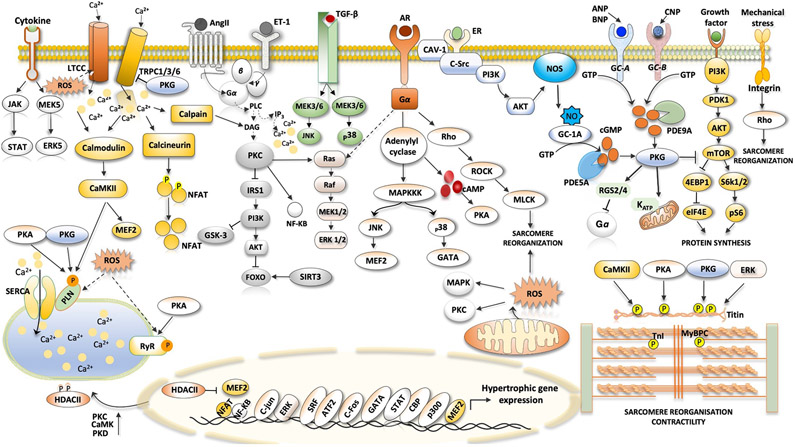
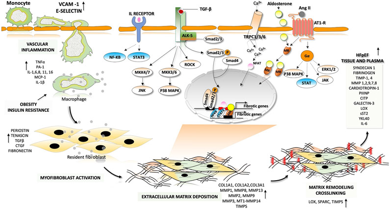
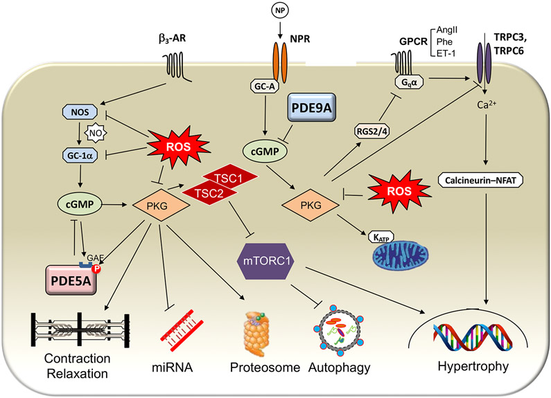
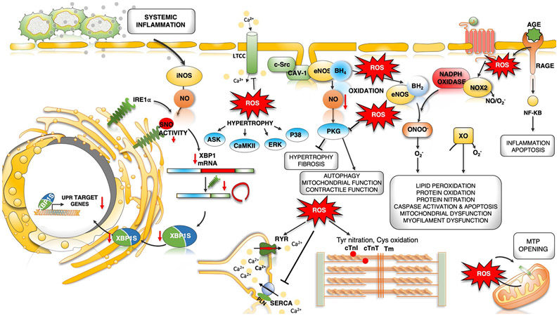
Heart failure with preserved ejection fraction (HFpEF) affects half of all patients with heart failure worldwide, is increasing in prevalence, confers substantial morbidity and mortality, and has very few effective treatments. HFpEF is arguably the greatest unmet medical need in cardiovascular disease. Although HFpEF was initially considered to be a haemodynamic disorder characterized by hypertension, cardiac hypertrophy and diastolic dysfunction, the pandemics of obesity and diabetes mellitus have modified the HFpEF syndrome, which is now recognized to be a multisystem disorder involving the heart, lungs, kidneys, skeletal muscle, adipose tissue, vascular system, and immune and inflammatory signalling. This multiorgan involvement makes HFpEF difficult to model in experimental animals because the condition is not simply cardiac hypertrophy and hypertension with abnormal myocardial relaxation. However, new animal models involving both haemodynamic and metabolic disease, and increasing efforts to examine human pathophysiology, are revealing new signalling pathways and potential therapeutic targets. In this Review, we discuss the cellular and molecular pathobiology of HFpEF, with the major focus being on mechanisms relevant to the heart, because most research has focused on this organ. We also highlight the involvement of other important organ systems, including the lungs, kidneys and skeletal muscle, efforts to characterize patients with the use of systemic biomarkers, and ongoing therapeutic efforts. Our objective is to provide a roadmap of the signalling pathways and mechanisms of HFpEF that are being characterized and which might lead to more patient-specific therapies and improved clinical outcomes.


Figures

Fig. 1 ∣
Fig. 1 ∣. Signalling pathways in cardiac hypertrophy.
Ventricular hypertrophy in heart failure with preserved ejection fraction, particularly in association with hypertension and neurohormonal stress, can involve many pathways identified in other hypertrophic syndromes. The figure shows the major pathways in cardiomyocytes that are thought to stimulate pathological muscle growth of the heart. The hormones angiotensin II (AngII), endothelin 1 (ET-1) and catecholamines bind to their cognate receptors, which are coupled to heterotrimeric G proteins to activate downstream signalling, such as the phospholipase C (PLC)–protein kinase C (PKC) axis. Activated PKC inhibits the insulin receptor substrate 1 (IRS1)–RACα serine/threonine-protein kinase (AKT)–forkhead box protein (FOXO) signalling pathway. β1-Adrenergic receptor (AR) stimulated protein kinase A (PKA) raises cytosolic Ca2+ levels by phosphorylation of Ca2+-handling proteins. Transient receptor potential channel 1 (TRPC1), TRPC3 and TRPC6 have been linked to pathological hypertrophy through elevated NFAT signalling. TRPC1 and TRPC6 are also mechanosensitive. Integrin transmembrane receptors also transduce intracellular hypertrophic signalling by activating downstream effectors such as Rho. Transforming growth factor-β (TGFβ) signalling and receptors transmitting signals through Gq-protein-coupled receptors promote activation of Rho-associated protein kinase (ROCK), extracellular-signal-regulated kinase (ERK) and p38 mitogen-activated protein kinase (MAPK), which contributes to pathological cardiac hypertrophy and fibrosis. Growth factor-mediated stimulation of mechanistic target of rapamycin (mTOR) signalling is linked to induction of protein synthesis and inhibition of autophagy. Hypertrophy is also associated with depressed cGMP–protein kinase G (PKG) signalling (both nitric oxide (NO)-mediated and natriuretic-peptide-mediated) and increased phosphodiesterase type 5A (PDE5A) and PDE9A expression. Many of these kinases can affect sarcomeric proteins, altering myofilament Ca2+ sensitivity and passive stiffness. Cytokines augment cardiac hypertrophy through their receptors (such as the IL-6 receptor). 4EBP1, eukaryotic translation initiation factor 4E-binding protein 1; AC, adenylyl cyclase; ANP, atrial natriuretic peptide; AT1R, angiotensin II receptor type 1; ATF2, cAMP-dependent transcription factor ATF2; BNP, B-type natriuretic peptide; CAMKII, Ca2+/calmodulin-dependent protein kinase II; CAV1, caveolin 1; CBP, CREB-binding protein; CNP, C-type natriuretic peptide; cTnI, cardiac troponin I; DAG, diacylglycerol; eIF4E, eukaryotic translation initiation factor 4E; ER, oestrogen receptor; FOS, proto-oncogene c-Fos; GATA4, transcription factor GATA4; HDAC, histone deacetylase; IP3, inositol trisphosphate; JAK, Janus kinase; JNK, JUN N-terminal kinase; JUN, proto-oncogene c-Jun; KATP, ATP-dependent K+ channel; LTCC, L-type Ca2+ channel; MAPKKK, mitogen-activated protein kinase kinase kinase; MEF2, myocyte enhancer factor 2; MEK, MAPK/ERK kinase; MLCK, myosin light chain kinase; MYBPC, cardiac myosin-binding protein C; NFAT, nuclear factor of activated T cells; NF-κB, nuclear factor-κB; NOS3, endothelial nitric oxide synthase; NPR-A, natriuretic peptide receptor A; NPR-B, natriuretic peptide receptor B; P, phosphate; p300, histone acetyltransferase p300; PDK1, 3-phosphoinositide-dependent protein kinase 1; PI3K, phosphoinositide 3-kinase; PKD, protein kinase D; PLN, cardiac phospholamban; pS6, ribosomal protein S6 kinase; RAF, RAF proto-oncogene serine/threonine-protein kinase; RGS2, regulator of G-protein signalling 2; RGS4, regulator of G-protein signalling 4; ROS, reactive oxygen species; RYR2, ryanodine receptor 2; S6K1, ribosomal protein S6 kinase-β1; S6K2, ribosomal protein S6 kinase-β2; SERCA2A, sarcoplasmic/endoplasmic reticulum Ca2+ ATPase; sGC, soluble guanylyl cyclase; SIRT3, mitochondrial NAD-dependent protein deacetylase sirtuin 3; SR, sarcoplasmic reticulum; SRC, proto-oncogene tyrosine-protein kinase SRC; SRF, serum response factor; STAT, signal transducer and activator of transcription; TGFBR, transforming growth factor-β receptor.
Fig. 2 ∣
Fig. 2 ∣. Fibrotic–inflammatory remodelling in heart failure with preserved ejection fraction.
In patients with heart failure with preserved ejection fraction, endothelial cells produce factors that induce inflammation and recruit monocytes for transendothelial migration. These factors include interleukins (IL-1, IL-6 and IL-8), colony-stimulating factors (granulocyte-colony stimulating factor (G-CSF), macrophage colony-stimulating factor (M-CSF) and granulocyte-macrophage colony-stimulating factor (GM-CSF) and chemotactic factors (C-C motif chemokine 2 (CCL2)). Pro-inflammatory cytokines such as tumour necrosis factor (TNF), IL-1 and IL-6 are involved in the initiation and propagation of inflammatory signals. This inflammatory signalling stimulates monocytes and endothelial cells to initiate weak interactions and the subsequent rolling of the monocytes over the endothelial cells, mediated by adhesion molecules (such as vascular cell adhesion protein 1 (VCAM1) and E-selectin). Finally, monocytes migrate through the intercellular clefts between the endothelial cells to the underlying tissue. Macrophage-derived mediators and pro-inflammatory cytokines drive the transformation of quiescent fibroblasts into proliferative and matrix-synthesizing active myofibroblasts. The communication between inflammatory cells and resident fibroblasts occurs through direct cell–cell interactions and through paracrine signalling. Sustained activation of myofibroblasts produces structural extracellular matrix (ECM) proteins and matricellular proteins. Crosslinking of collagen molecules by matrix-crosslinking enzymes, such as lysyl oxidase (LOX), prevents the enzymatic degradation of collagen, leading to an increase in collagen content and stiffness. The dynamic alterations in the composition of the ECM and the prolonged deposition of ECM modulates cardiac function. Both canonical transforming growth factor-β (TGFβ)– mothers against decapentaplegic homologue 2 (SMAD2)–SMAD3 signalling and non-canonical TGFβ signalling through Rho-associated protein kinase (ROCK), extracellular-signal-regulated kinase (ERK) and p38 contribute to the development of pathological hypertrophy and fibrosis. Phosphorylation and activation of ERK, p38, Janus kinase (JAK) and signal transducer and activator of transcription (STAT) by activation of G-protein-coupled receptors also leads to the development of fibrosis. Activation of the calcineurin–nuclear factor of activated T cells (NFAT) pathway by cardiac transient receptor potential channels (TRPCs) also has a major role in fibrotic remodelling. Mineralocorticoid receptors (MRs) are ligand-activated transcription factors that induce transcription of profibrotic genes upon aldosterone binding. AngII, angiotensin II; AT1R, angiotensin II receptor type 1; CITP, carboxy-terminal propeptide of procollagen type I; COL, collagen; CTGF, connective tissue growth factor; JNK, JUN N-terminal kinase; MAPK, mitogen-activated protein kinase; MMP, matrix metalloproteinase; MEK, MAPK/ERK kinase; NF-κB, nuclear factor-κB; P, phosphate; PIIINP, amino-terminal propeptide of procollagen type III; sST2, soluble protein ST2; TGFBR1, transforming growth factor-β receptor type 1; TIMP, metalloproteinase inhibitor; YKL40, chitinase-3-like protein 1.
Fig. 3 ∣
Fig. 3 ∣. Components of the cGMP–PKG signalling systems and their cellular effectors.
cGMP is generated by one of two different cyclases: guanylyl cyclase A (GC-A), which is coupled to the natriuretic peptide (NP) receptor (NPR), and soluble guanylyl cyclase (sGC), which is the target of nitric oxide (NO). Each relative pool of cGMP has a different primary targeting phosphodiesterase (PDE): PDE5A for GC-1α-derived cGMP and PDE9A for GC-A-derived cGMP. Increased cGMP levels in turn activate protein kinase G (PKG). PKG is the primary effector protein for many cGMP-mediated effects. Phosphorylation of sarcomeric proteins enhances relaxation and diastolic compliance and blunts β3-adrenergic receptor (β3-AR)-stimulated contractility. PKG stimulation, controlled by PDE5A but not PDE9A, reverses the abnormally altered expression of many microRNAs that is associated with hypertrophic remodelling induced by pressure stress. PKG activation also increases proteasome activity and stimulates autophagy, the latter coupled to the suppression of mechanistic target of rapamycin complex 1 (mTORC1) signalling. NP-stimulated PKG coupled to NPR-A (for which both atrial NP and B-type NP are ligands) has less of an effect on sarcomeres or microRNA levels, but augments autophagy. PKG stimulated by either NO or NPs activates regulator of G-protein signalling 2 (RGS2) and RGS4 to suppress Gq-coupled receptor signalling and phosphorylates transient receptor potential channel 3 (TRPC3) and TRPC6 to suppress hypertrophy and fibrosis. AngII, angiotensin II; ET-1, endothelin 1; GAF, GAF domain; GPCR, G-protein-coupled receptor; KATP, ATP-dependent K+ channel; NFAT, nuclear factor of activated T cells; NOS, nitric oxide synthase; P, phosphate; Phe, phenylephrine; ROS, reactive oxygen species; TSC1, hamartin; TSC2, tuberin.
Fig. 4 ∣
Fig. 4 ∣. Dysregulated oxidative and nitrosative stress in HFpEF pathogenesis.
Increased microvascular inflammation and pro-inflammatory cytokine levels result in increased expression of inducible nitric oxide synthase (NOS2) in cardiomyocytes. NOS2-derived nitric oxide (NO) mediates S-nitrosylation (SNO) of the unfolded protein response (UPR) regulator IRE1α, leading to a progressive decline in IRE1α-mediated generation of the spliced form of X-box-binding protein 1 (XBP1), known as XBP1s. Reduced XBP1s levels lead to decreased XBP1s-dependent expression of UPR target genes, compromised UPR and endoplasmic reticulum (ER) function, and prolonged ER stress. IRE1α activity and XBP1s levels are reduced in the hearts of patients with heart failure with preserved ejection fraction (HFpEF). Oxidative stress and increased reactive oxygen species (ROS) formation can directly modulate cardiac redox status by reacting with NO to decrease its bioavailability. Oxidative stress uncouples endothelial nitric oxide synthase (NOS3) by oxidation and depletion of its cofactor tetrahydrobiopterin (BH4) to dihydrobiopterin (BH2). NOS3-derived NO has antihypertrophic and antifibrotic effects primarily by activation of cGMP–protein kinase G (PKG) signalling. Uncoupled NOS3 generates oxidant species promoting protein tyrosine nitration, cysteine oxidation and lipid peroxidation, damaging proteins, lipids and DNA. Receptor-induced activation of NADPH oxidase 2 (NOX2) and mitochondrial redox mismatch are other major sources of ROS and stimulate mitochondrial transition pore (MTP) opening, Ca2+ overload and mitochondrial dysfunction. Advanced glycation end products (AGEs) that bind to cell surface receptors for AGEs (RAGEs) can stimulate NADPH oxidase, thereby increasing the production of ROS and aggravating inflammation by activation of the nuclear factor-κB (NF-κB) pathway. ROS can directly or indirectly activate various kinases, resulting in hypertrophy. Post-translational redox modifications of protein kinases (such as Ca2+/calmodulin-dependent protein kinase II (CAMKII), protein kinase A and PKG), sarcoplasmic reticulum (SR) proteins (ryanodine receptor 2 (RYR2) and sarcoplasmic/endoplasmic reticulum Ca2+ ATPase 2A (SERCA2A)) and myofilament proteins (cardiac troponin I (cTnI), cardiac troponin T (cTnT), tropomyosin (Tm) and cardiac myosin binding protein C can alter their activity and contribute to hypertrophy and altered excitation–contraction coupling. AngII, angiotensin II; ASK, apoptosis signal-regulating kinase; AT1R, angiotensin II receptor type 1; CAV1, caveolin 1; ERK, extracellular-signal-regulated kinase; LTCC, L-type Ca2+ channel; O2, superoxide anion; ONOO, peroxynitrite; PLN, cardiac phospholamban; SRC, proto-oncogene tyrosine-protein kinase SRC; XO, xanthine oxidoreductase.
Fig. 5 ∣
Fig. 5 ∣. Metabolic flexibility and HFpEF.
Heart failure involves alterations in multiple metabolic pathways that can alter mitochondrial ATP production and substrate utilization, and ultimately myocardial energy reserve and efficiency. Obesity, which is very common in patients with heart failure with preserved ejection fraction (HFpEF), induces systemic fatty acid oversupply from adipose tissue that is re-routed to peripheral organs, including the heart. Mismatch between cardiac fatty acid uptake and fatty acid β-oxidation (FAO) leads to intramyocardial accumulation of diglycerides and ceramides, uncoupling their oxidative phosphorylation. Hypoxia facilitates the metabolic shift towards glycolysis with a subsequent increase in lactate and pyruvate accumulation owing to impaired activity of pyruvate dehydrogenase (PDH). A compensatory increase in anaplerosis diverts the products of the glycolysis towards the hexosamine biosynthetic pathway and the pentose phosphate pathway. Several metabolism-modulating pharmaceuticals are being examined in HFpEF, as in other forms of heart failure. Ranolazine is a partial inhibitor of FAO, which reciprocally increases glucose oxidation and PDH activity. Etomoxir inhibits carnitine palmitoyl transferase 1 (CPT1), which controls the access of long-chain fatty acids (LCFAs) to the mitochondria for FAO. Elamipretide selectively targets the electron transport chain to increase energy efficiency. ACC, acetyl-CoA carboxylase; ACS, acetyl-CoA synthetase; AMPK, AMP-activated protein kinase; CAT, carnitine acyltransferase; CD36, fatty acid translocase; Cr, creatine; DAG, diacylglycerol; GLUT, glucose transporter; LDH, lactate dehydrogenase; LPL; lipoprotein lipase, mCK, muscle creatine kinase; PCr, phosphocreatine; PDK, pyruvate dehydrogenase kinase; PGC1α, peroxisome proliferator-activated receptor-γ coactivator 1α; Pi, inorganic phosphate; PPARα, peroxisome proliferator-activated receptor-α; SIRT1, NAD-dependent protein deacetylase sirtuin 1; TAG, triacylglycerol; TCA, tricarboxylic acid; TG, triglyceride; VLDL, very low density lipoprotein.

Similar articles

Cited by

References

    1. Gladden JD, Chaanine AH & Redfield MM Heart Failure with Preserved Ejection Fraction. Annual review of medicine 69, 65–79, doi:10.1146/annurev-med-041316-090654 (2018). - DOI - PubMed
    1. Lam CSP et al. Mortality associated with heart failure with preserved vs. reduced ejection fraction in a prospective international multi-ethnic cohort study. Eur Heart J 39, 1770–1780, doi:10.1093/eurheartj/ehy005 (2018). - DOI - PubMed
    1. Zakeri R & Cowie MR Heart failure with preserved ejection fraction: controversies, challenges and future directions. Heart 104, 377–384, doi:10.1136/heartjnl-2016-310790 (2018). - DOI - PubMed
    1. Shah SJ et al. Research Priorities for Heart Failure With Preserved Ejection Fraction: National Heart, Lung, and Blood Institute Working Group Summary. Circulation 141, 1001–1026, doi:10.1161/circulationaha.119.041886 (2020). - DOI - PMC - PubMed
    1. Dodek A, Kassebaum DG & Bristow JD Pulmonary edema in coronary-artery disease without cardiomegaly. Paradox of the stiff heart. N Engl J Med 286, 1347–1350, doi:10.1056/nejm197206222862507 (1972). - DOI - PubMed