

αα-Hub domains and intrinsically disordered proteins: A decisive combo
- PMID: 33361159
- PMCID: PMC7948954
- DOI: 10.1074/jbc.REV120.012928
αα-Hub domains and intrinsically disordered proteins: A decisive combo
Abstract
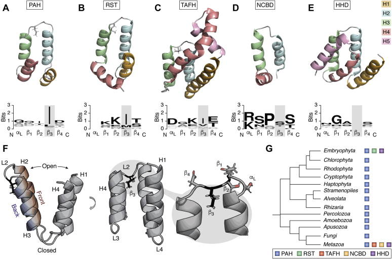
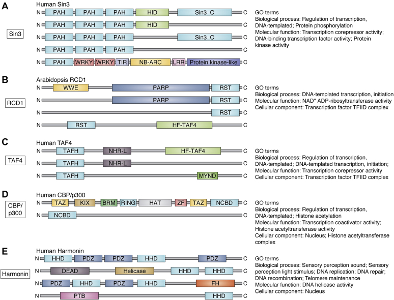
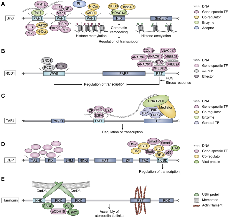
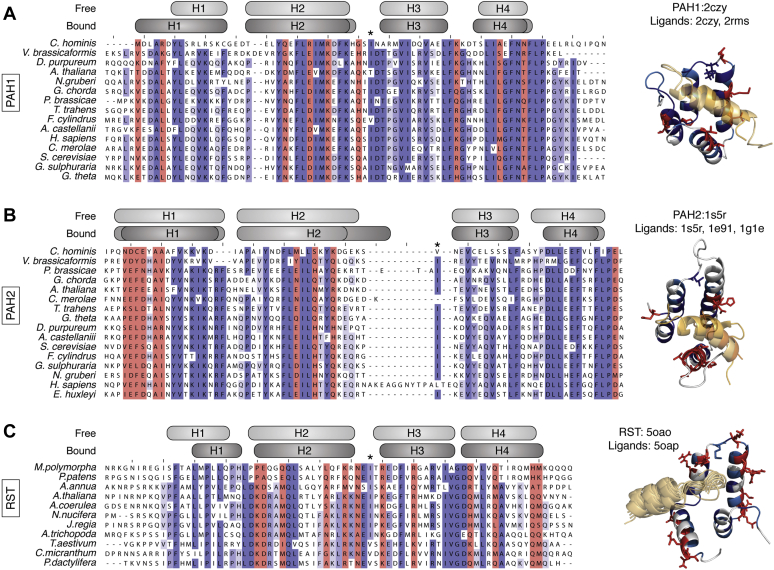
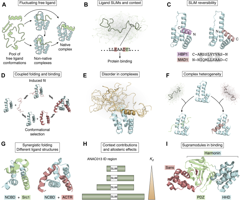
Hub proteins are central nodes in protein-protein interaction networks with critical importance to all living organisms. Recently, a new group of folded hub domains, the αα-hubs, was defined based on a shared αα-hairpin supersecondary structural foundation. The members PAH, RST, TAFH, NCBD, and HHD are found in large proteins such as Sin3, RCD1, TAF4, CBP, and harmonin, which organize disordered transcriptional regulators and membrane scaffolds in interactomes of importance to human diseases and plant quality. In this review, studies of structures, functions, and complexes across the αα-hubs are described and compared to provide a unified description of the group. This analysis expands the associated molecular concepts of "one domain-one binding site", motif-based ligand binding, and coupled folding and binding of intrinsically disordered ligands to additional concepts of importance to signal fidelity. These include context, motif reversibility, multivalency, complex heterogeneity, synergistic αα-hub:ligand folding, accessory binding sites, and supramodules. We propose that these multifaceted protein-protein interaction properties are made possible by the characteristics of the αα-hub fold, including supersite properties, dynamics, variable topologies, accessory helices, and malleability and abetted by adaptability of the disordered ligands. Critically, these features provide additional filters for specificity. With the presentations of new concepts, this review opens for new research questions addressing properties across the group, which are driven from concepts discovered in studies of the individual members. Combined, the members of the αα-hubs are ideal models for deconvoluting signal fidelity maintained by folded hubs and their interactions with intrinsically disordered ligands.
Keywords: IDP; SLiM; context; dynamics; hub; ligand binding; signaling; transcription.
Copyright © 2020 The Authors. Published by Elsevier Inc. All rights reserved.
Conflict of interest statement
Conflict of interest The authors declare that they have no conflicts of interest with the contents of this article.
Figures
Similar articles
-
αα-hub coregulator structure and flexibility determine transcription factor binding and selection in regulatory interactomes.J Biol Chem. 2022 Jun;298(6):101963. doi: 10.1016/j.jbc.2022.101963. Epub 2022 Apr 20. J Biol Chem. 2022. PMID: 35452682 Free PMC article.
-
Connecting the αα-hubs: same fold, disordered ligands, new functions.Cell Commun Signal. 2021 Jan 6;19(1):2. doi: 10.1186/s12964-020-00686-8. Cell Commun Signal. 2021. PMID: 33407551 Free PMC article.
-
Structure of Radical-Induced Cell Death1 Hub Domain Reveals a Common αα-Scaffold for Disorder in Transcriptional Networks.Structure. 2018 May 1;26(5):734-746.e7. doi: 10.1016/j.str.2018.03.013. Epub 2018 Apr 12. Structure. 2018. PMID: 29657132
-
Role of Intrinsic Protein Disorder in the Function and Interactions of the Transcriptional Coactivators CREB-binding Protein (CBP) and p300.J Biol Chem. 2016 Mar 25;291(13):6714-22. doi: 10.1074/jbc.R115.692020. Epub 2016 Feb 5. J Biol Chem. 2016. PMID: 26851278 Free PMC article. Review.
-
DSS1/Sem1, a Multifunctional and Intrinsically Disordered Protein.Trends Biochem Sci. 2016 May;41(5):446-459. doi: 10.1016/j.tibs.2016.02.004. Epub 2016 Mar 1. Trends Biochem Sci. 2016. PMID: 26944332 Review.
Cited by
-
Molecular switching in transcription through splicing and proline-isomerization regulates stress responses in plants.Nat Commun. 2024 Jan 18;15(1):592. doi: 10.1038/s41467-024-44859-2. Nat Commun. 2024. PMID: 38238333 Free PMC article.
-
Evolutionary fine-tuning of residual helix structure in disordered proteins manifests in complex structure and lifetime.Commun Biol. 2023 Jan 18;6(1):63. doi: 10.1038/s42003-023-04445-6. Commun Biol. 2023. PMID: 36653471 Free PMC article.
-
αα-hub coregulator structure and flexibility determine transcription factor binding and selection in regulatory interactomes.J Biol Chem. 2022 Jun;298(6):101963. doi: 10.1016/j.jbc.2022.101963. Epub 2022 Apr 20. J Biol Chem. 2022. PMID: 35452682 Free PMC article.
-
Enrichment patterns of intrinsic disorder in proteins.Biophys Rev. 2022 Nov 19;14(6):1487-1493. doi: 10.1007/s12551-022-01016-7. eCollection 2022 Dec. Biophys Rev. 2022. PMID: 36659984 Free PMC article. Review.
-
Harmonin homology domain-mediated interaction of RTEL1 helicase with RPA and DNA provides insights into its recruitment to DNA repair sites.Nucleic Acids Res. 2024 Feb 9;52(3):1450-1470. doi: 10.1093/nar/gkad1208. Nucleic Acids Res. 2024. PMID: 38153196 Free PMC article.
References
-
- Cumberworth A., Lamour G., Babu M.M., Gsponer J. Promiscuity as a functional trait: intrinsically disordered regions as central players of interactomes. Biochem. J. 2013;454:361–369. - PubMed
-
- Han J.J., Bertin N., Hao T., Goldberg D.S., Berriz G.F., Zhang L.V., Dupuy D., Walhout A.J.M., Cusick M.E., Roth F.P., Vidal M. Evidence for dynamically organized modularity in the yeast protein-protein interaction network. Nature. 2004;430:88–93. - PubMed
Publication types
MeSH terms
Substances
LinkOut - more resources
Full Text Sources
Other Literature Sources
Research Materials
