. 2021 Feb 15;81(4):885-897.
doi: 10.1158/0008-5472.CAN-19-3219. Epub 2020 Dec 22.

Cancer-Induced Muscle Wasting Requires p38β MAPK Activation of p300

Affiliations

Cancer-Induced Muscle Wasting Requires p38β MAPK Activation of p300

Thomas K Sin et al. Cancer Res. .

Abstract

Cancer-associated cachexia, characterized by muscle wasting, is a lethal metabolic syndrome without defined etiology or established treatment. We previously found that p300 mediates cancer-induced muscle wasting by activating C/EBPβ, which then upregulates key catabolic genes. However, the signaling mechanism that activates p300 in response to cancer is unknown. Here, we show that upon cancer-induced activation of Toll-like receptor 4 in skeletal muscle, p38β MAPK phosphorylates Ser-12 on p300 to stimulate C/EBPβ acetylation, which is necessary and sufficient to cause muscle wasting. Thus, p38β MAPK is a central mediator and therapeutic target of cancer-induced muscle wasting. In addition, nilotinib, an FDA-approved kinase inhibitor that preferentially binds p38β MAPK, inhibited p300 activation 20-fold more potently than the p38α/β MAPK inhibitor, SB202190, and abrogated cancer cell-induced muscle protein loss in C2C12 myotubes without suppressing p38α MAPK-dependent myogenesis. Systemic administration of nilotinib at a low dose (0.5 mg/kg/day, i.p.) in tumor-bearing mice not only alleviated muscle wasting, but also prolonged survival. Therefore, nilotinib appears to be a promising treatment for human cancer cachexia due to its selective inhibition of p38β MAPK. SIGNIFICANCE: These findings demonstrate that prevention of p38β MAPK-mediated activation of p300 by the FDA-approved kinase inhibitor, nilotinib, ameliorates cancer cachexia, representing a potential therapeutic strategy against this syndrome.


Figures

Figure 1.
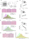
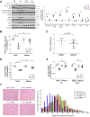
Figure 1.. Site-specific phosphorylation of 300 on Ser-12 induced by LLC is necessary and sufficient for the activation C/EBPβ and muscle wasting.
(A) LLC induces p300 phosphorylation on Ser-12 and this reaction is critical to p300-mediated acetylation and activation of C/EBPβ. C2C12 myoblasts were transfected with a plasmid encoding phosphorylation-defective mutant of p300, p300-S12A or p300-S89A, or empty vector as control in 3 independent experiments. After differentiation, myotubes were treated with LLC cell conditioned medium (LCM) or conditioned medium of non-tumorigenic NL20 cells for 2 h. Cell lysates were analyzed by Western blotting. (B) Overexpression of p300-S12A attenuates muscle weight loss in LLC tumor-bearing mice. Tibialis muscle (TA) of LLC tumor-bearing mice was transfected with the p300-S12A-encoding plasmid (n = 5). The contralateral TA was transfected with empty vector as control. After the development of cachexia, TA muscles were collected and weighed on day 21. Overexpression of p300-S12A was confirmed by Western blotting analysis against p300. (C) Overexpression of p300-S12A prevents the loss of myofiber mass in LLC tumor-bearing mice. H&E-stained TA cross sections of (B) were analyzed for myofiber cross-sectional area. (D) Overexpression of p300-S12A in TA attenuates muscle weight loss in KPC tumor-bearing mice (n = 5). (E) Overexpression of p300-S12A in TA prevents the loss of myofiber mass of KPC tumor-bearing mice (n = 5). (F) Overexpression of p300-S12D in TA causes loss of muscle weight in cancer-free mice (n = 5). (G) Overexpression of p300-S12D in TA causes loss of myofiber mass in cancer-free mice (n = 5). * indicates a statistically significant difference (p < 0.05) determined by one-way ANOVA (A), paired Student t-test (B, D and F) or Chi-square test (C, E and G).
Figure 2.
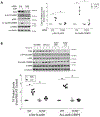
Figure 2.. TLR4 mediates LLC-induced p300 phosphorylation on Ser-12.
(A) TLR4 is critical to p300 phosphorylation on Ser-12 and C/EBPβ acetylation on Lys-39 in LCM-treated myotubes. C2C12 myoblasts were transfected with siRNA specific for TLR4 or scrambled control siRNA (n = 3). Differentiated myotubes were treated with LCM for 2 h. Cell lysates were analyzed by Western blotting. (B) TLR4 is required for p300 phosphorylation on Ser-12 and C/EBPβ acetylation on Lys-39 in the muscle of LLC tumor-bearing mice. Wild type (WT) and TLR4−/− mice were implanted with LLC cells or injected with PBS as control (n = 6). In 21 days TA muscle were analyzed by Western blotting. * indicates a statistically significant difference (p < 0.05) determined by one-way ANOVA.
Figure 3.
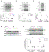
Figure 3.. Multiple types of cancer induce p300 phosphorylation on Ser-12 through p38β MAPK.
(A) LCM-induced Ser-12 phosphorylation of p300 in myotubes requires p38β MAPK. C2C12 myoblasts were transfected with siRNA specific for p38α MAPK or p38β MAPK, or scrambled control (n = 3). Differentiated myotubes were treated with LCM for 2 h, the cell lysates were analyzed by western blotting as indicated. (B) KCM induces Ser-12 phosphorylation of p300 in myotubes in a p38β MAPK-dependent manner. C2C12 myotubes transfected with p38β MAPK-specific or scrambled siRNA were treated with KCM for 2 h. The cell lysates were analyzed by western blotting as indicated (n = 3). (C) Overexpression of constitutively active p38β MAPK is sufficient to phosphorylate Ser-12 of p300. C2C12 myoblasts were transfected with a plasmid encoding either a constitutively active mutant of p38α MAPK or p38β MAPK. Differentiated myotubes were analyzed by Western blotting as indicated (n = 3). (D) LCM stimulates an interaction between p300 and p38β MAPK, but not p38α MAPK. C2C12 myotubes were transfected with either p38α or p38β MAPK-specific siRNAs, and then treated with LCM for 2 h. Immunoprecipitation of p38 MAPK from the cell lysates was performed with pre-immune IgG as a control. Precipitates were then analyzed by Western blotting to verify the knockdown effects and coprecipitation of p300. (E) Activation of p300 in the TA muscle of LLC tumor-bearing mice requires p38β MAPK. LLC cells were implanted to p38β MAPK muscle-specific knockout mice (p38β mKO) and p38β MAPK-floxed mice (p38βf/f) (n = 6). In 21 days mice were euthanized and TA lysates were analyzed by Western blotting as indicated. * signifies a statistically significant difference (p < 0.05) determined by one-way ANOVA.
Figure 4.
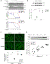
Figure 4.. Selective inhibition of p38β MAPK by nilotinib abrogates LLC-induced myotube catabolism without inhibiting myogenesis.
(A) Nilotinib is ~20-fold more potent than SB202190 in the inhibition of LLC-induced activation of p300. C2C12 myotubes were pre-treated with either nilotinib or SB202190 (SB) at indicated doses for 30 mins followed by 2 h of LCM treatment (n = 3). Activation of p300 and C/EBPβ were analyzed by Western blotting as indicated. (B) Nilotinib abrogates LCM-induced loss of MHC in myotubes. Myotubes pre-treated with 500 nM of nilotinib or DMSO were incubated with LCM for 72 hrs. Cell lysates were analyzed for MHC levels by Western blotting (n = 3). (C) Nilotinib abolishes LLC-induced loss of myotube mass. Myotubes treated as described in (B) were subjected to immunofluorescence staining of MHC. Diameter of myotubes was measured. (D) Nilotinib does not suppress myoblast differentiation at the dose that antagonizes LLC-induced myotube atrophy. C2C12 myoblasts were cultured with differentiation medium containing 10 μM SB202190 (SB), 500 nM nilotinib or DMSO for the indicated time periods. MHC content in the cell lysates were analyzed by Western blotting at indicated times (n = 3). * signifies a statistically significant difference (p < 0.05) determined by one-way ANOVA.
Figure 5.
Figure 5.. Nilotinib ameliorates muscle wasting by attenuating the catabolic response in LLC tumor-bearing mice.
Nilotinib (0.5 mg/kg/day) or DMSO was administered intraperitoneally to mice 7 days after LLC cell implantation for 14 days. Mice were euthanized on day 21. The tumor was isolated and weighed, and then the net body weight was measured. Muscle samples were collected immediately for analyses of muscle wasting (n = 5). (A) Nilotinib abrogates p300 activation and catabolic response in LLC tumor-bearing mice. Catabolic markers in TA muscle lysates were analyzed by Western blotting. (B) Nilotinib prevents body weight loss in LLC-bearing mice. (C) Nilotinib does not affect LLC tumor growth. (D) Nilotinib preserves skeletal muscle function in LLC tumor-bearing mice. Grip strength was measured on the day of euthanasia. (E) Nilotinib attenuates skeletal muscle weight loss in LLC tumor-bearing mice. (F) Nilotinib protects against the loss of myofiber mass in LLC tumor-bearing mice. H&E-stained TA cross sections were analyzed for the myofiber cross-sectional area. * signifies a statistically significant difference (p < 0.05) determined by one-way ANOVA (A-E) or Chi-square test (F).
Figure 6.
Figure 6.. Nilotinib prolongs survival of mice bearing pancreatic cancer by impeding the development of muscle wasting.
Five days after orthotopic implantation of KPC cells to mice, nilotinib (0.5 mg/kg/day) or DMSO were administered intraperitoneally until all tumor-bearing mice reached predetermined end point (n = 7). (A) Nilotinib prolongs survival of mice bearing KPC tumor. Survival of KPC tumor-bearing mice was recorded and analyzed using the Kaplan–Meier survival curve. (B) Nilotinib impedes the loss of muscle strength in mice bearing KPC tumor. Forelimb grip strength of the mice was monitored over the course of the survival study. Data were analyzed by 2-way ANOVA. * signifies a statistically significant difference (p < 0.05).
Figure 7.
Figure 7.
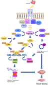
A graphic illustration of the central role of p38β MAKP in mediating muscle catabolism in response to TLR4 activation based on data from the current and previous studies.

Similar articles

Cited by

References

    1. Fearon KC, Glass DJ, Guttridge DC. Cancer cachexia: mediators, signaling, and metabolic pathways. Cell metabolism 2012;16:153–66 - PubMed
    1. Fearon K, Strasser F, Anker SD, Bosaeus I, Bruera E, Fainsinger RL, et al.Definition and classification of cancer cachexia: an international consensus. Lancet Oncol 2011;12:489–95 - PubMed
    1. Andreyev HJ, Norman AR, Oates J, Cunningham D. Why do patients with weight loss have a worse outcome when undergoing chemotherapy for gastrointestinal malignancies? Eur J Cancer 1998;34:503–9 - PubMed
    1. Baracos VE, Martin L, Korc M, Guttridge DC, Fearon KCH. Cancer-associated cachexia. Nat Rev Dis Primers 2018;4:17105 - PubMed
    1. Siegel RL, Miller KD, Jemal A. Cancer statistics, 2020. CA Cancer J Clin 2020;70:7–30 - PubMed

Publication types

MeSH terms

Substances