

Cryo-electron tomography structure of Arp2/3 complex in cells reveals new insights into the branch junction
- PMID: 33353942
- PMCID: PMC7755917
- DOI: 10.1038/s41467-020-20286-x
Cryo-electron tomography structure of Arp2/3 complex in cells reveals new insights into the branch junction
Abstract
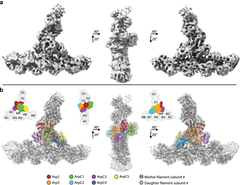
The actin-related protein (Arp)2/3 complex nucleates branched actin filament networks pivotal for cell migration, endocytosis and pathogen infection. Its activation is tightly regulated and involves complex structural rearrangements and actin filament binding, which are yet to be understood. Here, we report a 9.0 Å resolution structure of the actin filament Arp2/3 complex branch junction in cells using cryo-electron tomography and subtomogram averaging. This allows us to generate an accurate model of the active Arp2/3 complex in the branch junction and its interaction with actin filaments. Notably, our model reveals a previously undescribed set of interactions of the Arp2/3 complex with the mother filament, significantly different to the previous branch junction model. Our structure also indicates a central role for the ArpC3 subunit in stabilizing the active conformation.
Conflict of interest statement
The authors declare no competing interests.
Figures
Similar articles
-
Mechanism of actin filament branch formation by Arp2/3 complex revealed by a high-resolution cryo-EM structureof the branch junction.Proc Natl Acad Sci U S A. 2022 Dec 6;119(49):e2206722119. doi: 10.1073/pnas.2206722119. Epub 2022 Nov 29. Proc Natl Acad Sci U S A. 2022. PMID: 36442092 Free PMC article.
-
Structure of Arp2/3 complex at a branched actin filament junction resolved by single-particle cryo-electron microscopy.Proc Natl Acad Sci U S A. 2022 May 31;119(22):e2202723119. doi: 10.1073/pnas.2202723119. Epub 2022 May 27. Proc Natl Acad Sci U S A. 2022. PMID: 35622886 Free PMC article.
-
The structural basis of actin filament branching by the Arp2/3 complex.J Cell Biol. 2008 Mar 10;180(5):887-95. doi: 10.1083/jcb.200709092. Epub 2008 Mar 3. J Cell Biol. 2008. PMID: 18316411 Free PMC article.
-
Peering deeply inside the branch.J Cell Biol. 2008 Mar 10;180(5):853-5. doi: 10.1083/jcb.200802062. Epub 2008 Mar 3. J Cell Biol. 2008. PMID: 18316414 Free PMC article. Review.
-
Signalling to actin assembly via the WASP (Wiskott-Aldrich syndrome protein)-family proteins and the Arp2/3 complex.Biochem J. 2004 May 15;380(Pt 1):1-17. doi: 10.1042/BJ20040176. Biochem J. 2004. PMID: 15040784 Free PMC article. Review.
Cited by
-
Actin-Associated Proteins and Small Molecules Targeting the Actin Cytoskeleton.Int J Mol Sci. 2022 Feb 14;23(4):2118. doi: 10.3390/ijms23042118. Int J Mol Sci. 2022. PMID: 35216237 Free PMC article. Review.
-
Mitochondrial Dynamics: Working with the Cytoskeleton and Intracellular Organelles to Mediate Mechanotransduction.Aging Dis. 2023 Oct 1;14(5):1511-1532. doi: 10.14336/AD.2023.0201. Aging Dis. 2023. PMID: 37196113 Free PMC article. Review.
-
CK-666 and CK-869 differentially inhibit Arp2/3 iso-complexes.EMBO Rep. 2024 Aug;25(8):3221-3239. doi: 10.1038/s44319-024-00201-x. Epub 2024 Jul 15. EMBO Rep. 2024. PMID: 39009834 Free PMC article.
-
Specialization of actin isoforms derived from the loss of key interactions with regulatory factors.EMBO J. 2022 Mar 1;41(5):e107982. doi: 10.15252/embj.2021107982. Epub 2022 Feb 18. EMBO J. 2022. PMID: 35178724 Free PMC article.
-
Mechanism of actin filament branch formation by Arp2/3 complex revealed by a high-resolution cryo-EM structureof the branch junction.Proc Natl Acad Sci U S A. 2022 Dec 6;119(49):e2206722119. doi: 10.1073/pnas.2206722119. Epub 2022 Nov 29. Proc Natl Acad Sci U S A. 2022. PMID: 36442092 Free PMC article.
References
Publication types
MeSH terms
Substances
LinkOut - more resources
Full Text Sources
Other Literature Sources
Molecular Biology Databases
Miscellaneous
