

Organotypic Modeling of the Tumor Landscape
- PMID: 33330508
- PMCID: PMC7732527
- DOI: 10.3389/fcell.2020.606039
Organotypic Modeling of the Tumor Landscape
Abstract
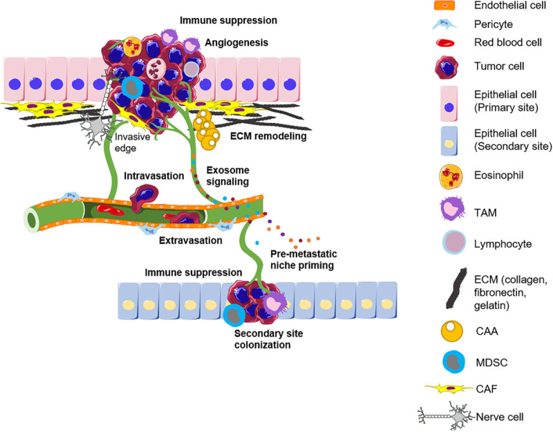
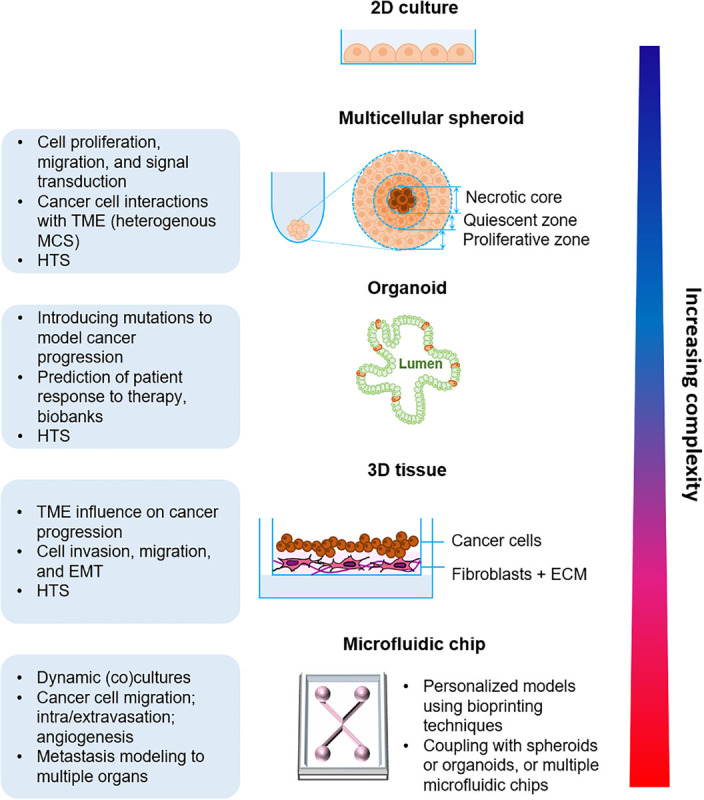
Cancer is a complex disease and it is now clear that not only epithelial tumor cells play a role in carcinogenesis. The tumor microenvironment is composed of non-stromal cells, including endothelial cells, adipocytes, immune and nerve cells, and a stromal compartment composed of extracellular matrix, cancer-associated fibroblasts and mesenchymal cells. Tumorigenesis is a dynamic process with constant interactions occurring between the tumor cells and their surroundings. Even though all connections have not yet been discovered, it is now known that crosstalk between actors of the microenvironment drives cancer progression. Taking into account this complexity, it is important to develop relevant models to study carcinogenesis. Conventional 2D culture models fail to represent the entire tumor microenvironment properly and the use of animal models should be decreased with respect to the 3Rs rule. To this aim, in vitro organotypic models have been significantly developed these past few years. These models have different levels of complexity and allow the study of tumor cells alone or in interaction with the microenvironment actors during the multiple stages of carcinogenesis. This review depicts recent insights into organotypic modeling of the tumor and its microenvironment all throughout cancer progression. It offers an overview of the crosstalk between epithelial cancer cells and their microenvironment during the different phases of carcinogenesis, from the early cell autonomous events to the late metastatic stages. The advantages of 3D over classical 2D or in vivo models are presented as well as the most promising organotypic models. A particular focus is made on organotypic models used for studying cancer progression, from the less complex spheroids to the more sophisticated body-on-a-chip. Last but not least, we address the potential benefits of these models in personalized medicine which is undoubtedly a domain paving the path to new hopes in terms of cancer care and cure.
Keywords: 3D model; cancer; metastasis; therapies; tumor dissemination; tumor microenvironment.
Copyright © 2020 Haykal, Nahmias, Varon and Martin.
Figures
Similar articles
-
Organotypic 3D Models of the Ovarian Cancer Tumor Microenvironment.Cancers (Basel). 2018 Aug 9;10(8):265. doi: 10.3390/cancers10080265. Cancers (Basel). 2018. PMID: 30096959 Free PMC article. Review.
-
Fabrication Method of a High-Density Co-Culture Tumor-Stroma Platform to Study Cancer Progression.Methods Mol Biol. 2021;2258:241-255. doi: 10.1007/978-1-0716-1174-6_16. Methods Mol Biol. 2021. PMID: 33340365
-
Development of primary human pancreatic cancer organoids, matched stromal and immune cells and 3D tumor microenvironment models.BMC Cancer. 2018 Mar 27;18(1):335. doi: 10.1186/s12885-018-4238-4. BMC Cancer. 2018. PMID: 29587663 Free PMC article.
-
Modeling the epithelial-mesenchymal transition process in a 3D organotypic cervical neoplasia.Acta Biomater. 2020 Oct 15;116:209-222. doi: 10.1016/j.actbio.2020.09.006. Epub 2020 Sep 7. Acta Biomater. 2020. PMID: 32911106
-
The matrix environmental and cell mechanical properties regulate cell migration and contribute to the invasive phenotype of cancer cells.Rep Prog Phys. 2019 Jun;82(6):064602. doi: 10.1088/1361-6633/ab1628. Epub 2019 Apr 4. Rep Prog Phys. 2019. PMID: 30947151 Review.
Cited by
-
Extracellular Vesicles in Breast Cancer: From Intercellular Communication to Therapeutic Opportunities.Pharmaceutics. 2024 May 14;16(5):654. doi: 10.3390/pharmaceutics16050654. Pharmaceutics. 2024. PMID: 38794316 Free PMC article. Review.
-
Organoids at the PUB: The Porcine Urinary Bladder Serves as a Pancreatic Niche for Advanced Cancer Modeling.Adv Healthc Mater. 2022 Jun;11(11):e2102345. doi: 10.1002/adhm.202102345. Epub 2022 Feb 18. Adv Healthc Mater. 2022. PMID: 35114730 Free PMC article.
-
Exploring New Dimensions of Tumor Heterogeneity: The Application of Single Cell Analysis to Organoid-Based 3D In Vitro Models.Adv Healthc Mater. 2023 Oct;12(26):e2300903. doi: 10.1002/adhm.202300903. Epub 2023 Aug 27. Adv Healthc Mater. 2023. PMID: 37589373 Free PMC article. Review.
-
Tumor spheroids and organoids as preclinical model systems.Med Genet. 2021 Dec 3;33(3):229-234. doi: 10.1515/medgen-2021-2093. eCollection 2021 Sep. Med Genet. 2021. PMID: 38835698 Free PMC article.
-
Toward improved models of human cancer: Two perspectives.APL Bioeng. 2021 Feb 19;5(1):010402. doi: 10.1063/5.0042324. eCollection 2021 Mar. APL Bioeng. 2021. PMID: 33644625 Free PMC article. No abstract available.
References
-
- Alimperti S., Mirabella T., Bajaj V., Polacheck W., Pirone D. M., Duffield J., et al. (2017). Three-dimensional biomimetic vascular model reveals a RhoA, Rac1, and N-cadherin balance in mural cell–endothelial cell-regulated barrier function. Proc. Natl. Acad. Sci. U.S.A. 114 8758–8763. 10.1073/pnas.1618333114 - DOI - PMC - PubMed
Publication types
LinkOut - more resources
Full Text Sources
Miscellaneous