. 2020 Dec 15;29(158):200001.
doi: 10.1183/16000617.0001-2020. Print 2020 Dec 31.

Lung involvement in monogenic interferonopathies

Affiliations

Lung involvement in monogenic interferonopathies

Salvatore Cazzato et al. Eur Respir Rev. .

Abstract

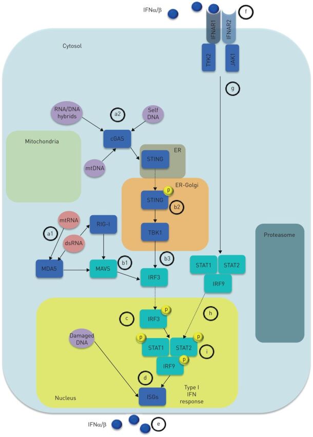
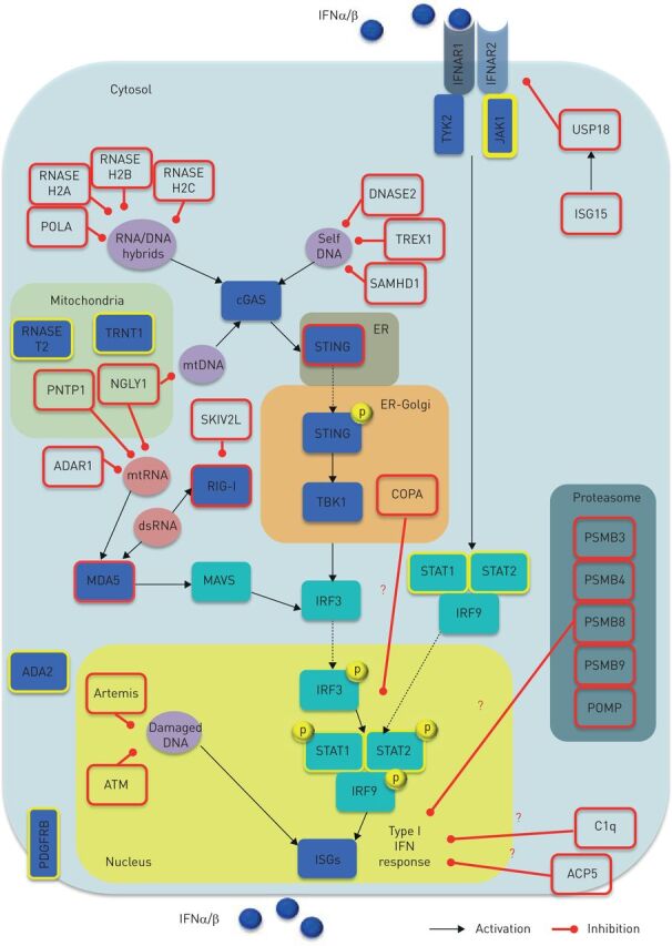
Monogenic type I interferonopathies are inherited heterogeneous disorders characterised by early onset of systemic and organ specific inflammation, associated with constitutive activation of type I interferons (IFNs). In the last few years, several clinical reports identified the lung as one of the key target organs of IFN-mediated inflammation. The major pulmonary patterns described comprise children's interstitial lung diseases (including diffuse alveolar haemorrhages) and pulmonary arterial hypertension but diagnosis may be challenging. Respiratory symptoms may be either mild or absent at disease onset and variably associated with systemic or organ specific inflammation. In addition, associated extrapulmonary clinical features may precede lung function impairment by years, and patients may display severe/endstage lung involvement, although this may be clinically hidden during the long-term disease course. Conversely, a few cases of atypical severe lung involvement at onset have been reported without clinically manifested extrapulmonary signs. Hence, a multidisciplinary approach involving pulmonologists, paediatricians and rheumatologists should always be considered when a monogenic interferonopathy is suspected. Pulmonologists should also be aware of the main pattern of presentation to allow prompt diagnosis and a targeted therapeutic strategy. In this regard, promising therapeutic strategies rely on Janus kinase-1/2 (JAK-1/2) inhibitors blocking the type I IFN-mediated intracellular cascade.


Conflict of interest statement

Conflict of interest: S. Cazzato has nothing to disclose. Conflict of interest: A. Omenetti has nothing to disclose. Conflict of interest: C. Ravaglia has nothing to disclose. Conflict of interest: V. Poletti has nothing to disclose.

Figures

FIGURE 1
FIGURE 1
Type I interferon (IFN) response. Any nuclear cell is capable of producing type I IFNs in response to viral-derived or endogenous nucleic acids, sensed by distinct cytosolic PPRs, that signal through cytosolic adapters and downstream signals causing activation of IRFs. IRFs translocate to the nucleus and induce ISG expression resulting in type I IFN response and IFN production. a1) Detection of cytosolic dsRNA and mtRNA largely relies on members of the RLR family including MDA5 (also known as IFN-induced helicase C domain containing protein 1, IFIH1) and RIG-I. b1) MDA5 and RIG-I mediate RNA sensing through the cytosolic adapter MAVS/IPS1. Thus, upon engaging nucleic acids, RLRs undergo conformational changes thereby allowing interaction with their adaptor proteins leading to activation of IRFs that c) translocate to the nucleus thus inducing d) IFNαβ transcription. Less defined is the DNA sensing machinery. a2) Cytoplasmic dsDNA seems to interact with cGAS, leading to the production of cGAMP, which eventually engages the adapter molecule STING (also known as TMEM173) STING, located in the ER. b2) Once activated, STING translocates to the ER–Golgi compartment, where the signal is propagated through the phosphorylation of the TBK1. b3) Finally, TBK1 leads to c) IRF-3 activation that d) induces type I IFN response following IRF-3 nuclear translocation. Once released, e) type I IFNs (blue circles) may act via autocrine/paracrine manner f) by binding a single heterodimeric type I IFN transmembrane receptor composed of the subunits IFNAR1 and IFNAR2. Upon engaging one of the IFNR subunits, type I IFNs cause dimerisation of IFNAR1 and IFNAR2, leading to g) activation of the JAKs: TYK2 and JAK-1. Activated TYK2 and JAK1 h) drive the phosphorylation and subsequent translocation to the cell nucleus of the STAT family members (i.e. STAT1 and STAT2) and IRF9, resulting in formation of STAT1-STAT2-IRF9 ternary complex ISGF3. The final outcome is type I IFN response refuelling, consisting of ISG expression and IFN production. cGAS: cyclic GMP-AMP synthase; dsDNA: double-stranded DNA; ER: endothelial reticulum; IFNAR1: IFNα receptor-1; IFNAR2: IFNα receptor-2; ISG: IFN stimulated gene; IPS1: interferon-β promoter stimulator 1; IRF: IFN regulatory factor; JAK-1: Janus kinase-1; JAK: Janus kinase; MAVS: mitochondrial antiviral signalling; MDA5: melanoma differentiation-associated protein 5; mtDNA: mitochondrial DNA; mtRNA: mitochondrial RNA; PPR: pattern recognition receptor; RIG-I: retinoic acid-inducible gene I; RLR: RIG-I-like receptors; STAT: signal trasducer activator of transcription; STING: stimulator of interferon genes; TBK1, TANK-binding kinase 1; TYK2: tyrosine-protein kinase 2.
FIGURE 2
FIGURE 2
Type I interferon (IFN) activity regulation and monogenic interferonopathies. Both inappropriate activation (e.g. triggered by self-nucleic acids) and impaired negative regulation of the type I IFN system can give rise to interferonopathies (red border boxes). Nucleic acid-driven inflammation may originate either from disruption in the sensing machinery or PPR downstream mediators, as well as impairment of their processing, metabolism (including autophagy) and repair. In turn, aberrant activity of deoxyribonuclease, ribonuclease, DNA polymerase, RNA helicase, RNA editing machinery, polynucleotide phosphorylase, N-deglycosylation, dsDNA break repairs and ER-localised UPR may account for overproduction of type I IFNs. Furthermore, aberrancies in the proteasome component have been proposed to induce type I IFN signalling through indirect effects upon nucleic acid species processing. Overall, aberrant type I IFN enhancement may be due to either: 1) abnormal stimulation (e.g. increased accumulation or change in composition of endogenous nucleic acids); 2) aberrant sensing (e.g. constitutive activation or enhanced sensitivity of PPRs, leading to a change in threshold at which endogenous nucleic acids are sensed); 3) perpetuated activation (e.g. increased sensitivity or constitutive activation of IFN-inducing mediators other than PPRs); or 4) impaired negative regulation (e.g. unrestrained signalling due to defective negative feedback). A key negative regulator of this loop is represented by the ISG15, which causes induction of USP18 that, in turn, inhibits IFNAR2 activity, thus providing negative feedback to restrain an appropriate type I IFN pathway activity. Genes causing monogenic interferonopathies are depicted by red border boxes. Red question marks indicate unknown mechanisms. Yellow border boxes indicate genes suggested to cause type I IFN signalling aberrancies, but putative mechanisms are provisional. Black arrows indicate activation; red lines address inhibitory activity. ACP5: acid phosphatase 5; ADA2: adenosine deaminase 2; ADAR1: adenosine deaminase, RNA-specific, 1; ATM: ataxia-telangiectasia mutated gene; C1q: complement component C1Q; cGAS: cyclic GMP-AMP synthase; COPA: coatomer protein complex, subunit-α; dsRNA: double-stranded RNA; ER: endothelial reticulum; IFNAR1: IFNα receptor-1; IFNAR2: IFNα receptor-2; ISG: IFN stimulated gene; IRF: IFN regulatory factor; JAK-1: Janus kinase-1; JAKs: Janus kinases; MAVS: mitochondrial antiviral signalling; MDA5: melanoma differentiation-associated protein 5; mtRNA: mitochondrial RNA; NGLY1: N-glycanase 1; PDGFRB: platelet-derived growth factor receptor β; PNTP1: polyribonucleotide nucleotidyltransferase 1; POLA: POLA DNA polymerase-α; POMP: proteasome maturation protein; PSMB: proteasome subunit β; RIG-I: retinoic acid-inducible gene I; SKIV2L: superkiller viralicidic activity 2-like alias SK12 like RNA helicase; STAT: signal trasducer activator of transcription; STING: stimulator of interferon genes; TBK1, TANK-binding kinase 1; TRNT1: tRNA nucleotidyl transferase 1; TYK2: tyrosine-protein kinase 2; UPR: unfolded protein response; USP18: ubiquitin-specific peptidase 18.
FIGURE 3
FIGURE 3
Patterns of lung involvement in monogenic interferonopathies. interstitial lung disease (ILD), diffuse alveolar haemorrhage (DAH) and pulmonary arterial hypertension (PAH) as major patterns of lung manifestation in monogenic interferonopathies. SAVI, COPA, Aicardi–Goutières syndrome, DNAse II deficiency and CANDLE/PRASS are depicted by specific coloured boxes, listing putative genes so far identified in patients displaying severe pulmonary manifestation during monogenic interferonopathies. CANDLE: chronic atypical dermatosis with lipodystrophy and elevated temperatures; COPA: coatomer protein complex, subunit-α; PRASS: proteasome-associated auto-inflammatory syndrome; SAVI: STING-associated vasculopathy with onset in infancy; STING: stimulator of interferon genes.

Similar articles

  • Overview of STING-Associated Vasculopathy with Onset in Infancy (SAVI) Among 21 Patients.
    Frémond ML, Hadchouel A, Berteloot L, Melki I, Bresson V, Barnabei L, Jeremiah N, Belot A, Bondet V, Brocq O, Chan D, Dagher R, Dubus JC, Duffy D, Feuillet-Soummer S, Fusaro M, Gattorno M, Insalaco A, Jeziorski E, Kitabayashi N, Lopez-Corbeto M, Mazingue F, Morren MA, Rice GI, Rivière JG, Seabra L, Sirvente J, Soler-Palacin P, Stremler-Le Bel N, Thouvenin G, Thumerelle C, Van Aerde E, Volpi S, Willcocks S, Wouters C, Breton S, Molina T, Bader-Meunier B, Moshous D, Fischer A, Blanche S, Rieux-Laucat F, Crow YJ, Neven B. Frémond ML, et al. J Allergy Clin Immunol Pract. 2021 Feb;9(2):803-818.e11. doi: 10.1016/j.jaip.2020.11.007. Epub 2020 Nov 18. J Allergy Clin Immunol Pract. 2021. PMID: 33217613
  • [When to consider type I interferonopathy in adulthood?].
    David C, Frémond ML. David C, et al. Rev Med Interne. 2022 Jun;43(6):347-355. doi: 10.1016/j.revmed.2021.11.003. Epub 2022 Feb 15. Rev Med Interne. 2022. PMID: 35177256 French.
  • Lung Inflammation in STING-Associated Vasculopathy with Onset in Infancy (SAVI).
    David C, Frémond ML. David C, et al. Cells. 2022 Jan 18;11(3):318. doi: 10.3390/cells11030318. Cells. 2022. PMID: 35159128 Free PMC article. Review.
  • JAK1/2 inhibition with baricitinib in the treatment of autoinflammatory interferonopathies.
    Sanchez GAM, Reinhardt A, Ramsey S, Wittkowski H, Hashkes PJ, Berkun Y, Schalm S, Murias S, Dare JA, Brown D, Stone DL, Gao L, Klausmeier T, Foell D, de Jesus AA, Chapelle DC, Kim H, Dill S, Colbert RA, Failla L, Kost B, O'Brien M, Reynolds JC, Folio LR, Calvo KR, Paul SM, Weir N, Brofferio A, Soldatos A, Biancotto A, Cowen EW, Digiovanna JJ, Gadina M, Lipton AJ, Hadigan C, Holland SM, Fontana J, Alawad AS, Brown RJ, Rother KI, Heller T, Brooks KM, Kumar P, Brooks SR, Waldman M, Singh HK, Nickeleit V, Silk M, Prakash A, Janes JM, Ozen S, Wakim PG, Brogan PA, Macias WL, Goldbach-Mansky R. Sanchez GAM, et al. J Clin Invest. 2018 Jul 2;128(7):3041-3052. doi: 10.1172/JCI98814. Epub 2018 Jun 11. J Clin Invest. 2018. PMID: 29649002 Free PMC article.
  • The role of interferons type I, II and III in myositis: A review.
    Bolko L, Jiang W, Tawara N, Landon-Cardinal O, Anquetil C, Benveniste O, Allenbach Y. Bolko L, et al. Brain Pathol. 2021 May;31(3):e12955. doi: 10.1111/bpa.12955. Brain Pathol. 2021. PMID: 34043262 Free PMC article. Review.

Cited by

References

    1. Crow YJ. Type I interferonopathies: a novel set of inborn errors of immunity. Ann N Y Acad Sci 2011; 1238: 91–98. doi:10.1111/j.1749-6632.2011.06220.x - DOI - PubMed
    1. Crow YJ. Type I interferonopathies: Mendelian type I interferon up-regulation. Curr Opin Immunol 2015; 32: 7–12. doi:10.1016/j.coi.2014.10.005 - DOI - PubMed
    1. Rodero MP, Crow YJ. Type I interferon-mediated monogenic autoinflammation: the type I interferonopathies, a conceptual overview. J Exp Med 2016; 213: 2527–2538. doi:10.1084/jem.20161596 - DOI - PMC - PubMed
    1. Eleftheriou D, Brogan PA. Genetic interferonopathies: an overview. Best Pract Res Clin Rheumatol 2017; 31: 441–459. doi:10.1016/j.berh.2017.12.002 - DOI - PubMed
    1. Crow YJ, Lebon P, Casanova JL, et al. . A brief historical perspective on the pathological consequences of excessive type I interferon exposure in vivo. J Clin Immunol 2018; 38: 694–698. doi:10.1007/s10875-018-0543-6 - DOI - PubMed

Substances