Shift-field refinement of macromolecular atomic models
- PMID: 33263325
- PMCID: PMC7709196
- DOI: 10.1107/S2059798320013170
Shift-field refinement of macromolecular atomic models
Abstract
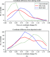
The aim of crystallographic structure solution is typically to determine an atomic model which accurately accounts for an observed diffraction pattern. A key step in this process is the refinement of the parameters of an initial model, which is most often determined by molecular replacement using another structure which is broadly similar to the structure of interest. In macromolecular crystallography, the resolution of the data is typically insufficient to determine the positional and uncertainty parameters for each individual atom, and so stereochemical information is used to supplement the observational data. Here, a new approach to refinement is evaluated in which a `shift field' is determined which describes changes to model parameters affecting whole regions of the model rather than individual atoms only, with the size of the affected region being a key parameter of the calculation which can be changed in accordance with the resolution of the data. It is demonstrated that this approach can improve the radius of convergence of the refinement calculation while also dramatically reducing the calculation time.
Keywords: computational methods; low resolution; refinement.
open access.
Figures


Similar articles
-
Macromolecular refinement by model morphing using non-atomic parameterizations.Acta Crystallogr D Struct Biol. 2018 Feb 1;74(Pt 2):125-131. doi: 10.1107/S205979831701350X. Epub 2018 Feb 1. Acta Crystallogr D Struct Biol. 2018. PMID: 29533238 Free PMC article.
-
Neutron crystallographic refinement with REFMAC5 from the CCP4 suite.Acta Crystallogr D Struct Biol. 2023 Dec 1;79(Pt 12):1056-1070. doi: 10.1107/S2059798323008793. Epub 2023 Nov 3. Acta Crystallogr D Struct Biol. 2023. PMID: 37921806 Free PMC article.
-
Structure Refinement at Atomic Resolution.Methods Mol Biol. 2017;1607:549-563. doi: 10.1007/978-1-4939-7000-1_22. Methods Mol Biol. 2017. PMID: 28573588 Review.
-
Low Resolution Refinement of Atomic Models Against Crystallographic Data.Methods Mol Biol. 2017;1607:565-593. doi: 10.1007/978-1-4939-7000-1_23. Methods Mol Biol. 2017. PMID: 28573589 Review.
-
Apparent instability of crystallographic refinement in the presence of disordered model fragments and upon insufficiently restrained model geometry.Acta Crystallogr D Biol Crystallogr. 2011 Nov;67(Pt 11):966-72. doi: 10.1107/S090744491103914X. Epub 2011 Oct 19. Acta Crystallogr D Biol Crystallogr. 2011. PMID: 22101823
Cited by
-
MUT-7 exoribonuclease activity and localization are mediated by an ancient domain.Nucleic Acids Res. 2024 Aug 27;52(15):9076-9091. doi: 10.1093/nar/gkae610. Nucleic Acids Res. 2024. PMID: 39188014 Free PMC article.
-
AlphaFold predictions are valuable hypotheses and accelerate but do not replace experimental structure determination.Nat Methods. 2024 Jan;21(1):110-116. doi: 10.1038/s41592-023-02087-4. Epub 2023 Nov 30. Nat Methods. 2024. PMID: 38036854 Free PMC article.
-
An intact S-layer is advantageous to Clostridioides difficile within the host.PLoS Pathog. 2023 Jun 29;19(6):e1011015. doi: 10.1371/journal.ppat.1011015. eCollection 2023 Jun. PLoS Pathog. 2023. PMID: 37384772 Free PMC article.
-
The CCP4 suite: integrative software for macromolecular crystallography.Acta Crystallogr D Struct Biol. 2023 Jun 1;79(Pt 6):449-461. doi: 10.1107/S2059798323003595. Epub 2023 May 30. Acta Crystallogr D Struct Biol. 2023. PMID: 37259835 Free PMC article.
-
Improved AlphaFold modeling with implicit experimental information.Nat Methods. 2022 Nov;19(11):1376-1382. doi: 10.1038/s41592-022-01645-6. Epub 2022 Oct 20. Nat Methods. 2022. PMID: 36266465 Free PMC article.
References
-
- Agarwal, R., Lifchitz, A. & Dodson, E. (1981). In Proceedings of the CCP4 Study Weekend. Refinement of Protein Structures, edited by P. A. Machin, J. W. Campbell & M. Elder. Warrington: Daresbury Laboratory.
-
- Blanc, E., Roversi, P., Vonrhein, C., Flensburg, C., Lea, S. M. & Bricogne, G. (2004). Acta Cryst. D60, 2210–2221. - PubMed
MeSH terms
Substances
Grants and funding
LinkOut - more resources
Full Text Sources

