Arginine off-kilter: guanidinium is not as planar as restraints denote
- PMID: 33263321
- PMCID: PMC7709202
- DOI: 10.1107/S2059798320013534
Arginine off-kilter: guanidinium is not as planar as restraints denote
Abstract
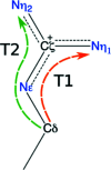
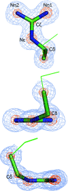
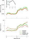
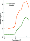
Crystallographic refinement of macromolecular structures relies on stereochemical restraints to mitigate the typically poor data-to-parameter ratio. For proteins, each amino acid has a unique set of geometry restraints which represent stereochemical information such as bond lengths, valence angles, torsion angles, dihedrals and planes. It has been shown that the geometry in refined structures can differ significantly from that present in libraries; for example, it was recently reported that the guanidinium moiety in arginine is not symmetric. In this work, the asymmetry of the Nϵ-Cζ-Nη1 and Nϵ-Cζ-Nη2 valence angles in the guanidinium moiety is confirmed. In addition, it was found that the Cδ atom can deviate significantly (more than 20°) from the guanidinium plane. This requires the relaxation of the planar restraint for the Cδ atom, as it otherwise causes the other atoms in the group to compensate by distorting the guanidinium core plane. A new set of restraints for the arginine side chain have therefore been formulated, and are available in the software package Phenix, that take into account the asymmetry of the group and the planar deviation of the Cδ atom. This is an example of the need to regularly revisit the geometric restraint libraries used in macromolecular refinement so that they reflect the best knowledge of the structural chemistry of their components available at the time.
Keywords: arginine; chemical restraints; guanidine; macromolecular refinement; planarity.
open access.
Figures


Similar articles
-
Accurate macromolecular crystallographic refinement: incorporation of the linear scaling, semiempirical quantum-mechanics program DivCon into the PHENIX refinement package.Acta Crystallogr D Biol Crystallogr. 2014 May;70(Pt 5):1233-47. doi: 10.1107/S1399004714002260. Epub 2014 Apr 26. Acta Crystallogr D Biol Crystallogr. 2014. PMID: 24816093 Free PMC article.
-
Geometry of guanidinium groups in arginines.Protein Sci. 2016 Sep;25(9):1753-6. doi: 10.1002/pro.2970. Epub 2016 Jul 4. Protein Sci. 2016. PMID: 27326702 Free PMC article.
-
Conformation-dependent restraints for polynucleotides: the sugar moiety.Nucleic Acids Res. 2020 Jan 24;48(2):962-973. doi: 10.1093/nar/gkz1122. Nucleic Acids Res. 2020. PMID: 31799624 Free PMC article.
-
An introduction to stereochemical restraints.Acta Crystallogr D Biol Crystallogr. 2007 Jan;63(Pt 1):58-61. doi: 10.1107/S090744490604604X. Epub 2006 Dec 13. Acta Crystallogr D Biol Crystallogr. 2007. PMID: 17164527 Free PMC article. Review.
-
Pound-wise but penny-foolish: How well do micromolecules fare in macromolecular refinement?Structure. 2003 Sep;11(9):1051-9. doi: 10.1016/s0969-2126(03)00186-2. Structure. 2003. PMID: 12962624 Review.
Cited by
-
Revisiting the concept of peptide bond planarity in an iron-sulfur protein by neutron structure analysis.Sci Adv. 2022 May 20;8(20):eabn2276. doi: 10.1126/sciadv.abn2276. Epub 2022 May 20. Sci Adv. 2022. PMID: 35594350 Free PMC article.
-
Ten things I `hate' about refinement.Acta Crystallogr D Struct Biol. 2021 Dec 1;77(Pt 12):1497-1515. doi: 10.1107/S2059798321011700. Epub 2021 Nov 30. Acta Crystallogr D Struct Biol. 2021. PMID: 34866607 Free PMC article.
-
Evidence of Gas Phase Glucosyl Transfer and Glycation in the CID/HCD-Spectra of S-Glucosylated Peptides.Int J Mol Sci. 2024 Jul 8;25(13):7483. doi: 10.3390/ijms25137483. Int J Mol Sci. 2024. PMID: 39000590 Free PMC article.
-
In situ ligand restraints from quantum-mechanical methods.Acta Crystallogr D Struct Biol. 2023 Feb 1;79(Pt 2):100-110. doi: 10.1107/S2059798323000025. Epub 2023 Jan 20. Acta Crystallogr D Struct Biol. 2023. PMID: 36762856 Free PMC article.
-
AQuaRef: Machine learning accelerated quantum refinement of protein structures.bioRxiv [Preprint]. 2024 Jul 21:2024.07.21.604493. doi: 10.1101/2024.07.21.604493. bioRxiv. 2024. PMID: 39071315 Free PMC article. Preprint.
References
-
- Beyer, H. (1981). Biom. J. 23, 413–414.
-
- Brünger, A. T. (1992). X-PLOR Version 3.1. A System for X-ray Crystallography and NMR. New Haven: Yale University Press.
-
- Bruno, I. J., Cole, J. C., Edgington, P. R., Kessler, M., Macrae, C. F., McCabe, P., Pearson, J. & Taylor, R. (2002). Acta Cryst. B58, 389–397. - PubMed
MeSH terms
Substances
Grants and funding
LinkOut - more resources
Full Text Sources

