. 2020 Nov 27;6(48):eabe1508.
doi: 10.1126/sciadv.abe1508. Print 2020 Nov.

Inactivation-mimicking block of the epithelial calcium channel TRPV6

Affiliations

Inactivation-mimicking block of the epithelial calcium channel TRPV6

Rajesh Bhardwaj et al. Sci Adv. .

Abstract

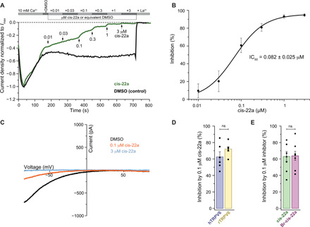
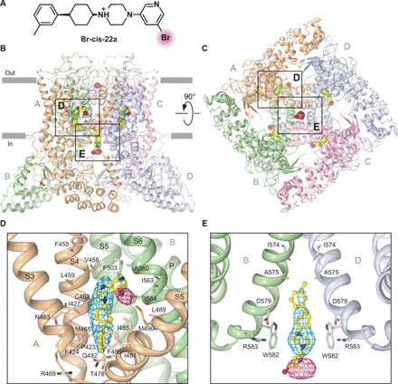
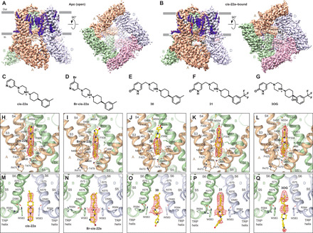
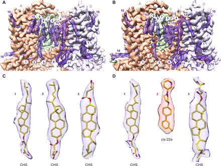
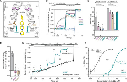
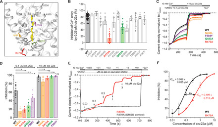
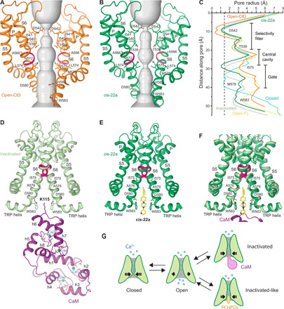
Epithelial calcium channel TRPV6 plays vital roles in calcium homeostasis, and its dysregulation is implicated in multifactorial diseases, including cancers. Here, we study the molecular mechanism of selective nanomolar-affinity TRPV6 inhibition by (4-phenylcyclohexyl)piperazine derivatives (PCHPDs). We use x-ray crystallography and cryo-electron microscopy to solve the inhibitor-bound structures of TRPV6 and identify two types of inhibitor binding sites in the transmembrane region: (i) modulatory sites between the S1-S4 and pore domains normally occupied by lipids and (ii) the main site in the ion channel pore. Our structural data combined with mutagenesis, functional and computational approaches suggest that PCHPDs plug the open pore of TRPV6 and convert the channel into a nonconducting state, mimicking the action of calmodulin, which causes inactivation of TRPV6 channels under physiological conditions. This mechanism of inhibition explains the high selectivity and potency of PCHPDs and opens up unexplored avenues for the design of future-generation biomimetic drugs.


Figures

Fig. 1
Fig. 1. Functional characterization of TRPV6 inhibition by PCHPDs.
(A) Representative whole-cell currents recorded from HEK 293 cells expressing hTRPV6 and normalized to their maximal value. Throughout the experiment, 10 mM Ca2+ solution was exchanged to the same solution containing the consecutively added dimethyl sulfoxide (DMSO), increasing concentrations of cis-22a (0.01 to 3 μM), and followed by La3+, as indicated by the gray bars. The arrows show time points when cis-22a was added. (B) Dose-response curve for hTRPV6 inhibition by cis-22a with calculated IC50 (means ± SEM; Hill coefficient, 1.25; n = 7). The inhibition was calculated relative to the current in DMSO. (C) Representative I-V curves before (DMSO) and after addition of cis-22a (0.1 and 3 μM). (D) Inhibition of hTRPV6- and rTRPV6-mediated currents (means ± SEM and individual values) by 0.1 μM cis-22a. (E) Inhibition of hTRPV6-mediated current (means ± SEM and individual values) by cis-22a and Br-cis-22a at 0.1 μM concentration.
Fig. 2
Fig. 2. Crystal structure of inhibitor-bound rTRPV6.
(A) Chemical structure of the inhibitor Br-cis-22a, with the bromine atom highlighted. (B and C) Side (B) and bottom (C) views of the rTRPV6 tetramer, with each subunit shown in different color. Molecules of Br-cis-22a are shown in yellow. Mesh shows omit (2.5σ, blue) and anomalous difference (3.0σ, pink) electron density. Boxed are regions expanded in (D) and (E). (D and E) Close-up views of the lipid-binding site 2 (LBS-2) (D) and the pore binding site (E). Only two of four TRPV6 subunits are shown in (E), with the front and back subunits removed for clarity.
Fig. 3
Fig. 3. Cryo-EM structures of hTRPV6 in the absence and presence of different PCHPD inhibitors.
(A and B) Side and bottom views of cryo-EM density for hTRPV6 in the absence of inhibitors (A) and in the presence of cis-22a (B), with each subunit shown in a different color and the inhibitor shown in red. Boxed regions in (B) are expanded in (H) and (M). (C to G) Chemical structures of different PCHPD inhibitors. (H to Q) Close-up views of the inhibitor-bound LBS-2 (H to L) and the inhibitor-bound pore binding site (M to Q). In (M) to (Q), only two of four TRPV6 subunits are shown, with the front and back subunits removed for clarity. Red mesh shows densities for different PCHPD inhibitors.
Fig. 4
Fig. 4. Nonprotein densities surrounding the TM domain of TRPV6.
(A and B) Side views of cryo-EM density for the TM domain of hTRPV6 in the absence of inhibitors (A) and in the presence of cis-22a (B). Protein density is nontransparent, with each hTRPV6 subunit shown in a different color. Fourteen well-resolved nonprotein densities per subunit of hTRPV6 are transparent, colored purple (lipids), or colored red (cis-22a) and fitted with CHS [sites 1 to 3 in (A) and sites 1 and 3 in (B)], cis-22a [site 2 in (B)], phosphatidylcholine (sites 4 and 11), or acyl chains (sites 5 to 10 and 12 to 14), shown as sticks. (C and D) Close-up views of density for sites 1 to 3 in the absence of inhibitors (A) and in the presence of cis-22a (B).
Fig. 5
Fig. 5. Functional analysis of the PCHPD pore binding site.
(A) Schematic view of the PCHPD pore binding site in hTRPV6 bound to cis-22a (yellow), with the surrounding residues shown as sticks. (B) Average percentage inhibition of Cd2+ entry into HEK 293 cells transiently expressing wild-type hTRPV6, I575A, and D580N pore mutants in response to a 10-min application of 0.25 μM cis-22a, measured using the FLIPR assay (n = 6; means ± SD). (C) Average amplitude of the whole-cell current (means ± SEM) recorded from HEK 293 cells expressing wild-type hTRPV6 and mutant channels I575A, D580N, D580K, and W583F, normalized to the current amplitude in DMSO (before application of cis-22a). Throughout the experiment, 10 mM Ca2+ solution was exchanged to the same solution containing the consecutively added DMSO, cis-22a (0.1 and 10 μM), and followed by Ca2+-free solution containing La3+, as indicated by the gray bars. (D) Percent inhibition of wild-type and mutant hTRPV6-mediated currents (means ± SEM and individual values) by 0.1 μM (left) and 10 μM (right) cis-22a. (E) Representative whole-cell currents recorded from HEK 293 cells expressing W583F mutant and normalized to the current amplitude in DMSO. Throughout the experiment, 10 mM Ca2+ solution was exchanged to the same solution containing the consecutively added DMSO, increasing concentrations of cis-22a (0.3 to 100 μM), and followed by La3+, as indicated by the gray bars. The arrows show time points when cis-22a was added. (F) Dose-response curves with calculated IC50 for wild-type (means ± SEM; Hill coefficient, 1.25; n = 7) and W583F mutant (means ± SEM; Hill coefficient, 1.5; n = 7) hTRPV6 channels.
Fig. 6
Fig. 6. Functional analysis of the LBS-2.
(A) Schematic view of the LBS-2 in hTRPV6 bound to cis-22a (yellow), with the surrounding residues shown as sticks. (B) Average percentage inhibition of Cd2+ entry into HEK 293 cells transiently expressing wild type hTRPV6 and LBS-2 mutants in response to 10-min application of 0.25 μM cis-22a, measured using the FLIPR assay (n ≥ 6 except for L460F where n = 3; means ± SD). Statistically significant difference in inhibition of the mutant compared to the wild-type channels is indicated by “*” (P < 0.001). (C) Averaged amplitude of whole-cell currents (means ± SEM) recorded from HEK 293 cells expressing wild-type hTRPV6 and mutant channels N464L, R470A, Q483A, and F504Y, normalized to the current amplitude in DMSO (before application of cis-22a). Throughout the experiment, 10 mM Ca2+ solution was exchanged to the same solution containing the consecutively added DMSO, cis-22a (0.1 and 10 μM), and followed by Ca2 +–free solution containing La3+, as indicated by the gray bars. (D) Percent inhibition of wild-type and mutant hTRPV6-mediated currents (means ± SEM and individual values) by 0.1 (left) and 10 (right) μM cis-22a. (E) Representative whole-cell currents recorded from HEK 293 cells expressing R470A mutant and normalized to the current amplitude in DMSO. Throughout the experiment, 10 mM Ca2+ solution was exchanged to the same solution containing the consecutively added DMSO, increasing concentration of cis-22a (0.03 to 10 μM), and followed by La3+, as indicated by the gray bars. The arrows show time points when cis-22a was added. (F) Dose-response curves for wild-type and R470A mutant hTRPV6 channels fitted by the logistic equation (means ± SEM; Hill coefficient, 1.25 and 0.98 for wild type and R470A, respectively).
Fig. 7
Fig. 7. Comparison of TRPV6 structures in the open state, inhibited by cis-22a and inactivated by CaM.
(A and B) Ion conduction pathway (gray) in the absence of inhibitors (A) and in the presence of cis-22a (B), with residues lining the selectivity filter and around the gate shown as sticks. Only two of four subunits are shown, with the front and back subunits removed for clarity. The π-bulge in the middle of the S6 helices is colored in pink. (C) Pore radius calculated using HOLE (62) for hTRPV6-CtD in the absence of inhibitors (orange) and in the presence of cis-22a (dark green) and for the hTRPV6-FL in the closed (cyan, hTRPV6-R470E; PDB ID: 6BOA), open (yellow, hTRPV6; PDB ID: 6BO8), and inactivated (light green, hTRPV6-CaM; PDB ID: 6E2F) states. (D to F) Pore domains in hTRPV6 structures inactivated by CaM (D), bound to cis-22a (E), and their superposition (F). Only two of four hTRPV6 subunits are shown, with the front and back subunits removed for clarity. Molecules of cis-22a and CaM are colored yellow and purple, respectively. Calcium ions are shown as blue spheres. Side chains of residues lining the binding sites for cis-22a, CaM and calcium, and CaM residue K115 are shown as sticks. The π-bulge in the middle of the S6 helices is colored in pink. (G) Cartoon illustrating the mechanism of TRPV6 inhibition by PCHPDs.

Similar articles

Cited by

References

    1. Clapham D. E., TRP channels as cellular sensors. Nature 426, 517–524 (2003). - PubMed
    1. Nilius B., Szallasi A., Transient receptor potential channels as drug targets: From the science of basic research to the art of medicine. Pharmacol. Rev. 66, 676–814 (2014). - PubMed
    1. Yelshanskaya M. V., Nadezhdin K. D., Kurnikova M. G., Sobolevsky A. I., Structure and function of the calcium-selective TRP channel TRPV6. J. Physiol., 10.1113/JP279024 (2020). - DOI - PMC - PubMed
    1. Bianco S. D., Peng J. B., Takanaga H., Suzuki Y., Crescenzi A., Kos C. H., Zhuang L., Freeman M. R., Gouveia C. H., Wu J., Luo H., Mauro T., Brown E. M., Hediger M. A., Marked disturbance of calcium homeostasis in mice with targeted disruption of the Trpv6 calcium channel gene. J. Bone Miner. Res. 22, 274–285 (2007). - PMC - PubMed
    1. Suzuki Y., Kovacs C. S., Takanaga H., Peng J. B., Landowski C. P., Hediger M. A., Calcium channel TRPV6 is involved in murine maternal-fetal calcium transport. J. Bone Miner. Res. 23, 1249–1256 (2008). - PMC - PubMed

MeSH terms

LinkOut - more resources