. 2021 Jan 1:452:247-264.
doi: 10.1016/j.neuroscience.2020.11.013. Epub 2020 Nov 24.

Lmx1a and Lmx1b are Redundantly Required for the Development of Multiple Components of the Mammalian Auditory System

Affiliations

Lmx1a and Lmx1b are Redundantly Required for the Development of Multiple Components of the Mammalian Auditory System

Victor V Chizhikov et al. Neuroscience. .

Abstract

The inner ear, projections, and brainstem nuclei are essential components of the auditory and vestibular systems. It is believed that the evolution of complex systems depends on duplicated sets of genes. The contribution of duplicated genes to auditory or vestibular system development, however, is poorly understood. We describe that Lmx1a and Lmx1b, which originate from the invertebrate Lmx1b-like gene, redundantly regulate development of multiple essential components of the mammalian auditory/vestibular systems. Combined, but not individual, loss of Lmx1a/b eliminated the auditory inner ear organ of Corti (OC) and disrupted the spiral ganglion, which was preceded by a diminished expression of their critical regulator Pax2. Innervation of the remaining inner ear vestibular organs revealed unusual sizes or shapes and was more affected compared to Lmx1a/b single-gene mutants. Individual loss of Lmx1a/b genes did not disrupt brainstem auditory nuclei or inner ear central projections. Combined loss of Lmx1a/b, however, eliminated excitatory neurons in cochlear/vestibular nuclei, and also the expression of a master regulator Atoh1 in their progenitors in the lower rhombic lip (RL). Finally, in Lmx1a/b double mutants, vestibular afferents aberrantly projected to the roof plate. This phenotype was associated with altered expression of Wnt3a, a secreted ligand of the Wnt pathway that regulates pathfinding of inner ear projections. Thus, Lmx1a/b are redundantly required for the development of the mammalian inner ear, inner ear central projections, and cochlear/vestibular nuclei.

Keywords: LIM-homeodomain transcription factors; auditory system; ear central projections; hindbrain; neurosensory development; roof plate.


Conflict of interest statement

Conflict of Interests: The authors declare no competing interests.

Figures

Fig. 1.
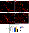
Fig. 1.. Auditory and vestibular inner ear innervation abnormalities of Lmx1a/b DKO embryos at e12.5.
Whole inner ear innervation in e12.5 embryos revealed by anti-acetylated α-Tubulin immunohistochemistry (green) combined with Hoechst nuclear stain (lilac). Posterior canal crista (PC) innervation appeared as a single branch in control (A), Lmx1a KO (B), and Lmx1b KO (C) embryos but consisted of two branches in Lmx1a/b DKO embryos (D). Innervation toward anterior/horizontal cristae (labeled as AC – anterior crista) and cochlear duct (CD) was clearly detectable in control, Lmx1a KO and Lmx1b KO embryos (A-C). Innervation toward AC was poorly developed and no CD innervation was detected in Lmx1a/b DKO embryos (D). VG - vestibular ganglion. Orientation is given in panel A. Scale bar: 100 μm.
Fig. 2.
Fig. 2.. Auditory and vestibular inner ear innervation abnormalities of Lmx1a/b DKO embryos at e14.5.
Anti-acetylated α-Tubulin immunostaining (green) in the inner ear of indicated genotypes. Some samples were co-stained with Hoechst nuclear stain (lilac) to visualize tissue. Panels A-D show innervation in the entire inner ear at low magnification; panels A’-D’ show auditory inner ear innervation at higher magnification; panels A”- D” and A”’- D”’ show vestibular inner ear innervation at higher magnification. (A-D’) Organ of Cori (OC) innervation was clearly detectable in control, Lmx1a KO and Lmx1b KO embryos (A-C, A’-C’) but no cochlear innervation was found in Lmx1a/b DKO embryos, in which a cochlear sac (COSac) formed (D, D’). Consistent with previously described partial transformation of the OC into an irregular vestibular epithelium (NICHOLS et al., 2008), innervation to an abnormal cochlear-vestibular organ (CVO) was detected in Lmx1a KO mice (B, B’). (A”-D”) In Lmx1a/b DKO mice, branches innervating anterior crista (AC) and horizontal crista (HC) appeared abnormally thin compared to control, Lmx1a KO and Lmx1b KO ears. Some innervation to utricle (U) and saccule (S) was present in Lmx1a KO, Lmx1b KO and Lmx1a/b DKO ears. (A”’-D”’) Innervation of posterior crista (PC) appeared extended in Lmx1a KO and Lmx1a/b DKO embryos, consisting of multiple small branches in Lmx1a KO and multiple large branches in Lmx1a/b DKO mutants. Scale bar: 100 μm for all images.
Fig. 3.
Fig. 3.. Lack of organ of Corti and spiral ganglion, and milder vestibular inner ear abnormalities in Lmx1a/b DKO embryos at e18.5.
(A-D) Dye tracing of afferents (red) and efferents (green) in the inner ear of indicated genotypes. In e18.5 Lmx1b KO inner ears, dye tracing revealed all auditory (organ of Corti - OC, spiral ganglion - SG) and vestibular (anterior canal crista - AC, horizontal canal crista - HC, posterior canal crista - PC, utricle - U, saccule - S) components. A reduced PC innervation and partially fused AC/HC innervation, however, was observed in Lmx1b KO embryos compared to controls (A, B). VG - vestibular ganglion. In Lmx1a KO embryos, innervation to AC/HC was partially fused, innervation to PC showed excessive branching, and innervation to saccule (S) was fused with OC, forming an abnormal cochlear-vestibular organ (CVO) (C). Lmx1a/b DKO ears lacked the innervation of OC and SG labeling. Innervation to AC/HC was partially fused, innervation to U was reduced, while innervation to PC was increased with excessive branching and abnormal ventral expansion of fibers (D). (E-F) Anti-acetylated α-Tubulin (green)/anti-Myo7 (magenta) double immunohistochemistry. (E) Low magnification image showing the entire e18.5 Lmx1a/b DKO inner ear. (E’, E”) High magnification of vestibular components of e18.5 Lmx1a DKO inner ear. Myo7a labeling of hair cells confirmed that vestibular sensory epithelia (AC, U, S) still formed in appropriate places in the Lmx1a/b DKO inner ear (E-E”). (F) High magnification image of OC in e18.5 control inner ear, showing properly arranged Myo7a+ outer hair cells (OHC) and inner hair cells (IHC), innervated by radial fibers (RF). (E”’) High magnification e18.5 Lmx1a/b DKO cochlear sac (COsac), which lacked OC based on the absence of both Myo7a labeling and tubulin+ RF. (G-J) Schematics summarizing inner ear abnormalities in Lmx1a/b single and double mutants. Orientation is given in panel A. Scale bar: 100 μm for all images.
Fig. 4.
Fig. 4.. Simultaneous but not individual loss of Lmx1a and Lmx1b reduces Pax2 expression in the inner ear.
Transverse sections of the e12 inner ear immunostained with an anti-Pax2 antibody. Pax2 was strongly expressed in the inner ear, including the developing cochlear duct (CD, arrowhead) in control (A), Lmx1a KO (B) and Lmx1b KO (C) embryos. Pax2 expression appeared reduced in the inner ear, including the developing CD, of Lmx1a/b DKO embryos (D, open arrowhead). Note that Pax2 was similarly expressed in the neural tube of Lmx1a/b DKO and control embryos (arrows in A and D) indicating that double loss of Lmx1a and Lmx1b reduced Pax2 expression specifically in the ear. (E) Quantification of Pax2 immunostaining intensity revealed no statistically significant difference between control, Lmx1a KO and Lmx1b KO inner ears (p>0.05), but a reduction of Pax2 expression in the inner ear of Lmx1a/b DKO embryos (asterisk) relative to control (p<0.001), Lmx1a KO (p=0.014) and Lmx1b KO (p<0.001) embryos. n=4 control, n=3 Lmx1a KO, n=3 Lmx1b KO, and n=4 Lmx1a/b DKO embryos. Scale bar: 200 μm.
Fig. 5.
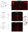
Fig. 5.. Inner ear central vestibular projections aberrantly target the hindbrain roof plate in e11.5 Lmx1a/b DKO embryos.
Whole mounted hindbrains (A-D”) and transverse hindbrain sections (A’-C’, E-H) from e11.5 embryos of indicated genotypes. Discrete sensory afferent projections were visualized by labeling distinct cranial nerves (indicated by Roman numbers) with lipophilic dyes. V - trigeminal nerve, dV - descending trigeminal tract, VII - facial nerve, VIII – inner ear vestibular nerve, IX - glossopharyngeal nerve, X - vagus nerve, XII - hypoglossal nerve. (A-C, A’-C’) In control, Lmx1a KO, and Lmx1b KO embryos, different afferent projections run parallel to the anterior-posterior axis of the hindbrain, do not significantly overlap with each other, and do not cross the dorsal midline occupied by the wide choroid plexus epithelium derived from the IVth ventricle roof plate. (D-D”) Dorsal whole mount view showing that in Lmx1a/b DKO embryos, inner ear vestibular (VIII), trigeminal (V), and solitary tract (ST) afferents extend aberrantly close to or cross the dorsal midline. Arrow (D-D”) shows location of the dorsal midline, which is heavily populated by inner ear vestibular (VIII) afferent fibers. D’ and D” are high power views of D. (A’-C’, E-H) Transverse hindbrain sections showing that similar to control, Lmx1a KO and Lmx1b KO embryos, in Lmx1a/b DKO hindbrain, the descending trigeminal tract (dV) was located ventral to inner ear vestibular (VIII) fibers. However, in contrast to control, Lmx1a KO and Lmx1b KO embryos (A’-C’), inner ear vestibular (VIII) fibers extended to and populated the dorsal midline roof plate (RP) covering the central canal in Lmx1a/b DKO littermates (E-H). Note the near normal location of facial branchial motor neurons (FBM) and ventral motor neurons (Vm) in the Lmx1a/b DKO (E, F) near the floor plate (FP), and solitary tract afferents crossing the roof plate (G). G is a high power view of E, without the magenta channel; panels F and H show two different sections from another embryo. Scale bar: 100 μm.
Fig. 6.
Fig. 6.. In e12.5 Lmx1a/b DKO mutants, inner ear central projections from left and right ears interdigitate but do not fuse at the hindbrain dorsal midline.
Dorsal view of whole mount hindbrain (A-B’) or transverse sections (C-I) of e12.5 embryos. Selected afferents were labeled with dye (A-C, E-G) or anti-Neurofilament/anti-acetylated α-Tubulin immunohistochemistry (D, H, I). Cranial nerves are indicated by Roman numbers. (A, C) Selected ipsilateral afferents labeled with dye (cranial nerves VII/VIII are green, nerves dV/VI are red, and nerves IX/X/XII are lilac. In control embryos, beyond r1-derived cerebellum (CB) (A), vestibular inner ear (cranial nerve VIII) and trigeminal (dV) projections do not come close to the dorsal midline. (B-G) Labeling of the trigeminal (lilac), inner ear (red) and facial/inner ear projection from the opposite side (green) in a Lmx1a/b DKO embryo. B’ shows higher magnification of B. Panels F and G show consecutive sections from the same embryo, Panel E shows a different embryo. All these nerves, after entering the brainstem, aberrantly extend across the roof plate (RP) that occupies the brainstem dorsal midline. Afferents from the two ears clearly interdigitate at the roof plate but do not fuse (B, B’ F, G). In the double mutant, afferents of the cranial nerve VII-derived solitary tract and trigeminal afferents cross the roof plate below the vestibular inner ear nerve VIII (F, G). In contrast to pathfinding errors of afferents that normally target the alar plate, facial branchial motor neurons (FBM) and inner ear efferents (IEE) were appropriately located in the basal plate of e12.5 Lmx1a/b DKO embryos (E). (D, H, I) In Lmx1a/b DKO mutants, fiber bundles crossing the roof plate can also be visualized using anti-Neurofilament (red)/anti-acetylated α-Tubulin (green) immunohistochemistry. Hoechst counterstaining is blue (H, I). Panels H and I show sections from two different embryos. No comparably located fibers were detected in control embryos (D). Note that because the IVth ventricle does not properly form in Lmx1a/b DKO embryos, the double mutant overall brainstem morphology is somewhat similar to the spinal cord (E-I). Scale bars: 1 mm (A), 100 μm (all other panels).
Fig. 7.
Fig. 7.. Anterior-posterior expansion of inner ear central projections is disrupted in e12.5 Lmx1a/b DKO embryos.
In all panels, anterior is left, posterior is right, dorsal is up, ventral is down. (A-D) Side view of dissected whole mount brains. Overall brainstem morphology is similar in control, Lmx1a KO and Lmx1b KO embryos (A-C). While the overall shape and size of the caudal brainstem show no obvious deviations in Lmx1a/b DKO embryos, their cerebellum (CB) and midbrain are reduced in size (D). (E-H) Anti-Neurofilament immunohistochemistry shows the presence and appropriate location of all cranial nerve roots (V-XII) in Lmx1a KO, Lmx1b KO and Lmx1a/b DKO embryos, suggesting that overall anterior-posterior hindbrain patterning is not grossly disrupted in these mutants. (I-P) Dye labeling of cranial nerve V (red), VII/VIII (green) and IX (lilac). Panels I-L show co-imaging of three different colors, while panels M-P – only green labeling to better visualize defects in vestibular (nerve VIII) projections. No gross deviation of overall projections in Lmx1a KO (J, N) and Lmx1b KO (K, O) embryos was detected compared to controls (I, M). In contrast, Lmx1a/b DKO embryos show limited posterior expansion of ear vestibular projections (nerve VIII) (L, P, arrow) and an abnormal trigeminal (nerve V) expansion into the topographical equivalent of r1/cerebellum (L). Scale bars: 1mm (A-D), 100 μm (E-P).
Fig. 8.
Fig. 8.. Ectopic expression of Wnt3a in the hindbrain roof plate in Lmx1a/b DKO embryos.
Transverse sections of e12.5 hindbrain stained with Wnt3a RNAscope in situ hybridization. Sections are taken at the level shown by dashed blue line in whole mount brain diagrams. Sections shown in panels A and B are taken at the level of otic vesicles (ov, r4). Section in panel C is posterior to otic vesicles. Tissue morphology on transverse sections is illustrated adjacent to each whole mount diagram. Note, that in contrast to control embryos, in Lmx1a/b DKO embryos, the hindbrain does not possess a large ventricle and shows a spinal cord-like morphology. In Lmx1a/b DKO embryos, the IVth ventricle roof plate (RP) still occupies the dorsal midline but does not differentiate into the choroid plexus epithelium (ChPe) that covers the IVth ventricle in control embryos (Mishima et al., 2009). (A) In control embryos, Wnt3a is highly expressed in the RL (arrowhead) adjacent to the ChPe but not in the ChPe itself. (B-D) In Lmx1a/b DKO embryos, at the level of otic vesicles, a smaller ectopic Wnt3a expression domain was located in the dorsal midline roof plate (RP, arrowhead) (B). At more posterior levels, Wnt3a expression was virtually absent, appearing as just a few dots in the roof plate (open arrowhead) (C). (D) Quantification of RNAscope in situ hybridization signal confirmed a significant reduction of Wnt3a expression in the posterior hindbrain roof plate relative to the roof plate at the otic vesicles level. n=3 Lmx1a/b DKO embryos, *** p<0.001. Scale bar: 200 μm.
Fig. 9.
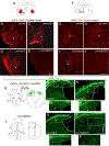
Fig. 9.. Simultaneous, but not individual, loss of Lmx1a and Lmx1b prevents the development of excitatory neurons in cochlear and vestibular nuclei.
Pax6 (B-E), Tbr1 (G-J), and Atoh1 (M-P’) immunostained transverse sections of the hindbrain at indicated stages. High magnification panels correspond to regions boxed in adjacent diagrams (A, F, K, L) or low magnification panels. (A-E) Numerous Pax6+ neurons were present in the cochlear nuclei (CN) of wild type control, Lmx1a KO, and Lmx1b KO mutants (arrowheads) but were not detected in Lmx1a/b DKO embryos. (F-J) Tbr1+ neurons were present in the superior vestibular nuclei (SuVe) of wild type control, Lmx1a KO, and Lmx1b KO mutants (arrowheads) but were not detected in Lmx1a/b DKO embryos. (K, L) Diagrams illustrating location of sections showing lower RL in control and Lmx1a/b mutant embryos (M-P). Sections were taken at the level of otic vesicles (ov, blue dashed line in whole mount diagrams in K, L). Tissue morphology is illustrated in right diagrams in K and L. (M-P’) Numerous Atoh1+ progenitors were present in the lower RL of wild type control, Lmx1a KO and Lmx1b KO mutants (arrowheads) but not in Lmx1a/b DKO embryos. Scale bar: 100 μm.
Fig. 10.
Fig. 10.. Simultaneous loss of Lmx1a and Lmx1b disrupts the inner ear, central projections and cochlear/vestibular brainstem nuclei.
(A, A’) Control mice have a large IVth ventricle covered by a choroid plexus. Specific nuclei develop in the alar and basal plate of the hindbrain. CN – cochlear nuclei, which develop in r2–5, VestN – vestibular nuclei, which extend from the caudal cerebellar vermis to the obex, SN – solitary tract nuclei, Vsn – viscerosensory (trigeminal) nuclei, FBM – facial branchial motor neurons, Vmn – ventral motor neurons. Each set of nuclei receives distinct sensory afferents. For example, auditory information is provided by spiral ganglion neuronal projections to cochlear nuclei. (B, B’) In Lmx1a/b DKO mice, excitatory neurons of cochlear and vestibular nuclei do not develop. Segregated projections to viscerosensory trigeminal nuclei (Vsn) and solitary tract nuclei (SN) were detected in these mice. Inner ear vestibular afferents project to and cross the roof plate in r4, with limited caudal extension. Similar to inner ear afferents, some solitary tract and trigeminal fibers project to the roof plate. In contrast, basal plate development and innervation (motor neurons) were not significantly affected in Lmx1a/b DKO mice. (A”, B”) Organ of Corti (OC), spiral ganglion (red oval near the ear in control mice) and spiral ganglion projections were not detected in Lmx1a/b DKO mice. In contrast, although altered in size or shape, vestibular inner ear components (AC, HC, U, S, PC and vetibular ganglion - pink circle near the ear) still develop in Lmx1a/b DKO embryos.

Similar articles

Cited by

References

    1. Abello G, Khatri S, Radosevic M, Scotting P, Giraldez F, Alsina B (2010) Independent regulation of Sox3 and Lmx1b by FGF and BMP signaling influences the neurogenic and non-neurogenic domains in the chick otic placode. Developmental Biology 339:166–178. - PubMed
    1. Boëda B, El-Amraoui A, Bahloul A, Goodyear R, Daviet L, Blanchard S, Perfettini I, Fath KR, Shorte S, Reiners J, Houdusse A, Legrain P, Wolfrum U, Richardson G, Petit C (2002) Myosin VIIa, harmonin and cadherin 23, three Usher I gene products that cooperate to shape the sensory hair cell bundle. EMBO J 21:6689–99. - PMC - PubMed
    1. Bouchard M, de Caprona D, Busslinger M, Xu P, Fritzsch B (2010) Pax2 and Pax8 cooperate in mouse inner ear morphogenesis and innervation. BMC Developmental Biology 10:89. - PMC - PubMed
    1. Butts T, Green MJ, Wingate RJ (2014) Development of the cerebellum: simple steps to make a ‘little brain’. Development 141:4031–4041. - PubMed
    1. Chang C-H, Tsai R-K, Tsai M-H, Lin Y-H, Hirobe T (2014) The roles of Frizzled-3 and Wnt3a on melanocyte development: in vitro studies on neural crest cells and melanocyte precursor cell lines. Journal of Dermatological Science 75:100–108. - PubMed

Publication types

Substances

LinkOut - more resources