Review
. 2020 Nov;10(11):200302.
doi: 10.1098/rsob.200302. Epub 2020 Nov 25.

Phosphoglycerate kinase: structural aspects and functions, with special emphasis on the enzyme from Kinetoplastea

Affiliations
Review

Phosphoglycerate kinase: structural aspects and functions, with special emphasis on the enzyme from Kinetoplastea

Maura Rojas-Pirela et al. Open Biol. 2020 Nov.

Abstract

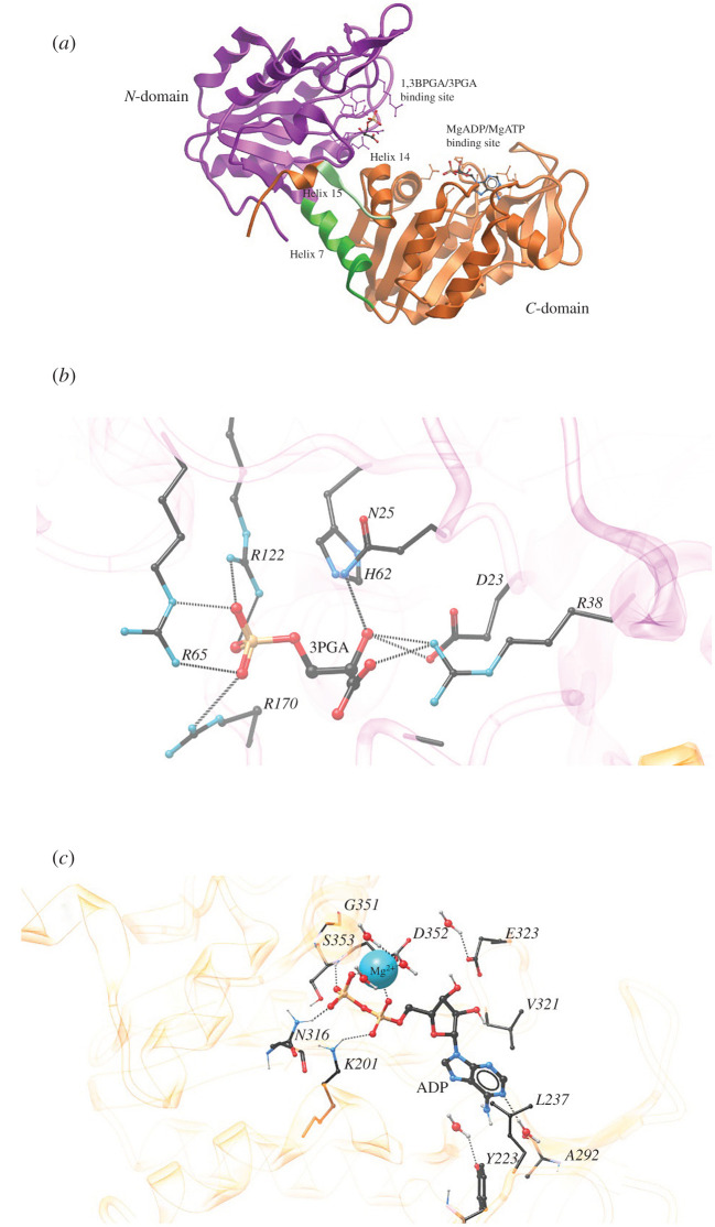
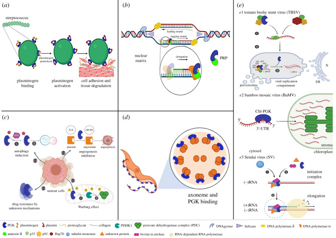
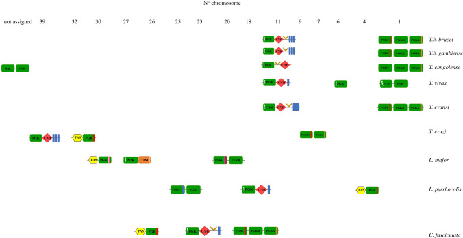
Phosphoglycerate kinase (PGK) is a glycolytic enzyme that is well conserved among the three domains of life. PGK is usually a monomeric enzyme of about 45 kDa that catalyses one of the two ATP-producing reactions in the glycolytic pathway, through the conversion of 1,3-bisphosphoglycerate (1,3BPGA) to 3-phosphoglycerate (3PGA). It also participates in gluconeogenesis, catalysing the opposite reaction to produce 1,3BPGA and ADP. Like most other glycolytic enzymes, PGK has also been catalogued as a moonlighting protein, due to its involvement in different functions not associated with energy metabolism, which include pathogenesis, interaction with nucleic acids, tumorigenesis progression, cell death and viral replication. In this review, we have highlighted the overall aspects of this enzyme, such as its structure, reaction kinetics, activity regulation and possible moonlighting functions in different protistan organisms, especially both free-living and parasitic Kinetoplastea. Our analysis of the genomes of different kinetoplastids revealed the presence of open-reading frames (ORFs) for multiple PGK isoforms in several species. Some of these ORFs code for unusually large PGKs. The products appear to contain additional structural domains fused to the PGK domain. A striking aspect is that some of these PGK isoforms are predicted to be catalytically inactive enzymes or 'dead' enzymes. The roles of PGKs in kinetoplastid parasites are analysed, and the apparent significance of the PGK gene duplication that gave rise to the different isoforms and their expression in Trypanosoma cruzi is discussed.

Keywords: Trypanosoma; domains; metabolism; moonlighting protein; phosphoglycerate kinase; protists.


Conflict of interest statement

We declare we have no competing interests.

Figures

Figure 1.
Figure 1.
Phylogenetic relationships of the eukaryotes. LECA is the ‘last eukaryotic common ancestor’ from which different eukaryotic supergroups evolved [6]. The main protistan organisms discussed in this paper are indicated by coloured dots.
Figure 2.
Figure 2.
Evolutionary relationships among Kinetoplastea. Outgroups for the construction of the tree (based on small subunit ribosomal RNA sequences) are D. papillatum and E. gracilis, which, together with the Kinetoplastea, belong to the clade Euglenozoa (figure 1). Figure based on [16].
Figure 3.
Figure 3.
Three-dimensional structure of phosphoglycerate kinase. (a) Ribbon representation of the overall structure of pig muscle PGK (PDB: 1HDI). In colour is highlighted the N-domain (violet); helix 7 or interdomain helix (green); the link between helix 14 and 15; amino acids 404–408 (light green), and the C-domain (orange). The substrate binding sites for 1,3BPGA/3PGA and MgADP/MgATP are indicated in both the N- and C-domain, respectively. The pig muscle ternary complex shown here exhibits an open conformation in comparison with the ternary complex of other PGK structures [46]. (b) Substrate 3PGA binding site at the N-terminal domain of pig muscle PGK. (c) The MgADP/MgATP binding site of B. stearothermophilus (bacterium later renamed to Geobacillus stearothermophilus) for the ligand MgADP (PDB: 1PHP). In both (b,c), interactions between amino acid residues and substrate through hydrogen bonds (dashed lines) are shown. Atom colour code: black (carbon); white (hydrogen); red (oxygen); blue (nitrogen), orange (phosphorus). Ion colour code: cyan (magnesium, Mg2+).
Figure 4.
Figure 4.
PGK moonlighting functions. (a) Cell invasion: PGK is located at the surface of some pathogenic bacteria where the processing of plasminogen to plasmin is promoted. (b) DNA replication: Together PGK and Annexin II protein constitute the Primer Recognition Particle (PRP) localized in the nuclear matrix. (c) Tumour growth and cancer progression: C.1. PGK is secreted from cancer cells. In the extracellular space, PGK allows the conversion of plasmin into angiostatin and the subsequent inhibition of angiogenesis. C.2. PGK phosphorylated at residue S203 is translocated into mitochondria to activate PDHK1 by phosphorylation (I). PDHK1 phosphorylates and inhibits the catalytic activity of the pyruvate dehydrogenase complex (PDC) and downstream metabolic reactions in mitochondria (II). C.3. Overexpression of PGK involved in drug resistance. C.4. PGK posttranslational modification (acetylation of residue K388) by ARD1 (I) induces PGK activation and subsequent phosphorylation of intermediate proteins that finally switch on the autophagy mechanism (II). Acetylation of other K residues contributes to increasing the glycolytic flux of tumour cells. (d) Functions associated with the flagellum. (e) Viral replication in TBSV: PGK and other host proteins are involved in viral mechanisms and able to bind to viral RNA. PGK also provides local ATP required in viral replication. (e.2) The bamboo mosaic virus (BaMV): chloroplast PGK (chlPGK) interacts with 3′-UTR of Viral RNA to direct the translocation of viral RNA from the cytosol to the chloroplast and allow its accumulation in the stroma. (e.3). In Sendai virus (SV): PGK participates in the initiation complex and stimulates viral gene transcription.
Figure 5.
Figure 5.
Genomic location of pgk genes in different species of Kinetoplastea. Determination of the chromosomal localization of identified pgk genes was performed using the Kinetoplastid Genomic Resource (TriTrypDB) [94]. Identification of domains and motifs present in PGK sequences was done using the following bioinformatics servers: PAS domain: Protein Blast (NCBI) [96], InterProScan (EMBL-EBI) [95,97] and Simple Modular Architecture Research Tool (SMART) [98]; CNB domain: NCBI, EMBL-EBI, SMART; HTH (Helix–Turn–Helix) motif: ExPaSy [99], GYM [100], iDNA-Prot [101]; Membrane helix: SMART [98]; Predicting transmembrane protein topology with a hidden Markov model (TMHMM prediction) [102] and Phobius [103]. Glycosomal localization prediction was based on recognition of a PTS1 consensus sequence as reported by Acosta et al. [104]. Domain symbology: PGK domain formula image, PGK domain having lost substrate (3PGA)-binding residues formula image, triosephosphate isomerase domain formula image, HTH domain formula image, transmembrane segment formula image, PAS domain formula image, cyclic-nucleotide binding-domain formula image. Import sequences and inserts identified in PGK isoenzymes through experimental and theoretical studies: putative PTS1 formula image, putative lysosome formula image, PGKA-like insert sequence formula image.
Figure 6.
Figure 6.
Kinetoplastid phylogenetic tree based on PGK sequences. PGK gene sequences were retrieved from the TriTrypDB, GeneDB and GenBank databases [94,123,360], and the predicted amino acid sequences aligned using Muscle (EMBL-EBI) [95]. Regions of uncertain alignment were omitted from the analysis leaving a total of 57 sequences (19 taxa) and 268 amino acid positions. The evolutionary history was inferred using the neighbour-joining (NJ) and the maximum parsimony (MP) methods [361], with similar results. The branches of the tree represent sequences, orthologous and paralogous, from different members of the Kinetoplastea phylum, with nodes depicted as filled blue ovals representing gene duplication events. Support for the nodes in the NJ and MP analyses was evaluated using the bootstrap procedure, and in each case, the bootstrap consensus tree inferred from 1000 replicates [362] was taken to represent the evolutionary history of the taxa analysed [362]. Branches corresponding to partitions reproduced in less than 50% bootstrap replicates are collapsed. The percentage of replicate trees in which the associated taxa clustered together in the bootstrap test (1000 replicates) are shown next to the branches [362]. For the NJ analysis, the evolutionary distances were computed using the p-distance method [363] and are given in the units of the number of amino acid differences per site. This analysis involved 58 amino acid sequences. All positions containing gaps and missing data were eliminated (complete deletion option). There were a total of 268 positions in the final dataset. For the MP analysis branches corresponding to partitions reproduced in less than 50% bootstrap replicates are collapsed. The percentages of replicate trees in which the associated taxa clustered together in the bootstrap test (1000 replicates) are shown next to the branches [361]. The MP tree was obtained using the subtree-pruning-regrafting (SPR) algorithm ([362], p. 126)] with search level 1 in which the initial trees were obtained by the random addition of sequences (10 replicates). This analysis involved 57 amino acid sequences. There were a total of 391 positions in the final dataset. Evolutionary analyses by NJ and MP were conducted in MEGA X [364].

Similar articles

Cited by

References

    1. Cicenas J, Zalyte E, Bairoch A, Gaudet P. 2018. Kinases and cancer. Cancers 10, 63 (10.3390/cancers10030063) - DOI - PMC - PubMed
    1. Rauch J, Volinsky N, Romano D, Kolch W. 2011. The secret life of kinases: functions beyond catalysis. Cell Commun. Signal. 9, 23 (10.1186/1478-811X-9-23) - DOI - PMC - PubMed
    1. Parsons M, Worthey EA, Ward PN, Mottram JC. 2005. Comparative analysis of the kinomes of three pathogenic trypanosomatids: Leishmania major, Trypanosoma brucei and Trypanosoma cruzi. BMC Genomics 6, 127 (10.1186/1471-2164-6-127) - DOI - PMC - PubMed
    1. Rojas-Pirela M, Rigden DJ, Michels PA, Cáceres AJ, Concepción JL, Quiñones W. 2018. Structure and function of Per-ARNT-Sim domains and their possible role in the life-cycle biology of Trypanosoma cruzi. Mol. Biochem. Parasitol. 219, 52–66. (10.1016/j.molbiopara.2017.11.002) - DOI - PubMed
    1. Jackson AP, et al. 2016. Kinetoplastid phylogenomics reveals the evolutionary innovations associated with the origins of parasitism. Curr. Biol. 26, 161–172. (10.1016/j.cub.2015.11.055) - DOI - PMC - PubMed

Publication types

Substances

LinkOut - more resources